INTRODUCTION
Cytochrome P450 is the most important enzyme in the
body and involved in the biotransformation of endog-enous substances and exogendog-enous chemicals (Dey et al., 1999). In particular, cytochrome P450 1A2 (CYP1A2) is
Immunohistological expression of cytochrome
P450 1A2 (CYP1A2) in the ovarian follicles of
prepubertal and pubertal rat
Jong-Chan Hwang
1, Byung-Joon Park
1, Hwan-Deuk Kim
1,2, Su-Min Baek
1, Seoung-Woo Lee
1,
Ryoung-Hoon Jeon
3, Min Jang
1, Seul-Gi Bae
1, Sung-Ho Yun
1, Jin-Kyu Park
1, Young-Sam Kwon
1,
Seung-Joon Kim
1and Won-Jae Lee
1,*
1College of Veterinary Medicine, Kyungpook National University, Daegu 41566, Korea
2Department of Veterinary Research, Daegu Metropolitan City Institute of Health & Environment, Daegu 42183, Korea 3College of Veterinary Medicine, Gyeongsang National University, Jinju 52828, Korea
Original Article
pISSN: 2671-4639 • eISSN: 2671-4663
https://doi.org/10.12750/JARB.35.4.329
JARB
Journal of Animal Reproduction and Biotechnology
Received December 3, 2020 Revised December 11, 2020 Accepted December 14, 2020 *Correspondence Won-Jae Lee E-mail: iamcyshd@knu.ac.kr ORCID https://orcid.org/0000-0003-1462-7798
ABSTRACT Cytochrome P450 1A2 (CYP1A2) is a member of the cytochrome P450 superfamily enzymes in mammals and plays a major role in metabolizing endogenous hormones in the liver. In recent days, CYP1A2 expression has been found in not only the liver but also other tissues including the pancreas and lung. However, little information is available regarding the expression of CYP1A2 in the ovary, in spite of the facts that the ovarian follicle growth and atresia are tightly associated with controls of endocrine hormonal networks. Therefore, the expression of CYP1A2 in the ovaries of prepubertal and pubertal rats was investigated to assess its expression pattern and puberty-related alteration. It was demonstrated that the expression level of CYP1A2 was significantly (p < 0.01) higher in the pubertal ovaries than prepubertal counterparts. At the ovarian follicle level in both groups, whereas CYP1A2 expression was less detectable in the primordial, primary and secondary follicles, the strongly positive expression of CYP1A2 was localized in the granulosa cell layers in the antral and pre-ovulatory follicles. However, the ratio of CYP1A2-positive ovarian follicle was significantly (p < 0.01) higher in the ovary of pubertal group (73.1 ± 3.1%) than prepubertal one (41.0 ± 10.5%). During the Immunofluorescence, expression of CYP1A2 was mainly localized in Fas-positive follicles, indicating the atretic follicles. In conclusion, these results suggested that CYP1A2 expression was mainly localized at the atretic follicular cells and affected by the onset of puberty. Further study is still necessary but we hypothesize that CYP1A2 expresses in the atretic follicles to metabolize residue of the reproductive hormones. These findings may have important implications for the fields of reproductive biology of animals.
Keywords: cytochrome P450 1A2, follicle atresia, ovary, puberty
a member of the cytochrome P450 superfamily enzymes in mammals as the mono-oxygenase and plays a major role in biotransformation of a wide range of exogenous chemicals, pollutants and drugs via hydroxylation, de-methylation, epoxidation, oxidation and quinol formation of compounds (Dey et al., 1999; Sugiyama et al., 2019; Lu et al., 2020). Furthermore, it primarily metabolizes endogenous hormones with respect to progesterone to 6β-hydroxyl progesterone by hydroxylation, estrone to 2- or 4-hydroxyestrone by hydrolysis and estradiol to es-triol by hydroxylation in humans (Mikhailova et al., 2006; Sugiyama et al., 2019; Lu et al., 2020). CYP1A subfamily contains only two functional isoforms as CYP1A1 and CY-P1A2, and it has been addressed that CYP1A2 is expressed mainly in the liver; whereas the expression of CYP1A1 is mainly distributed outside the liver and involves the me-tabolism for aromatic hydrocarbon, CYP1A2 is expressed in the liver and metabolizes the aromatic amines and heterocyclic compounds (Wijnen et al., 2007; Lee et al., 2012; Lu et al., 2020). However, in recent days, the ex-pression of CYP1A2 is identified in the pancreas and lung as well (Vukovic et al., 2016). Likewise, the detection of CYP1A2 expression in other tissues has been investigating to understand its function at each organ.
Puberty is regarded as the developmental stage when reproductive capacity is accomplished and sexual matura-tion completed under controls of complex endocrine net-works, known as the hypothalamic-pituitary-gonadal (HPG) axis; it has been known that several hormones such as kis-speptin, gonadotropin-releasing hormone (GnRH), follicle-stimulating hormone (FSH), luteinizing hormone (LH) and estradiol are tightly associated to the puberty-related phys-ical changes (Mayer et al., 2010; Uenoyama et al., 2018; Kim, 2019). In rodents, external signs of puberty with re-spect to vaginal opening (VO) and cornification of vaginal epithelial cells are highly associated with the mature ovary and first ovulation due to the rise in estradiol levels. There-fore, VO in the rodent is regarded as a reliable marker for the onset of puberty and its observation is widely used be-cause of non- or minimally-invasive procedures to assess the onset of puberty (Gaytan et al., 2017). Of particular, it is reported that VO in Spraque Dawley (SD) rat occurs on postnatal day (PND) 37-39, indirectly indicating the onset of puberty and the first ovulation (Rivest, 1991).
The follicular atresia is defined as the breakdown of the ovarian follicles including the oocyte, granulosa cells and
internal/external theca cells, and its apoptotic process is hormonally controlled throughout a female mammals’ life; the limited numbers of developing follicles, less than 1%, are allowed for ovulation but the rest, remaining 99%, undergo atresia at the various developing stages of ovar-ian follicle (Yu et al., 2004; Zhou et al., 2019). Among component of the ovarian follicle, survival or death of the granulosa cells plays an important role in normal follicle development with serving essential molecules for fol-licular growth/maintenance or follicle atresia related with hormonal imbalance between estrogen and progesterone in the follicular fluid, respectively (Yu et al., 2004; Li et al., 2016). In particular, deprivation of growth factors or activation of cell death molecules in regards to death li-gands/receptors, pro-apoptotic protein and cytokines are regarded as the main cause of apoptosis of the granulosa cells; the death molecules include Fas, Fas ligand (FasL), tumor necrosis factor (TNF)-α, TNF receptor (TNFR), TNF-α related apoptosis inducing ligand (TRAIL), TRAIL receptor (TRAILR) and Bcl-2 protein family (Matsuda et al., 2012; Zhou et al., 2019).
Although CYP1A2 is mainly expressed as a hepatic en-zyme for metabolism of endogenous hormones, it can potentially present in other tissues. Specially, little infor-mation is available regarding the expression of CYP1A2 in the female reproductive system, in spite of the fact that the ovarian cycle is tightly and directly related with re-productive hormones. Moreover, the alteration of ovarian CYP1A2 expression depending on the onset of puberty is not understood yet. Therefore, the primary objective of the present study is to clarify the expression of CYP1A2 in the prepubertal and pubertal ovaries of female SD rats with observing different ovarian follicle stages.
MATERIALS AND METHODS
Ethics statement
All procedures for animal experiments were approved by the Institutional Animal Care Use Committee at Kyung-pook National University (approval number: KNU2019-0159).
Chemicals and media
All chemicals and media were purchased from Thermo Fisher Scientific (Waltham, MA, USA), unless otherwise specified.
Animal housing and experiment groups
The 21-day-old (PND 21) female SD rats (mean body weight: 58.1 g; total n = 16) were housed in the plastic cages (40 × 32 × 17 cm; 3-4 animals/cage), maintained for temperature at 23 ± 2℃, humidity at 50-80% and ap-proximately 12 h light/dark cycle. The standard feed (Jeil Feed Co., Ltd., Daejeon, Korea) and tap water were pro-vided ad libitum. The SD rats were acclimatized for 3 days (PND 24). Then the rats were sacrificed at PND 24 or Day 51 as prepubertal group (n = 8) or pubertal group (n = 8), respectively.
Sacrifice of animals and sampling
The animals were anesthetized with isoflurane inhala-tion in the closed chamber and exsanguinated via the right atrium. The ovary and liver tissues of each group were collected, trimmed, snap-frozen into liquid nitro-gen (LN2) and stored in deep freezer (-80℃) for western
blotting (each n = 5) or fixed with 4% paraformaldehyde (Duksan chemical, Korea) for hematoxylin & eosin stain-ing (H&E) and immunohistochemistry (IHC) (each n = 3).
Body weight and vaginal opening assessment
Before sacrificing the animals, body weight (BW) and vaginal opening (VO) of all animals was assessed by us-ing a weighus-ing machine or recognizus-ing the vaginal hole over the urethra in accordance with the previous article, respectively (Rivest, 1991).
Western blotting for quantitative expression of CYP1A2
The western blotting was conducted in accordance with the previous article to assess CYP1A2 expression in the ovary and liver (positive control) (Lee et al., 2019). The snap-frozen ovaries and livers were homogenized, lysed with a radioimmunoprecipitation assay (RIPA) buffer supplemented a proteinase inhibitor and centrifugated at 14,000 rpm for 5 min at 4℃. The supernatants were carefully collected and quantified for the total amount of protein using a Bicinchoninic Acid Protein Assay Reagent Kit. The sodium dodecyl sulfate-polyacrylamide gel elec-trophoresis (SDS-PAGE) was conducted using 10 µg of total protein for protein separation. The gels were trans-ferred onto polyvinylidene difluoride (PVDF) membranes (Millipore, MA USA), followed by blocking the membranes with 3% bovine serum albumin (BSA) for 1 h at room tem-perature (RT). Thereafter, membranes were incubatedwith a mouse polyclonal anti-CYP1A2 antibody (1:500 dilution with 1% BSA; Santa Cruz Biotechnology, TX, USA) for overnight at 4℃. As a reference, a mouse polyclonal anti-Glyceraldehyde 3-phosphate dehydrogenase (anti-GAPDH; 1:1,000 dilution with 1% BSA) was involved. The membranes were then washed with tris buffer saline con-taining 0.1% tween-20 (TBST), incubated with a horserad-ish peroxidase conjugated goat anti-mouse IgG (1:3,000 dilution with TBST) for 1 h at RT and developed on X-ray films (AGFA, Belgium) using an enhanced chemilumines-cence (ECL) kit. Image J software (National Institutes of Health, USA) was employed for quantitative analysis of the intensities of developed bands. The expression level of CYP1A2 was relatively normalized against that of GAPDH.
Hematoxylin & eosin staining and immunohistochemistry
The preparation for histological observation was fol-lowed with the previous article (Kim, 2019). In brief, fixed ovaries from each group were dehydrated, embedded in paraffin and sectioned into 5 mm thick using a micro-tome (Leica Microsystems, Germany). The H&E staining was then conducted in a routine manner to observe the presence of the corpus lutea. In case of IHC, the slide sections were deparaffinized, treated with 0.01 M citrate buffer (pH 6.0) at 95℃ for 30 min for antigen retrieval, cooled at RT for 60 min, treated with 3% hydrogen per-oxide for 30 min, washed with phosphate buffered saline with triton-X (PBST) and blocked with 2% normal horse serum (Histostain-plus kit) for 1 h at RT. Thereafter, the slides were incubated with a mouse polyclonal anti-CYP1A2 antibody (1:50 dilution with 2% normal horse serum) at 4℃ for overnight, incubated with a biotinylated secondary antibody (Histostain-plus kit; 1:200 dilution with PBS) at RT for 90 min, treated with an ABC solution (Histostain-plus kit) at RT for 60 min, reacted with a 3,3’ -diaminobenzidine (DAB) kit (Vector Laboratories, CA, USA) and counterstained with hematoxylin; all slides were reacted with DAB for equal time. When the slides were observed using Moticam Pro 20A (Motic, Hong Kong), brownish color on the tissue was determined as positive expression of CYP1A2.Classification of ovarian follicles
The ovarian follicles in IHC were classified, in accor-dance with the previous article in regards to the primary follicles (a single layer of cuboidal granulosa cells),
sec-ondary follicles (surrounding of more than one layer of cuboidal granulosa cells without visible antrum), antral follicles (multiple layer of granulosa cells and 1-2 small spaces of antrum) and pre-ovulatory follicles (the largest follicle with cumulus granulosa cell layer surrounding the oocyte) (Kim, 2019).
Immunofluorescence
To identify the association between CYP1A2 expression and the follicle atresia, immunofluorescence with CYP1A2 and Fas, a common apoptosis inducer, was conducted. Preparation of slides until antigen retrieval step was con-ducted in a same manner with IHC. Then the slides were washed with PBST, blocked with 2% normal goat serum (Vector Laboratories) for 1 h at RT and incubated with a mouse polyclonal anti-CYP1A2 antibody (1:50 dilution with 2% normal goat serum) supplemented with a rab-bit polyclonal anti-Fas antibody (1:1,000 dilution with 2% normal goat serum) at 4℃ for overnight. The slides were washed with PBS, incubated with a goat anti-mouse IgG conjugated with Alexa Fluor 594 (1:1,000 dilution with PBS) supplemented with a goat anti-rabbit conjugated with Alexa Fluor 488 (1:1,000 dilution with PBS) at RT for 60 min, counterstained with 4′,6-diamidino-2-phenylin-dole (DAPI) for 5 min and mounted with VectaShield (Vec-tor Labora(Vec-tories). The slides were observed in fluorescent microscope (Moticam Pro 20A) at the proper wavelengths.
Statistical analysis
Student’s T-test or Kruskal-Wallis test with Bonferroni correction was applied using PASW Statistics 18 (SPSS Inc., Chicago, IL, USA). A p value of < 0.01 was consid-ered to be statistically significant.
RESULTS
Confirmation of prepuberty and puberty
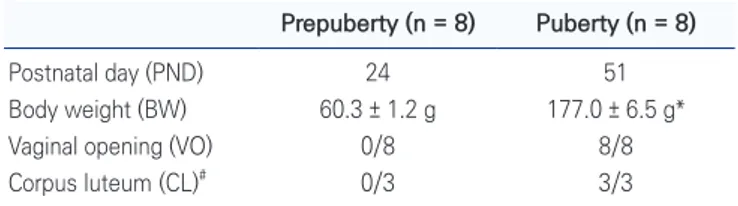
In accordance with previous article, SD rats in PND 24 or 51 of the present study were expected as prepubertal or pubertal periods, respectively (Rivest, 1991). In addi-tion, BW and VO were assessed prior to sacrificing ani-mals (Table 1). Prepubertal group (60.3 ± 1.2 g) exhibited significantly (p < 0.01) lower BW than pubertal group (177.0 ± 6.5 g). All animals in prepubertal group (0/8) did not present VO but all did in pubertal group (8/8), indirectly indicating the onset of puberty. In case of H&E staining, the corpus lutea, implying the ovulation after the onset of puberty, were not observable in prepubertal group (Fig. 1A) but revealed in pubertal group (red arrows in Fig. 1B). Therefore, it was confirmed that each group presented the puberty-related features in consistent with their ages.
Western blotting for quantitative assessment of
CYP1A2 expression
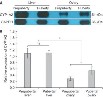
The western blotting was conducted to assess quan-titative expression of CYP1A2 in the ovaries; because CYP1A2 was mainly known as a hepatic enzyme, the liver tissue samples from prepubertal and pubertal groups were also employed in this assay as positive controls (Fig. 2A). Overall, the expression level of CYP1A2 in the ovaries was significantly (p < 0.01) weaker than that of the livers, regardless of the onset of puberty (Fig. 2B). However, in case of CYP1A2 expression in the ovary, there was a sig-nificant (p < 0.01) upregulation of CYP1A2 expression in pubertal group than prepubertal group (Fig. 2B).
Table 1. Confirmation of prepuberty and puberty
Prepuberty (n = 8) Puberty (n = 8)
Postnatal day (PND) 24 51 Body weight (BW) 60.3 ± 1.2 g 177.0 ± 6.5 g* Vaginal opening (VO) 0/8 8/8 Corpus luteum (CL)# 0/3 3/3
The values are described as Mean ± SD (BW) or positive number/total number of animals (VO and CL). *Superscript indicates significant difference between groups (p < 0.01). #Histological assessment is conducted in three
animals from each group (n = 3).
B A
Prepuberty Puberty
Fig. 1. Confirmation of prepuberty and puberty. Representative image of the prepubertal ovary (A) and pubertal ovary with the corpus lutea (red arrows) (B). Magnification: ×40; Bars: 1,000 µm.
Immunohistochemistry of CYP1A2 in prepubertal and
pubertal ovaries
The expressions of CYP1A2 in both groups were ana-lyzed by IHC (Fig. 3). Although the ovaries of both groups expressed CYP1A2 at the cells surrounding follicles, the patterns were dependent on the stage of developing folli-cle and the onset of puberty. At both groups, the primor-dial, primary and secondary follicles exhibited the nega-tive expression of CYP1A2 (red arrows in Fig. 3A and 3D).
The CYP1A2-positive cells were observed from antral to pre-ovulatory follicles (blue arrows in Fig. 3B, 3C, 3E and 3F), and they were localized at the granulosa cell layer. When the CYP1A2-positive follicles were counted (%), sig-nificantly (p < 0.01) higher number of follicles expressed CYP1A2 in pubertal group (73.1 ± 3.1%) than prepu-bertal group (41.0 ± 10.5) (Table 2). Collectively, these results suggested that CYP1A2 was strongly expressed at the granulosa cells in developing follicles (antral and pre-ovulatory follicles) and affected by the onset of puberty.
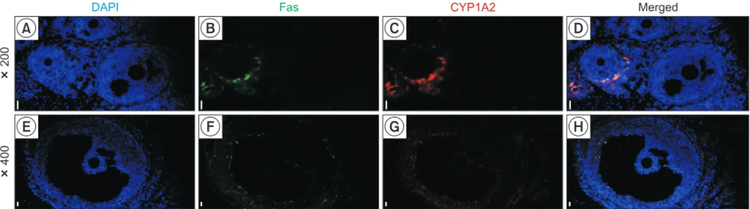
Immunofluorescence of CYP1A2 and Fas in the ovary
To clarify the relationship between the CYP1A2 expres-sion and atretic follicle, immunofluorescence assay of CYP1A2 and Fas was conducted in the ovary tissues (Fig. 4). As shown in Fig. 4A-4D, whereas the Fas-positive fol-licle, indicating the atretic folfol-licle, co-expressed CYP1A2 (follicle on the left), the Fas-negative follicle did not ex-press CYP1A2 (follicle on the right). In addition, the co-expression of Fas and CYP1A2 was evident in the granu-losa and theca cell layers of pre-ovulatory follicle stage (Fig. 4E-4H). Based on these findings, it was concluded that CYP1A2 specifically expressed in the atretic follicle.
Prepuberty
Puberty
Primary/Secondary Antral Pre-ovulatory
A B C
D E F
Fig. 3. Immunohistological expression of CYP1A2 in prepubertal and puber-tal ovaries. The negative expressions of CYP1A2 at the primordial, primary and secondary follicles in both groups (red arrows in A and D). The CYP1A2-positive cells, localized at the granulosa cells, from antral follicles (blue arrows in Fig. B and E) to pre-ovulatory follicles (blue arrows in C and F). Magnification: ×400 (A, B, D and E) or ×100 (C and F); Bars: 100 µm (A, B, D and E) or 1,000 µm (C and F).
Table 2. Counting for CYP1A2-positive follicles in prepubertal and pubertal ovaries
Prepuberty (n = 3) Puberty (n = 3)
CYP1A2-positive follicle (%) 41.0 ± 10.5 73.1 ± 3.1* The values are described as Mean ± SD. *Superscript indicates significant difference between groups (p < 0.01).
Prepubertal liver Pubertal liver Prepubertal ovary Pubertal ovary 1.8 1.6 1.4 1.2 1.0 0.8 0.6 0.4 0.2 0.0 Relative expression of CYP1A2 * CYP1A2 GAPDH 51 kDa 36 kDa Prepuberty Puberty Liver Ovary Puberty Prepuberty A B ns *
Fig. 2. Western blotting for assessment of CYP1A2 expression in prepubertal and pubertal group. Representative image of CYP1A2 expression in the ovary and liver (A). Graph for the expression level of CYP1A2 was presented as mean ± SEM (B). *p < 0.01 or ns indicated a significant difference or non-significance, respectively.
DISCUSSION
It has been addressed that CYP1A2 is mainly expressed at the liver and plays a role in biotransformation of ex-ogenous substances and metabolism of endex-ogenous hor-mones (Mikhailova et al., 2006; Sugiyama et al., 2019). However, recent articles suggest that CYP1A2 is also expressed in other tissues such as the pancreas and lung (Vukovic et al., 2016). Therefore, we studied the localiza-tion of CYP1A2 expression depending on the onset of puberty of rats. Here, we demonstrated that CYP1A2 ex-pression was mainly localized at the atretic follicular cells and affected by the onset of puberty.
The expression of CYP1A2 in the ovary has shown con-troversial and opposed results. Especially, some articles reported absence of CYP1A2 in the ovary. Via in situ hy-bridization, CYP1A2 mRNA was measurable in the liver at intact mouse and 3-methylcholanthrene (3MC; an Ah receptor ligand) could induce CYP1A2 mRNA expression in the liver, lung and duodenum; even though the ovaries were included as specimen in this study design, the ex-pression of CYP1A2 in the ovary was negative (Dey et al., 1999). In addition, when the ovarian expressions of CYP isoforms were investigated in rats under diestrus or proes-trus cycle, CYP1A2, CYP1A1, CYP2B1, CYP2C12, CYP2E1, CYP3A1, CYP3A2 and CYP4A1 were not detectable in the ovarian microsomes regardless of estrus cycles (Lee et al., 2012). In contrast, several articles have reported opposed results, detection of CYP1A2 expression in the ovary tis-sue. Positive expressions of CYP1A2 in ovaries and testes of domestic cats were identified using polymerase chain reaction (PCR) (Sugiyama et al., 2019). Moreover, the
ovarian surface epithelium (OSE) of mouse was positive for CYP1A2, CYP1A1, CYP1B1 and CYP2C29 expression via PCR assay (Symonds et al., 2006). In the present study, we aimed to clarify localization of CYP1A2 expression in prepubertal and pubertal ovaries in rats. In agreement with the latter two articles, expressions of CYP1A2 in the ovary of rat were identified (Fig. 2 and 3, and Table 2). Especially, the intensity and localization of CYP1A2 expression were dependent on the onset of puberty and the stage of folliculogenesis; the expressions were nega-tive at primordial, primary and secondary follicles at both prepubertal and pubertal groups, but strongly positive in the granulosa cells of antral and pre-ovulatory follicles. The reasons for the different findings in the present study compared to former two studies are still unknown, but such variations may be caused by differences of inducing substance, species, strain, age, gender and assay method (Dey et al., 1999; Symonds et al., 2006). For instance, hu-man-mouse difference in the tissue specificity of CYP1A2 expression was observed when 3MC induced CYP1A2 ex-pression in mouse lung but did not in human lung (Omie-cinski et al., 1990; Dey et al., 1999). In addition, while CYP1A2 was detectable in the brain and colon of human, it was negative at the same set of tissues in mouse (Mer-curio et al., 1996; Dey et al., 1999).
Numerous articles have contributed to reveal the clas-sification of CYP family (e.g., CYP1, CYP2), its subfamily (e.g., CYP1A, CYP1B) and their isoform (e.g., CYP1A1, CYP1A2), and it is available to approach more than 18,000 sequences of CYPs today (Guengerich, 2013; Mc-Donnell and Dang, 2013). Therefore, now it is important to reveal the function of each CYP in the body to
under-200
400
DAPI Fas CYP1A2 Merged
A B C D
E F G H
Fig. 4. Immunofluorescence of CYP1A2 and Fas in the ovary. The nucleus, Fas-positive cells and CYP1A2-positive cells were immunologi-cally stained with DAPI (A and E, blue), Alexa Fluor 488 (B and F, green) and Alexa Fluor 594 (C and G, red), respectively. Magnification: × 200 (A-D) or ×400 (E-H); Bars: 100 µm.
stand maintenance of homeostasis, pharmacogenetics, carcinogenesis, molecular epidemiology and bioremedia-tion. Especially, the study for function of CYPs has mostly been focused on the liver as the primary site of enzymati-cal metabolism of endogenous and exogenous substances (McDonnell and Dang, 2013). However, several CYP iso-forms were detectable in not only the liver but also other organs such as the lung, prostate, adrenal gland, placenta and kidney (Nishimura et al., 2003). In case of CYP1A2, its expression was identified in the pancreas and lung as well as the ovary during the present study (Fig. 2 and 3) (Vukovic et al., 2016). Aside from CYP1A2, there have been several reports described expression and alteration of various CYP subfamilies or isoforms in the ovary. It was demonstrated that mouse OSE expressed CYP1A1, CYP1B1, CYP1A2 and CYP2C29, which are capable of metabolizing estrogen; in particular, the expression of CYP1B1 was elevated under estrogen treatment but CY-P1A1 did not (Symonds et al., 2006). Whereas CYCY-P1A1, CYP1A2, CYP2B1, CYP2C12, CYP2E1, CYP3A1, CYP3A2 and CYP4A1 were not detectable, CYP1B1 was expressed in the ovarian microsomes from rat (Lee et al., 2012). In addition, the ovarian CYP1B1 of rat was increasingly ex-pressed during evening of proestrus than other days, pos-sibly due to metabolism of serum estradiol (Dasmahapatra et al., 2002). In human, several types of CYP subfamilies and isoforms (CYP1A1, CYP2A, CYP2B, CYP2F1, CYP2R1, CYP2U1, CYP3A5, CYP3A7, CYP3A43, CYP4Z1, CYP26A1 and CYP51) were immunoreactively positive in the healthy ovary (Downie et al., 2005). When ovarian frac-tions derived follicles were assessed by PCR, all follicles expressed CYP2E1, CYP2A and CYP2B even if there were follicle size-dependent differences (Cannady et al., 2003). In case of granulosa cell of pig, the expression of CYP1A1 was extensively induced after treatment of 3MC or dexa-methasone (Leighton et al., 1995). Likewise, it is true that the expression of CYP is abundant across various tissues including the ovary and the expression pattern is depen-dent on the role of organs and several factors.
During the ovarian folliculogenesis, follicular cells in the primordial, primary, secondary and antral follicles mor-phologically are observed as one layer of flat granulosa cells, one layer of cuboidal granulosa cells, more than one layer of cuboidal granulosa cell without antrum and mul-tiple layers of proliferating granulosa cells with presence of theca cells and small antrum, respectively. And fully
formed antrum with the mural granulosa cells and theca cells is identified in the pre-ovulatory follicles (Kim, 2019; Gershon and Dekel, 2020). During this folliculogenesis, it has been well clarified that only the limited numbers of developing follicles can ovulate but the rest undergo atresia at the various developing stages of ovarian follicle, initiated from apoptosis of the granulosa cell (Yu et al., 2004; Li et al., 2016; Zhou et al., 2019). While the granu-losa cell layer is aligned along the follicular basal lamina in the developing healthy follicles, apoptotic granulosa cells is appeared from even early stage of follicle atresia; the apoptosis of granulosa cells is gradually extended to the whole follicle and reached to the theca cell layer, and finally the majority of granulosa cell layer was severely disrupted with elimination of follicle (Matsuda et al., 2012). The profile for expression of proteins extracted from the granulosa cells of healthy and atretic follicles in the porcine ovary presented that 399 from 4,591 proteins were differentially expressed; granulosa cells from atretic follicles exhibited upregulation of proteolysis, protein destabilization, phagocytosis and engulfment (Shan et al., 2020). In another study using the porcine ovary, 450 differentially expressed genes were identified between healthy and atretic follicles (Zhang et al., 2018). More-over, atretic granulosa cells increasingly expressed Fas, FasL, TNF-α, TNFR and pro-apoptotic protein (BAK), and decreased insulin-like growth factor (IGF) (Yu et al., 2004; Matsuda et al., 2012; Zhang et al., 2018). Furthermore, granulosa cells in the atretic follicles in goats exhibited higher number of apoptotic body (51 ± 2%) than those from healthy developing follicles (13 ± 2%) and slightly atretic follicles (32 ± 2%) by terminal deoxynucleotidyl transferase-mediated dUTP nick end labeling (TUNEL), and the concentration of estrogen in the follicular fluid was lower in atretic follicles than others (Yu et al., 2004). In case of rodents, granulosa cells and theca cells of atretic follicles positively presented Fas during prepu-bertal period from PND 17 to 35, and 192 upregulated and 116 downregulated gene expressions were observed between the transcriptomes from cyclophosphamide-induced atretic follicles and controls (Takagi et al., 2007; Lin et al., 2018). Collectively, it has been proven that the granulosa cells are undergone for several alterations dur-ing follicular atresia. In the present study, the expression of CYP1A2 was strongly localized in proliferated granu-losa cell layers in not early follicles (primordial, primary
and secondary follicles) but the developing follicles (antral and pre-ovulatory follicles) and dependent on the puberty (Fig. 2 and 3, and table 2). In addition, we found that Fas and CYP1A2 were co-expressed in an atretic follicle (Fig. 4), indicating change of the ovarian steroidogenesis path-way in the atretic follicles. Further study is still necessary but now we hypothesize that the atretic follicles may ex-press CYP1A2 to metabolize residue of hormones because atretic follicles do not require the reproductive hormones for follicle development.
In conclusion, the present study hypothesized that CYP1A2 expressed in the ovary because ovarian cycle is tightly and directly related with reproductive hormones. Collectively, we demonstrated that CYP1A2 expression was localized at the granulosa cells in the atretic follicles, and affected by the onset of puberty. These findings may have important implications for the fields of reproductive biology.
CONFLICTS OF INTEREST
No potential conflict of interest relevant to this article was reported.
ACKNOWLEDGEMENTS
This work was supported by a grant from the National Re-search Foundation (NRF) of Korea, funded by the govern-ment of the Republic of Korea (NRF-2020R1F1A1076723).
AUTHOR CONTRIBUTIONS
Conceptualization: W.J.L., S.J.K., Y.S.K. Funding Acquisition: W.J.L. Animal works: J.C.H., B.J.P, H.D.K. Molecular works: J.C.H., S.M.B., S.W.L., J.K.P., S.G.B., R.H.J, M.J.Writing original draft: J.C.H.
Review & editing: W.J.L., S.J.K., S.H.Y.
AUTHOR’S POSITION AND ORCID NO.
JC Hwang, MS Candidate, https://orcid.org/0000-0002-1741-3405 BJ Park, MS, https://orcid.org/0000-0003-1901-0869 HD Kim, PhD Candidate, https://orcid.org/0000-0003-0917-9863 SM Baek, PhD Candidate, https://orcid.org/0000-0002-7222-6186 SW Lee, PhD Candidate, https://orcid.org/0000-0002-7678-9242 RH Jeon, PhD, https://orcid.org/0000-0003-3174-1197 M Jang, Assistant Professor,https://orcid.org/0000-0002-2188-1906 SG Bae, Assistant Professor,
https://orcid.org/0000-0001-9487-5665 SH Yun, Assistant Professor,
https://orcid.org/0000-0002-9027-3859 JK Park, Associate Professor,
https://orcid.org/0000-0003-4876-1055 YS Kwon, Professor,
https://orcid.org/0000-0002-6489-0327 SJ Kim, Professor,
https://orcid.org/0000-0002-8521-8898 WJ Lee, Assistant Professor,
https://orcid.org/0000-0003-1462-7798
REFERENCES
Cannady EA, Dyer CA, Christian PJ, Sipes IG, Hoyer PB. 2003. Expression and activity of cytochromes P450 2E1, 2A, and 2B in the mouse ovary: the effect of 4-vinylcyclohexene and its diepoxide metabolite. Toxicol. Sci. 73:423-430.
Dasmahapatra AK, Trewin AL, Hutz RJ. 2002. Estrous cycle-reg-ulated expression of CYP1B1 mRNA in the rat ovary. Comp. Biochem. Physiol. B Biochem. Mol. Biol. 133:127-134. Dey A, Jones JE, Nebert DW. 1999. Tissue- and cell type-specific
expression of cytochrome P450 1A1 and cytochrome P450 1A2 mRNA in the mouse localized in situ hybridization. Bio-chem. Pharmacol. 58:525-537.
Downie D, McFadyen MC, Rooney PH, Cruickshank ME, Parkin DE, Miller ID, Telfer C, Melvin WT, Murray GI. 2005. Profil-ing cytochrome P450 expression in ovarian cancer: identi-fication of prognostic markers. Clin. Cancer Res. 11:7369-7375.
Gaytan F, Morales C, Leon S, Heras V, Barroso A, Avendaño MS, Vazquez MJ, Castellano JM, Roa J, Tena-Sempere M. 2017. Development and validation of a method for precise dating of female puberty in laboratory rodents: the puberty ovarian maturation score (Pub-Score). Sci. Rep. 7:46381.
Gershon E and Dekel N. 2020. Newly identified regulators of ovarian folliculogenesis and ovulation. Int. J. Mol. Sci. 21:4565.
at the half-century mark. J. Biol. Chem. 288:17063-17064. Kim SJ. 2019. The chronic and unpredictable stress suppressed
kisspeptin expression during ovarian cycle in mice. J. Anim. Reprod. Biotechnol. 34:40-49.
Lee HJ, Jeon RH, Park BJ, Jang SJ, Lee SL, Rho GJ, Kim SJ, Lee WJ. 2019. Differentiation inductions altered telomere length and telomerase activity in human dental pulp-derived mes-enchymal stem cell. J. Anim. Reprod. Biotechnol. 34:93-99. Lee SY, Oh SJ, Yun KU, Kim HM, Kim BH, Lee K, Kim SK. 2012.
Expression of hepatic and ovarian cytochrome P450 during estrous cycle in rats. Arch. Toxicol. 86:75-85.
Leighton JK, Canning S, Guthrie HD, Hammond JM. 1995. Ex-pression of cytochrome P450 1A1, an estrogen hydroxylase, in ovarian granulosa cells is developmentally regulated. J. Steroid Biochem. Mol. Biol. 52:351-356.
Li J, Gao H, Tian Z, Wu Y, Wang Y, Fang Y, Lin L, Han Y, Wu S, Haq I, Zeng S. 2016. Effects of chronic heat stress on granu-losa cell apoptosis and follicular atresia in mouse ovary. J. Anim. Sci. Biotechnol. 7:57.
Lin J, Zheng J, Zhang H, Chen J, Yu Z, Chen C, Xiong Y, Liu T. 2018. Cytochrome P450 family proteins as potential bio-markers for ovarian granulosa cell damage in mice with premature ovarian failure. Int. J. Clin. Exp. Pathol. 11:4236-4246.
Lu J, Shang X, Zhong W, Xu Y, Shi R, Wang X. 2020. New insights of CYP1A in endogenous metabolism: a focus on single nucleotide polymorphisms and diseases. Acta Pharm. Sin. B 10:91-104.
Matsuda F, Inoue N, Manabe N, Ohkura S. 2012. Follicular growth and atresia in mammalian ovaries: regulation by survival and death of granulosa cells. J. Reprod. Dev. 58:44-50.
Mayer C, Acosta-Martinez M, Dubois SL, Wolfe A, Radovick S, Boehm U, Levine JE. 2010. Timing and completion of pu-berty in female mice depend on estrogen receptor alpha-signaling in kisspeptin neurons. Proc. Natl. Acad. Sci. U. S. A. 107:22693-22698.
McDonnell AM and Dang CH. 2013. Basic review of the cyto-chrome p450 system. J. Adv. Pract. Oncol. 4:263-268.
Mercurio MG, Shiff SJ, Galbraith RA, Sassa S. 1995. Expression of cytochrome P450 mRNAs in the colon and the rectum in normal human subjects. Biochem. Biophys. Res. Commun. 210:350-355.
Mikhailova ON, Gulyaeva LF, Prudnikov AV, Gerasimov AV, Krasilnikov SE. 2006. Estrogen-metabolizing gene polymor-phisms in the assessment of female hormone-dependent
cancer risk. Pharmacogenomics J. 6:189-193.
Nishimura M, Yaguti H, Yoshitsugu H, Naito S, Satoh T. 2003. Tissue distribution of mRNA expression of human cyto-chrome P450 isoforms assessed by high-sensitivity real-time reverse transcription PCR. Yakugaku Zasshi 123:369-375. Omiecinski CJ, Redlich CA, Costa P. 1990. Induction and
de-velopmental expression of cytochrome P450IA1 messenger RNA in rat and human tissues: detection by the polymerase chain reaction. Cancer Res. 50:4315-4321.
Rivest RW. 1991. Sexual maturation in female rats: hereditary, developmental and environmental aspects. Experientia 47:1027-1038.
Shan X, Yu T, Yan X, Wu J, Fan Y, Guan X, Fang F, Lin Y, Zhang Y, Li Y, Liu Y. 2020. Proteomic analysis of healthy and atretic porcine follicular granulosa cells. J. Proteomics 31:104027. Sugiyama S, Uno Y, Amano T, Kitazawa T, Teraoka H. 2019.
Ge-netic diversity of cytochrome P450 1A2 with different meta-bolic activities in domestic cats. J. Vet. Med. Sci. 81:980-982. Symonds DA, Miller KP, Tomic D, Flaws JA. 2006. Effect of
me-thoxychlor and estradiol on cytochrome p450 enzymes in the mouse ovarian surface epithelium. Toxicol. Sci. 89:510-514.
Takagi K, Yamada T, Miki Y, Umegaki T, Nishimura M, Sasaki J. 2007. Histological observation of the development of folli-cles and follicular atresia in immature rat ovaries. Acta Med. Okayama 61:283-298.
Uenoyama Y, Inoue N, Maeda KI, Tsukamura H. 2018. The roles of kisspeptin in the mechanism underlying reproductive functions in mammals. J. Reprod. Dev. 64:469-476.
Vukovic V, Ianuale C, Leoncini E, Pastorino R, Gualano MR, Amore R, Boccia S. 2016. Lack of association between poly-morphisms in the CYP1A2 gene and risk of cancer: evidence from meta-analyses. BMC Cancer 16:83.
Wijnen PA, Op den Buijsch RA, Drent M, Kuijpers PM, Neef C, Bast A, Bekers O, Koek GH. 2007. Review article: the preva-lence and clinical relevance of cytochrome P450 polymor-phisms. Aliment. Pharmacol. Ther. 26 Suppl 2:211-219. Yu YS, Sui HS, Han ZB, Li W, Luo MJ, Tan JH. 2004. Apoptosis
in granulosa cells during follicular atresia: relationship with steroids and insulin-like growth factors. Cell Res. 14:341-346.
Zhang J, Liu Y, Yao W, Li Q, Liu H, Pan Z. 2018. Initiation of fol-licular atresia: gene networks during early atresia in pig ova-ries. Reproduction 156:23-33.
Zhou J, Peng X, Mei S. 2019. Autophagy in ovarian follicular de-velopment and atresia. Int. J. Biol. Sci. 15:726-737.