저작자표시-비영리-변경금지 2.0 대한민국 이용자는 아래의 조건을 따르는 경우에 한하여 자유롭게 l 이 저작물을 복제, 배포, 전송, 전시, 공연 및 방송할 수 있습니다. 다음과 같은 조건을 따라야 합니다: l 귀하는, 이 저작물의 재이용이나 배포의 경우, 이 저작물에 적용된 이용허락조건 을 명확하게 나타내어야 합니다. l 저작권자로부터 별도의 허가를 받으면 이러한 조건들은 적용되지 않습니다. 저작권법에 따른 이용자의 권리는 위의 내용에 의하여 영향을 받지 않습니다. 이것은 이용허락규약(Legal Code)을 이해하기 쉽게 요약한 것입니다. Disclaimer 저작자표시. 귀하는 원저작자를 표시하여야 합니다. 비영리. 귀하는 이 저작물을 영리 목적으로 이용할 수 없습니다. 변경금지. 귀하는 이 저작물을 개작, 변형 또는 가공할 수 없습니다.
A Doctoral Dissertation
Association between plasma copeptin levels and
neurological outcomes in patients presenting with
poor-grade subarachnoid hemorrhage
Jong-Kook Rhim
Department of Medicine
GRADUATE SCHOOL
JEJU NATIONAL UNIVERSITY
1
ABSTRACT
The prognostic value of copeptin in subarachnoid hemorrhage (SAH) has been reported, but its role in poor-grade SAH is limited. I investigated the associations between copeptin and outcomes in poor-grade SAH patients who are at a high risk of delayed cerebral ischemia (DCI) and poor outcomes. I serially measured plasma copeptin on days 1, 3, 5, 7, and 9 using ELISA in 63 patients to determine the association between copeptin and DCI, and the 3-month outcome. I also compared the diagnostic accuracy of copeptin and transcranial Doppler ultrasonography (TCD) in severe vasospasm related to DCI using area under receiver operating characteristic curve (AUROC). The results are presented as the median and 25th - 75th percentile. Plasma copeptin increased and peaked on day 7 and then declined. The differences were prominent on days 5, 7, and 9 between the two groups [DCI, n=23 (36.5%); non-DCI, n=40 (63.5%)]: day 5, 423.70 (391.63-468.90) in DCI vs. 337.0 (317.75-347.50) in non-DCI; day 7, 498.40 (471.75-513.43) in DCI vs. 380.30 (338.20-397.35) in non-DCI; and day 9, 420.30 (401.28-459.75) in DCI vs. 360.70 (322.70-390.40) in non-DCI. Copeptin on day 7 was most strongly correlated with poor neurological outcomes (p=0.021). Changes in plasma copeptin were associated with better diagnostic accuracy than TCD (AUROC difference, 0.162, 95% CI: 0.008-0.315) for severe vasospasm. The serial sampling of plasma copeptin is a feasible option to identify patients with high-risk DCI and poor outcome. Copeptin levels can facilitate risk stratification of poor-grade SAH patients with vasospasm related to DCI better than TCD.
2
CONTENTS
ABSTRACT……….….1 CONTENTS………..2 LIST OF FIGURES………..………3 LIST OF TABLES……….…...4 INTRODUCTION….………..……….5MATERIALS AND METHODS……….6
RESULTS…...………...9 DISCUSSION……….…18 CONCLUSION……….….21 FUNDING SOURCES……….……..22 REFERENCE……….……23 SUPPLEMENTAL DATA.……….……...28 ABSTRACT IN KOREAN……….……...29
3
LIST OF FIGURES
Figure 1. Serial changes in plasma copeptin levels of patients with poor-grade SAH according to DCI ………..…....19
Figure 2. The receiver operator characteristics curve for plasma copeptin level in DCI.…...…21
Figure 3. Association between plasma copeptin and neurological outcomes at 3 months after SAH.……….……….…...……...………23
Figure 4. A comparison of ROC curves between plasma copeptin changes and TCD velocity for the detection of severe vasospasm seen on cerebral angiography….………...…25
4
LIST OF TABLES
Table 1. Clinical and radiologic characteristics in patients with delayed cerebral ischemia (DCI) in poor-grade subarachnoid hemorrhage ……….………….………….16
5
1. Introduction
Delayed cerebral ischemia (DCI) is a clinical condition caused by an ischemic process resulting in secondary or delayed neurological deterioration in aneurysmal subarachnoid hemorrhage (aSAH) (Kumar G et al., 2016). DCI is the most important preventable cause of mortality and poor neurological outcome (Francoeur et al., 2016). Most neurological specialists try to detect impending DCI as soon as possible and do screen DCI routinely with transcranial Doppler ultrasonography (TCD). TCD is described as a reasonable method to monitor for development of arterial vasospasm but how TCD is interpreted and how it should influence treatment decisions are not clear (Hollingworth et al., 2019). Especially in poor-graded SAH, prompt medical management for DCI should be started and another ways or boosting tools to find vasospasm should be considered.
Arginine vasopressin (AVP) is a cerebral vasoconstrictor, which acts on the V1 receptor (Esposito et al., 2011). Copeptin is a 39-amino acid glycopeptides located at the C-terminal portion of pre-provasopressin (pre-proAVP). Pre-proAVP comprises a signal peptide, AVP, neurophysin Ⅱ and copeptin (Jochberger et al., 2006, Katan et al., 2007). Although its function is largely unknown, copeptin is regarded as a surrogate marker for vasopressin release due to the instability of vasopressin (Zuo et al., 2019). Studies have suggested a prognostic value of copeptin for neurological outcomes in SAH patients. However, most studies have focused on the single measurements correlated with the diagnosis itself (SAH vs. healthy control group, cerebral infarction or intracranial hemorrhage) or functional outcomes irrespective of their clinical status at admission or surgical methods associated with outcomes (Zuo et al., 2019). Here, we hypothesized that serial measurements of plasma copeptin are more effective in screening for DCI and neurological outcomes in poor-grade SAH patients.
6
2. Materials and Methods
2.1. Study population
The derivation cohort was based on two databases, the Jeju national university hospital stroke database and Chuncheon Sacred Heart Hospital (CSH) stroke database, between March 2016 and August 2019. The cohort is a prospective and observational project in the regional centers of Jeju Self-governing Province and the district of Chuncheon city, the capital city of Gangwon Province in Korea(Cho et al., 2018, Jeon et al., 2019, Kim et al., 2018, Kim et al., 2019). This database included SAH patients with the following conditions: 1) spontaneous SAH in adults (≥ 18 years old); 2) saccular aneurysm; 3) poor-grade SAH patients with dense hemorrhage greater than 1mm thickness; and 4) SAH patients who underwent endovascular coil embolization. The exclusion criteria were as follows: 1) non-saccular aneurysms such as fusiform or dissecting aneurysms; 2) traumatic or infectious aneurysms; 3) minimal or thin SAH on initial radiologic tests; and 4) treatment with surgical clipping. To avoid inherent bias of DCI development due to differences in initial levels of hemorrhage, the degree of early brain injury and retraction during surgical clipping, we only included poor-grade SAH patients who presented with Hunt and Hess grade 4 and thick amounts of hemorrhage compared with Fisher scale grade III in initial CT. Primary outcomes involved associations between plasma copeptin level and DCI development and 3-month poor neurological outcomes. DCI was diagnosed according to new neurological changes (e.g. dysphasia, motor weakness, sensory change, and decreased level of consciousness) associated with severe cerebral vasospasm observed on cerebral angiography (Kim et al., 2018). The secondary outcome included a comparison of diagnostic accuracy between plasma copeptin and TCD to determine the role of severe vasospasm in DCI associated with cerebral angiography as the reference value. Severe
7
vasospasm found on TCD was defined by a mean flow velocity higher than 200 cm/s in the middle cerebral artery (MCA) or 85 cm/s in the basilar artery (BA) (Samagh et al., 2019, Sviri
et al., 2006, Vora et al., 1999). Nimodipine (20 μg/kg/hour; Samjin Pharmaceutical, Seoul,
Korea) was administered intravenously to prevent vasospasm (Jeon et al., 2018). Poor-grade SAH was defined as Glasgow Coma Scale (GCS) score less than 13 or Hunt and Hess grade 4 and 5 (de Oliveira Manoel et al., 2016). Poor outcome was defined as a modified Rankin scale (mRS) score of ≥ 3 or Glasgow Outcome Scale (GOS) of ≤ 4 at 3 months after SAH (Cho et
al., 2019). This study was approved by the Institutional Review Board (No. 2016-3, 2017-9,
2018-6, and 2019-5) and written informed consent was received from the patients or their relatives.
2.2 Enzyme-linked immunosorbent assay (ELISA)
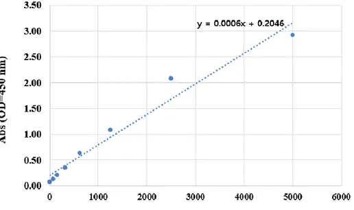
The levels of copeptin in blood plasma were determined using an ELISA assay Kit (CUSABIO, Wuhan, china) according to the manufacturer’s instructions. The blood samples were directly placed into serum separator tubes (SST) and allowed to clot overnight at 4℃, before centrifugation at 1000 x g for 15 minutes. The plasma samples were removed and stored at –70℃ in deep freezer until assayed. The optical density values were measured at 450 nm using a GloMAX Discover System (Promega, WI, USA).
2.3. Statistical analyses
Continuous variables are expressed as the mean and ± standard deviation (SD). A chi-square or Student’s t-test was used. To evaluate the difference in continuous copeptin by DCI, repeated variance measurements (ANOVA), Mauchly's Test of Sphericity and a lower-bound
8
correction were used (Jeon et al., 2012). Comparative analysis of plasma copeptin was
performed using Mann-Whitney U test and the results were shown as medians and 25th – 75th percentiles (Chou et al., 2017). In poor-grade SAH patients, the predicted value of severe vasospasm contributing to DCI was determined using the area under the receiver operator characteristic curve (AUROC) between plasma copeptin and TCD (Jeon et al., 2018). Statistical analyses showed the statistical significance as p<0.05 using SPSS V.21 (SPSS, Illinois, USA) and MedCalc (www.Medcalc.org).
9
3. Results
3.1. Patients’ clinical characteristics
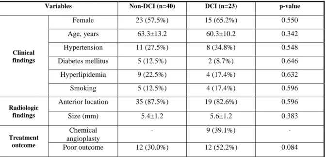
A total of 63 poor-grade SAH patients were consecutively enrolled and their plasma copeptin levels were measured on days 1, 3, 5, 7 and 9 after ictus. Among them, 23 patients (36.5%) had DCI during the follow-up period. Differences in the number of variables such as female gender, age, hypertension, diabetes mellitus, hyperlipidemia, smoking, aneurysm location and size did not reach any statistical significance. DCI patients (n=12, 52.2%) were more likely to exhibit poor clinical outcome at 3 months after ictus than non-DCI patients (n=12, 30.0%), but the differences were not statistically significant (p=0.084). Nine out of 23 DCI patients (39.1%) underwent chemical angioplasty to reverse severe cerebral vasospasm (Table 1).
10
Table 1. Clinical and radiologic characteristics in patients with delayed cerebral ischemia (DCI) in poor-grade subarachnoid hemorrhage (SAH)
Variables Non-DCI (n=40) DCI (n=23) p-value
Clinical findings Female 23 (57.5%) 15 (65.2%) 0.550 Age, years 63.3±13.2 60.3±10.2 0.342 Hypertension 11 (27.5%) 8 (34.8%) 0.548 Diabetes mellitus 5 (12.5%) 2 (8.7%) 0.646 Hyperlipidemia 9 (22.5%) 4 (17.4%) 0.632 Smoking 5 (12.5%) 4 (17.4%) 0.596 Radiologic findings Anterior location 35 (87.5%) 19 (82.6%) 0.596 Size (mm) 5.4±1.2 5.6±1.2 0.383 Treatment outcome Chemical angioplasty - 9 (39.1%) - Poor outcome 12 (30.0%) 12 (52.2%) 0.084
a Data show the numbers of subjects (percentage) for discrete and categorical variables and mean ± standard deviation.
11
3.2. Association between plasma copeptin and DCI
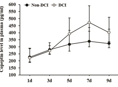
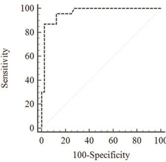
Serial plasma copeptin levels in poor-grade SAH patients with or without DCI are described in Figure 1. The plasma copeptin level increased and peaked on day 7 after ictus and decreased thereafter. Mauchly’s test of sphericity and Huynh-Feldt sphericity correction revealed significance (p<0.001). These results indicated significant differences in the plasma copeptin levels between the two groups. More specifically, the differences in copeptin level were prominent on days 5, 7, and 9: day 5, 423.70 (391.63-468.90) in DCI vs. 337.0 (317.75-347.50) in DCI patients; day 7, 498.40 (471.75-513.43) in DCI vs. 380.30 (338.20-397.35) in DCI patients; and day 9, 420.30 (401.28-459.75) in DCI vs. 360.70 (322.70-390.40) in non-DCI patients. Using the rate of increase in plasma copeptin with the first-day results as a reference value, we created an ROC curve (AUC=0.964, 95% CI: 0.883-0.995). A rate of plasma copeptin increase > 71.7% yielded the most favorable outcome with a sensitivity of 95.65% (95% CI: 78.1%-99.9%) and a specificity of 87.50% (95% CI: 73.2%-95.8%) (Figure 2).
12
Figure 1. Serial changes in plasma copeptin levels of patients with poor-grade SAH according to DCI.
Plasma copeptin levels were serially checked on days 1, 3, 5, 7, and 9 after ictus. DCI patients showed a significant increase in copeptin level compared with non-DCI patients on days 5, 7, and 9. The bar indicates the median value and the 25th – 75th percentile.
13
Figure 2. The ROC curve for plasma copeptin level in DCI.
The area under the receiver operator characteristics curve is 0.964. The increased rate of plasma copeptin levels higher than 71.7% revealed a sensitivity of 95.65% (95% CI: 78.1%-99.9%) and a specificity of 87.50% (95% CI: 73.2%-95.8%). CI= confidence interval.
14
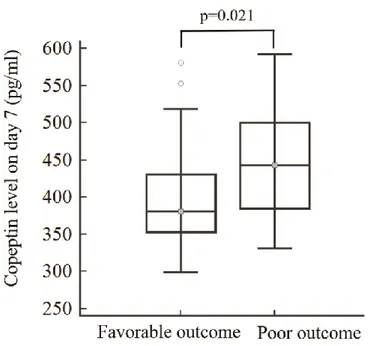
3.3. Plasma copeptin and neurologic outcome
The association between plasma copeptin and neurological outcomes at 3 months was evaluated. Copeptin levels on day 7 in patients with poor outcome were significantly elevated than in those with favorable outcome (442.90 (385.00-500.05) in patients with poor outcome vs. 380.60 (352.98-430.73) in those with favorable outcome; p=0.021) (Figure 3). Plasma copeptin levels on days 1, 3, 5, and 9 were higher in patients with poor outcomes than in those with favorable outcome, but were not statistically significant: day 1, 251.50 (237.00-268.90) in patients with poor outcome vs. 239.40 (226.63-255.60) in those with favorable outcome (p=0.170); day 3, 289.50 (275.95-301.45) in those with poor outcome vs. 286.90 (279.50-291.48) in those with favorable outcome (p=0.400); day 5, 374.45 (338.70-417.50) in patients with poor outcome vs. 340.40 (318.08-387.40) in those with favorable outcome (p=0.053); and day 9, 400.40 (355.50-422.50) in patients with poor outcome vs. 389.70 (332.35-400.68) in those with favorable outcome (p=0.128).
15
Figure 3. Association between plasma copeptin and neurological outcomes at 3 months after SAH.
The copeptin level on day 7 was most strongly correlated with poor neurological outcomes (p=0.021). Copeptin levels measured on days 1, 3, 5, and 9 were higher in patients with poor outcomes than those with favorable outcome, but were not statistically significant. The bar indicates the median value and 25th – 75th percentile.
16
3.4. Diagnostic comparison of plasma copeptin and TCD velocity
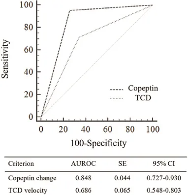
We further evaluated the diagnostic accuracy of plasma copeptin changes (>70%) and compared with TCD (MCA velocity > 200 cm/s or BA > 85 cm/s) in patients with severe angiographic vasospasm progressing to DCI. Seven out of 63 poor-grade SAH patients (11.1%) were excluded for diagnostic comparison due to poor bone window. Accordingly, 56 patients were enrolled in the analysis. For the identification of severe angiographic vasospasm, TCD showed a sensitivity of 71.4% (95% CI: 47.7%-87.8%), specificity of 65.7% (47.8%-80.3%), and negative predictive value of 79.3% (59.7%-91.3%). As shown in Figure 4, plasma copeptin changes yielded better diagnostic accuracy than TCD with a difference between areas of 0.162 (95% CI: 0.008-0.315; p<0.039) in cases of severe vasospasm presenting with poor-grade SAH.
17
Figure 4. A comparison of ROC curves between plasma copeptin changes and TCD velocity for the detection of severe vasospasm seen on cerebral angiography.
The difference between AUROC curves was 0.162 (95% CI: 0.008-0.315; p=0.039). AUROC, Area under the ROC curve; CI, confidence interval; SE, standard error.
18
4. Discussion
In this study, I found that the serial measurement of plasma copeptin facilitates risk stratification in DCI and identify poor outcomes in patients presenting with poor-grade SAH. Plasma copeptin levels on days 5, 7, and 9 were used to successfully predict DCI in poor-grade SAH. In particular, the prognostic performance of plasma copeptin was better than TCD in case of severe vasospasm.
Copeptin, a 39-amino acid glycopeptide, has been investigated as a direct marker of AVP secretion in various cerebrovascular diseases as well as myocardial infarction, pulmonary diseases, shock, and sepsis (Dobsa et al., 2013). Aksu et al. compared copeptin levels among patients with cerebral infarction, intracranial hemorrhage, SAH and healthy volunteers in the emergency room (Aksu et al., 2016). Copeptin levels were significantly higher in patients with cerebrovascular diseases compared with healthy volunteers, but not significantly different among patients with infarction, hemorrhage or SAH. Zouetal et al. reported that initial copeptin levels at admission were related to SAH severity and poor functional outcome at 3 months (Zuo
et al., 2019). In their study, mortality rate and poor outcomes were increased by 6% and 9%,
respectively, with every 1 pmol/l increase in plasma copeptin level. Zheng et al. also showed that SAH patients with symptomatic vasospasm carried higher copeptin levels than those without symptomatic vasospasm (Zheng et al., 2017). The copeptin concentration was strongly correlated with the World Federation of Neurological Surgeons subarachnoid hemorrhage scale (WFNS) score or the National Institutes of Health Stroke Scale (NIHSS), suggesting its role as a robust indicator of neurological outcomes following SAH. Nevertheless, in daily practice, copeptin was not used to monitor DCI or predict neurological outcomes because previous studies focused on the association between single measurements of copeptin and the initial
19
diagnosis at admission or clinical outcomes without considering inherent biases such as SAH severity and treatment methods.
SAH outcomes and DCI development are strongly correlated with initial brain injury, clinical severity, and degree of hemorrhage at admission (Kramer et al., 2008). Good-grade SAH patients with lesser hemorrhage showed a lower incidence of DCI and a lower mortality than poor-grade SAH patients with thick hemorrhage (Komotar et al., 2009). Accordingly, clinicians may have greater interest in improving outcomes in poor-grade SAH patients than in SAH patients with a favorable grade. Copeptin levels alleviate even mild-to-moderate stress situations (Dobsa et al., 2013), suggesting dynamic secretion requiring serial monitoring in DCI patients. In this study, the plasma copeptin level increased and peaked on day 7 and then decreased. The copeptin level on day 1 or day 3 did not differ significantly between the two groups: day 1, 252.30 (220.18-277.75) in the DCI vs. 239.50 (227.30-254.90) in the non-DCI patients; day 3, 290.70 (271.40-304.93) in the DCI vs. 284.80 (279.50-291.25) in the non-DCI patients. Fernandez et al. also showed varying CSF levels of serial copeptin between DCI (n=10) and non-DCI (n=6) patients (Fernandez et al., 2018). In their study, copeptin levels in DCI patients continued to rise between days 6 and 10 after ictus, but copeptin levels in non-DCI patients peaked on days 7 or 8 and decreased thereafter. Therefore, the copeptin level measured at admission or within 24 hours after ictus cannot accurately predict outcomes in SAH patients.
The rapid recognition and treatment of DCI starting from day 3 of post-ictus has a profound effect on prognosis. Most neurointensivists and neurosurgeons prescribe preventive medication for DCI according to Fisher grade and begin a triple therapy if there is a neurological change and no other reasons are found through minimally rapid examinations. These minimum tests are laboratory results, brain CT, TCD for hydrocephalus, rebleeding, electrolyte imbalance, etc. In a state of strong suspicion of vasospasm, TCD can provide important clues to deciding treatment. However, inadequate TCD levels are showed in seven patients in our cohort (11.1%)
20
and TCD level is not obtained due to poor temporal window. Therefore, the serial measurement of copeptin was less than TCD (AUROC difference = 0.162, p <0.039) in patients presenting with poor-grade SAH.
Although the mechanism of copeptin is not known in DCI or worse neurological outcomes, copeptin may be a remarkable marker in stress, especially SAH. The hypothalamic-pituitary-adrenalaxis is disrupted by the initial surge of intracranial pressure in acute SAH and pituitary deficiency can occur in up to one-third of SAH patients (Vespa et al., 2011). Copeptin is co-synthesized with arginine vasopressin in the hypothalamus and is released into the portal circulation of the neurohypophysis (Zuo et al., 2019). Inappropriate AVP is released and induce sodium imbalance by diminishing water permeability of the brain. Greater differences between the copeptin concentrations of DCI and non-DCI patients started to occur from days 6–7 after aSAH (Fernandez et al., 2019). Also, administration of intracisternal AVP antiserum prior to SAH prevented acute vasospasm (Dobsa et al., 2013).
The study limitations are as follows. First, I serially measured the plasma copeptin level until 9 days after ictus. Although most DCI can occur between days 4 and 8 after SAH (Francoeur
et al., 2016), it can be developed more than 10 days later. Second, I only included SAH patients
who underwent coil embolization. Mees et al. reported that DCI was more common after surgical clipping than after coil embolization, although the impact of DCI on neurological outcomes did not differ between the two treatment groups (Dorhout Mees et al., 2012). Accordingly, the results of this study may not be suitable for prediction of DCI in SAH patients with surgical clipping.
21
5. Conclusion
Serial measurements of plasma copeptin enable screening and diagnosis of patients with high-risk DCI and poor neurological outcomes following SAH. Copeptin can be used to facilitate risk stratification of severe vasospasm in poor-grade SAH patients than TCD. Further studies involving larger cohorts of SAH patient populations are required to reveal the diagnostic accuracy and to establish the optimal cut-off value depending on various clinical conditions.
22
FUNDING SOURCES
This work was supported by a research grant from Jeju National University Hospital development fund in 2016.
23
References
1. Aksu F, Gurger M, Yilmaz M, Atescelik M, Yildiz M, Ilhan N, Ilhan S, Goktekin MC. 2016.
Copeptin Levels in Cerebral Infarction, Intracranial Hemorrhage and Subarachnoid Hemorrhage. Clin Lab 62: 2387-2393.
2. Cho SS, Kim SE, Kim HC, Kim WJ, Jeon JP. 2019. Clazosentan for Aneurysmal Subarachnoid
Hemorrhage: An Updated Meta-Analysis with Trial Sequential Analysis. World Neurosurg 123: 418-424.
3. Cho YD, Kim SE, Lim JW, Choi HJ, Cho YJ, Jeon JP. 2018. Protected versus Unprotected
Carotid Artery Stenting: Meta-Analysis of the Current Literature. J Korean Neurosurg Soc 61: 458-466.
4. Chou SH, Lan J, Esposito E, Ning M, Balaj L, Ji X, Lo EH, Hayakawa K. 2017. Extracellular
Mitochondria in Cerebrospinal Fluid and Neurological Recovery After Subarachnoid Hemorrhage. Stroke 48: 2231-2237.
5. de Oliveira Manoel AL, Goffi A, Marotta TR, Schweizer TA, Abrahamson S, Macdonald RL. 2016. The critical care management of poor-grade subarachnoid haemorrhage. Crit Care 20: 21.
6. Delgado TJ, Arbab MA, Warberg J, Svendgaard NA. 1988. The role of vasopressin in acute
cerebral vasospasm. Effect on spasm of a vasopressin antagonist or vasopressin antiserum. J Neurosurg 68: 266-273.
24 diseases. Biochem Med (Zagreb) 23: 172-190.
8. Dorhout Mees SM, Kerr RS, Rinkel GJ, Algra A, Molyneux AJ. 2012. Occurrence and impact
of delayed cerebral ischemia after coiling and after clipping in the International Subarachnoid Aneurysm Trial (ISAT). J Neurol 259: 679-683.
9. Esposito P, Piotti G, Bianzina S, Malul Y, Dal Canton A. 2011. The syndrome of inappropriate
antidiuresis: pathophysiology, clinical management and new therapeutic options. Nephron Clin Pract 119: c62-73.
10. Fernandez SJ, Barakat I, Ziogas J, Frugier T, Stylli SS, Laidlaw JD, Kaye AH, Adamides AA. 2018. Association of copeptin, a surrogate marker of arginine vasopressin, with cerebral
vasospasm and delayed ischemic neurologic deficit after aneurysmal subarachnoid hemorrhage. J Neurosurg: 1-7.
11. Francoeur CL, Mayer SA. 2016. Management of delayed cerebral ischemia after subarachnoid
hemorrhage. Crit Care 20: 277.
12. Hollingworth M, Jamjoom AAB, Bulters D, Patel HC. 2019. How is vasospasm screening
using transcranial Doppler associated with delayed cerebral ischemia and outcomes in aneurysmal subarachnoid hemorrhage?. Acta Neurochir (Wien) 161:385-392.
13. Hong EP, Kim BJ, Cho SS, Yang JS, Choi HJ, Kang SH, Jeon JP. 2019. Genomic Variations
in Susceptibility to Intracranial Aneurysm in the Korean Population. J Clin Med 8.
14. Jeon JP, Kim C, Oh BD, Kim SJ, Kim YS. 2018. Prediction of persistent hemodynamic
25 Neurol Neurosurg 164: 127-131.
15. Jeon JS, Sheen SH, Hwang G, Kang SH, Heo DH, Cho YJ. 2012. Intravenous magnesium
infusion for the prevention of symptomatic cerebral vasospasm after aneurysmal subarachnoid hemorrhage. J Korean Neurosurg Soc 52: 75-79.
16. Jochberger S, Morgenthaler NG, Mayr VD, Luckner G, Wenzel V, Ulmer H, Schwarz S, Hasibeder WR, Friesenecker BE. 2006. Copeptin and arginine vasopressin concentrations in
critically ill patients. J Clin Endocrinol Metab 91: 4381-4386.
17. Kagawa M, Nagao S, Bemana I. 1996. Arginine vasopressin receptor antagonists for treatment
of vasogenic brain edema: an experimental study. J Neurotrauma 13: 273-279.
18. Katan M, Morgenthaler NG, Dixit KC, Rutishauser J, Brabant GE, Muller B, Christ-Crain M. 2007. Anterior and posterior pituitary function testing with simultaneous insulin tolerance test
and a novel copeptin assay. J Clin Endocrinol Metab 92: 2640-2643.
19. Kim BJ, Kim Y, Kim SE, Jeon JP. 2018. Study of Correlation Between Hp alpha1 Expression of
Haptoglobin 2-1 and Clinical Course in Aneurysmal Subarachnoid Hemorrhage. World Neurosurg 117: e221-e227.
20. Kim CH, Jeon JP, Kim SE, Choi HJ, Cho YJ. 2018. Endovascular Treatment with Intravenous
Thrombolysis versus Endovascular Treatment Alone for Acute Anterior Circulation Stroke: A Meta-Analysis of Observational Studies. J Korean Neurosurg Soc 61: 467-473.
21. Kim HC, Rhim JK, Ahn JH, Park JJ, Moon JU, Hong EP, Kim MR, Kim SG, Lee SH, Jeong JH, Choi SW, Jeon JP. 2019. Machine Learning Application for Rupture Risk Assessment in
26
22. Komotar RJ, Schmidt JM, Starke RM, Claassen J, Wartenberg KE, Lee K, Badjatia N, Connolly Jr ES, Mayer SA. 2009. Resuscitation and critical care of poor-grade subarachnoid
hemorrhage. Neurosurgery 64: 397-410.
23. Kramer AH, Hehir M, Nathan B, Gress D, Dumont AS, Kassell NF, Bleck TP. 2013. A
comparison of 3 radiographic scales for the prediction of delayed ischemia and prognosis following subarachnoid hemorrhage. J Neurosurg 109: 199-207.
24. Kumar G, Shahripour RB, Harrigan MR. 2016. Vasospasm on transcranial Doppler is
predictive of delayed cerebral ischemia in aneurysmal subarachnoid hemorrhage: a systematic review and meta-analysis. J Neurosurg 124: 1257-1264.
25. Samagh N, Bhagat H, Jangra K. 2019. Monitoring cerebral vasospasm: How much can we rely
on transcranial Doppler. J Anaesthesiol Clin Pharmacol 35: 12-18.
26. Sviri GE, Ghodke B, Britz GW, Douville CM, Haynor DR, Mesiwala AH, Lam AM, Newell DW. 2006. Transcranial Doppler grading criteria for basilar artery vasospasm. Neurosurgery 59:
360-366.
27. Vespa P. 2011. Participants in the International Multi-Disciplinary Consensus Conference on the
Critical Care Management of Subarachnoid H: SAH pituitary adrenal dysfunction. Neurocrit Care 15: 365-368.
28. Vora YY, Suarez-Almazor M, Steinke DE, Martin ML, Findlay JM. 1999. Role of transcranial
Doppler monitoring in the diagnosis of cerebral vasospasm after subarachnoid hemorrhage. Neurosurgery 44: 1237-1247.
27
29. Zheng YK, Dong XQ, Du Q, Wang H, Yang DB, Zhu Q, Che ZH, Shen YF, Jiang L, Hu W, Wang KY, Yu WH. 2017. Comparison of plasma copeptin and multiple biomarkers for assessing
prognosis of patients with aneurysmal subarachnoid hemorrhage. Clin Chim Acta 475: 64-69.
30. Zuo Z, Ji X. 2019. Prognostic value of copeptin in patients with aneurysmal 2subarachnoid
28
SUPPLEMENTAL DATA
29
국문 초록
뇌지주막하 출혈에서 copeptin이 예후인자로 보고되어 왔지만 중증의 뇌지주막하 출혈에서의 역할은 제한적이다. 그리하여 예후가 좋지 않고 지연성 뇌허혈 고위험인 중증 뇌지주막하 출혈환자에서 치료 결과와 copeptin과의 연관관계를 조사했다. 혈장 copeptin을 출혈 당일, 3일째, 5일째, 7일째 그리고 9일째 연속적으로 채취하여 ELISA로 측정하였고 총 63명의 환자들에 대해 copeptin과 지연성 뇌허혈 그리고 3개월째 치료 결과 사이의 관계를 알아보았다. 또한 지연성 뇌허혈과 연관된 중증 뇌혈관 연축에 대해 copeptin과 두개경 초음파 도플러의 진단 정확도를 the area under ROC curve (AUROC)를 이용하여 비교했고 결과는 중앙값 그리고 25-75번째 백분위 수로 정리하였다. 혈장 copeptin은 출혈 당시 증가하기 시작하여 7일째 최고로 증가하다가 감소한다. 지연성 뇌허혈 그룹 [DCI, n=23 (36.5%)]과 지연성 뇌허혈이 없는 그룹 [non-DCI, n=40 (63.5%)]을 비교하였을 때, 5일째 혈장 copeptin 양이 DCI 그룹은 423.70 (391.63 ~ 468.90), non-DCI 그룹은 337.0 (317.75-347.50); 7일째에는 DCI 그룹에서는 498.40 (471.75-513.43), non-DCI 그룹에서는 380.30 (338.20-397.35); 그리고 9일째 DCI 그룹에서는 420.30 (401.28-459.75), non-DCI 그룹에서는 360.70 (322.70-390.40)으로 측정되었다. 7일째 copeptin 값이 예후가 좋지 않은 신경학적 결과와 가장 밀접한 관계를 보였고 (p=0.021) 혈장 copeptin의 변화는 중증의 혈관 연축 시기에 두개경 도플러 초음파보다 더 좋은 진단 정확성을30
보여줬다 (AUROC difference, 0.162, 95% CI: 0.008-0.315). 이것으로 보아 연속적 혈장 copeptin 검사는 고위험 지연성 뇌허혈 환자 그리고 나쁜 예후를 가진 환자를 구분할 수 있는 진단 방법이라 생각하며 지연성 뇌허혈에 의한 혈관 연축을 보이고 좋지 않은 예후를 가진 환자에게 위험 요인에 대한 치료 계획을 세우는 방법으로는 두개경 도플러 초음파보다는 copeptin 검사가 더 좋은 방법이라 생각할 수 있다.