2011년도 추계학술발표회 논문요약집 대한방사선방어학회
100_http://www.karp.or.kr
Investigation of the HU-density conversion method and
comparison of dose distribution for dose calculation on
MV cone beam CT images
Min-Joo Kima,b, Seu-Ran Leea,b and Tae-Suk Suha,b a
Department of Biomedical Engineering, The Catholic University of Korea b
Research Institute of Biomedical Engineering, The Catholic University of Korea
E-mail: smiling_mjkim@catholic.ac.kr 중심어 : Dose calculation, Megavoltage Cone-beam CT image
Introduction
Modern radiation therapy techniques, such as Image-guided radiation therapy (IGRT), Adaptive radiation therapy (ART) has become a routine clinical practice on linear accelerators for the increase the tu-mor dose conformity and improvement of normal tissue sparing at the same time. For these highly developed techniques, megavoltage cone beam computed tomog-raphy (MVCBCT) system produce volu-metric images at just one rotation of the x-ray beam source and detector on the bottom of conventional linear accelerator for real-time application of patient con-dition into treatment planning. MV CBCT image scan be directly registered to a ref-erence CT data set which is usually kilo-voltage fan-beam computed tomog-raphy (kVFBCT) on treatment planning system and the registered image scan be used to adjust patient set-up error. However, to use MV CBCT images in ra-diotherapy, reliable electron density (ED)
distribution are required. Patients scatter-ing, beam hardening and softening effect caused by different energy application be-tween kVCT, MV CBCT can cause cup-ping artifacts in MV CBCT images and distortion of Houns field Unit (HU) to ED conversion.
The goal of this study, for reliable appli-cation of MV CBCT images into dose cal-culation, MV CBCT images was modified to correct distortion of HU to ED using the relationship of HU and ED from kV FBCT and MV CBCT images.
Materials and Methods
Data acquisition and Image processing To obtain modification level, the image data sets were acquired using MV CBCT (MVision, Artiste, Siemens Medical Solutions, Forchheim, Germany) and ref-erence kilovoltage CT (SOMATOM Sensation 64, Siemens Medical Solutions, Forchheim, Germany) with 12 different density plugs for the density correction.
2011년도 추계학술발표회 논문요약집 대한방사선방어학회
제1분과(의료 및 생물)_ 101
Then MV CBCT voxel intensities were converted to relative electron density to water using the calibration curves. Treatment planning and evaluation A realistic patient-equivalent phantom (Rando phantom, Alderson Research Laboratories, Stamford, CT, USA) images were acquired to evaluate the CT den-sity-HU conversion method in dose calcu-lation result on treatment planning system. Moreover, to evaluate the dose distribution using difference map, these realistic patient-equivalent phantom im-ages were modified using affine trans-formation to make each slice has same po-sition before import to treatment planning. Treatment planning was performed in CorePlan radiation treatment planning system. Dose distribution was exported from CorePlan to compare the dose dis-tribution between based on kV CT and MV CBCT images. Finally, each dose dis-tribution was used to acquire dose differ-ence map and to calculate percentage dif-ference map.
결과 및 고찰
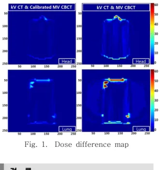
The HU-density conversion was per-formed on MV CBCT image set using Dose difference map was showing in Figure 1. Finally, percentage differences above 3% were reduced depending on ap-plying density calibration method. As a result, total error could be reduced to un-der 3%.
Fig. 1. Dose difference map
결 론
The present study demonstrates that dose calculation accuracy using MV CBCT image set can be improved my ap-plying HU-density conversion method. The dose calculation and comparison of dose distribution from MV CBCT image set with/without HU-density conversion method was performed. An advantage of this study compared to other approaches is that HU-density conversion method can be easily applied into any CT dataset in radiation treatment planning system using CT-density table within brief time.
참 고 문 헌
1. J Hatton, B McCurdy, PB Greer. Cone beam computerized tomography: the ef-fect of calibration of the Hounsfield unit number to electron density on dose cal-culation accuracy for adaptive radiation therapy. Physics in Medicine and Biology 54, N329-346(2009)