Clinical Practice Guideline of Acute
Respiratory Distress Syndrome
Young-Jae Cho, M.D., M.P.H.
1,*, Jae Young Moon, M.D., Ph.D.
2,*, Ein-Soon Shin, Ph.D., M.P.H.
3, Je
Hyeong Kim, M.D., Ph.D.
4, Hoon Jung, M.D., Ph.D.
5, So Young Park, M.D.
6, Ho Cheol Kim, M.D.
7,
Yun Su Sim, M.D.
8, Chin Kook Rhee, M.D., Ph.D.
9, Jaemin Lim, M.D.
10, Seok Jeong Lee, M.D.
11,
Won-Yeon Lee, M.D.
11, Hyun Jeong Lee, M.D.
12, Sang Hyun Kwak, M.D., Ph.D.
12, Eun Kyeong Kang, M.D.,
Ph.D.
13, Kyung Soo Chung, M.D.
14and Won-Il Choi, M.D., Ph.D.
15, The Korean Society of Critical Care
Medicine and the Korean Academy of Tuberculosis and Respiratory Diseases Consensus Group
1Division of Pulmonary and Critical Care Medicine, Department of Internal Medicine, Seoul National University Bundang Hospital,Seoul National University College of Medicine, Seongnam, 2Division of Pulmonary and Critical Care Medicine, Department of
Internal Medicine, Chungnam National University Hospital, Daejeon, 3Research Agency for Clinical Practice Guidelines, Korean
Academy of Medical Sciences Research Center, Seoul, 4Division of Pulmonary and Critical Care Medicine, Department of Internal
Medicine, Korea University Ansan Hospital, Korea University College of Medicine, Ansan, 5Department of Pulmonary and Critical
Care Medicine, Inje University Ilsan Paik Hospital, Goyang, 6Department of Pulmonary and Critical Care Medicine, Kyung Hee
University Medical Center, Seoul, 7Department of Internal Medicine, Gyeongsang National University Changwon Hospital,
Gyeongsang National University School of Medicine, Changwon, 8Division of Pulmonary, Allergy, and Critical Care Medicine,
Department of Internal Medicine, Hallym University Kangnam Sacred Heart Hospital, Seoul, 9Division of Pulmonary and Critical
Care Medicine, Department of Medicine, Seoul St. Mary’s Hospital, College of Medicine, The Catholic University of Korea, Seoul,
10Division of Pulmonary and Critical Care Medicine, Department of Medicine, Gangneung Asan Hospital, University of Ulsan College
of Medicine, Gangneung, 11Division of Pulmonary, Allergy, and Critical Care Medicine, Department of Internal Medicine, Yonsei
University Wonju College of Medicine, Wonju, 12Department of Anesthesiology and Pain Medicine, Chonnam National University
Medical School , Gwangju, 13Department of Pediatrics, Dongguk University Ilsan Hospital, Goyang, 14Division of Pulmonology,
Department of Internal Medicine, Severance Hospital, Institute of Chest Diseases, Yonsei University College of Medicine, Seoul,
15Department of Internal Medicine, Keimyung University Dongsan Hospital, Daegu, Korea
There is no well-stated practical guideline for mechanically ventilated patients with or without acute respiratory distress syndrome (ARDS). We generate strong (1) and weak (2) grade of recommendations based on high (A), moderate (B) and low (C) grade in the quality of evidence. In patients with ARDS, we recommend low tidal volume ventilation (1A) and prone position if it is not contraindicated (1B) to reduce their mortality. However, we did not support high-frequency oscillatory ventilation (1B) and inhaled nitric oxide (1A) as a standard treatment. We also suggest high positive end-expiratory pressure (2B), extracorporeal membrane oxygenation as a rescue therapy (2C), and neuromuscular blockage for 48 hours after starting mechanical ventilation (2B). The application of recruitment maneuver may reduce mortality (2B), however, the use of systemic steroids cannot reduce mortality (2B). In mechanically ventilated patients, we recommend light sedation (1B) and low tidal volume even without ARDS (1B) and suggest lung protective ventilation strategy during the operation to lower the incidence of lung complications including ARDS (2B). Early tracheostomy in mechanically ventilated patients can be performed only in limited patients (2A). In conclusion, of 12 recommendations, nine were in the management of ARDS, and three for mechanically ventilated patients.
Keywords: Practice Guideline; Respiratory Distress Syndrome, Adult; Respiratory Distress Syndrome, Acute; Ventilators, Mechanical; Respiration, Artificial
Copyright © 2016
The Korean Academy of Tuberculosis and Respiratory Diseases. All rights reserved.
Address for correspondence: Won-Il Choi, M.D., Ph.D.
Department of Internal Medicine, Keimyung University School of Medicine, 56 Dalseong-ro, Jung-gu, Daegu 41931, Korea
Phone: 82-53-250-7572, Fax: 82-53-250-8379, E-mail: wichoi@dsmc.or.kr
*Young-Jae Cho and Jae Young Moon contributed equally to this work.
Received: Jun. 17, 2016, Revised: Jun. 27, 2016, Accepted: Aug. 16, 2016
Introduction
Since the first description of acute respiratory distress syn-drome (ARDS) as a series of 12 patients in 1967 by Ashbaugh et al.1, it still remains a major public health problem that incurs
high health care costs and causes major mortality in the in-tensive care unit (ICU) despite improvements in outcomes in the last two decades. ARDS refers to the occurrence of severe hypoxemia that was not corrected by oxygen treatment and is characterized by heterogeneous acute lung inflammation with increased permeability of the alveolar-capillary membrane, resulting in the development of exudate within the alveolar space, damage due to activated neutrophils and cytokines, and abnormalities of surfactant and the coagulation system2.
The definition also recently changes as the Berlin criteria3,
which was modified to the original American-European Con-sensus Conference definitions4 and novel clinical trial designs
in ARDS may anticipate a new era of successful therapies. Although over the past decades, there has been a remark-able development in the therapeutic approach and manage-ment of critically ill patients with ARDS, the mortality of pa-tients with ARDS is unacceptably high, up to 40%5. In Korea, it
has been reported that 79 patients with ARDS were admitted to the ICUs of 28 university hospitals all over the country with-in 1 month, July 2009, and 45 of those patients died, resultwith-ing in a mortality rate of 57%6. Also, until now there is no
well-stated clinical practice guideline for intensivists about ARDS, especially focused on the critical care including applying me-chanical ventilation until now.
Herein, we report the recommendations and suggestions of how to manage mechanically ventilated patients with or with-out ARDS.
Methods
1. Selection of panel membersThe board members of the Korean Society of Critical Care Medicine (KSCCM) appointed the editor for the new guidelines addressing ARDS management. The panels of the guideline committee were recruited from the members of the KSCCM and the Korean Academy of Tuberculosis and Lung Diseases (KATRD). The KSCCM and KATRD approved all panelists, and all of them applied voluntarily to the positions. The 16 panelists include intensivists, anesthesiologists, pul-monologists, pediatricians, and methodologists. All of the pan-elists were required to disclose any conflicts of interest (COI) about the topics. None of the panelists has any COI with the related topic.
2. Selection of topics and key questions
The board members of the KSCCM and KATRD agreed with the development of guidelines on ARDS management. During the 2014 KSCCM conference, we surveyed important topics related to ARDS management from the KSCCM mem-bers. Initially, 20 topics were collected from the survey. Then, panel members selected 12 topics, with a consensus. All of the panels agreed with the final topics. For each topic, we de-veloped standardized questions using the PICO (population, intervention, comparator, outcome) format.
3. Guideline development
There was no guideline for ARDS management available during the beginning of the guideline development meeting, and we tried to develop de novo guideline. This guideline is based on Korean AGREE II as an assessment tool, which was published by the Korean Ministry of Health and Welfare and the Korean Academy of Medical Sciences. We ask literature search for a specialist. The National Library of Medicine’s medical subject headings (MeSH) keyword nomenclature was used with PICO. We searched the literature using Med-line (1948 to July 2014). Searches were limited to literature written in English or the Korean language. We searched all of the possible investigation methods, including retrospective cohort studies and case series. We also searched both original investigations and systemic reviews. We assessed the quality of systemic reviews and original investigations carefully.
4. Selection and assessment of study
The keywords and search formula were based on the PICO elements of the standardized questions and the study design, which is documented in the Korean version (http://www. ksccm.org or http://www.lungkorea.org). Selection of studies was conducted by the specialist of the area. First title screen-ing was completed, then abstract and full-text screenscreen-ing. If a paper was selected for risk of bias assessment, we abstracted the data based on the following characteristics: study design, participants, intervention, control, outcomes, funding, and COI. We assessed the risk of bias using the Cochrane Risk of Bias Tool in randomized trials. We also performed meta-analyses especially in two PICOs, the effect of recruitment maneuvers and neuromuscular blockers. However, the results of the meta-analysis were the same as previously published meta-analyses.
5. Assessing quality of evidence
The quality of evidence is categorized as high (A level), moderate (B level), or low (includes very low) (C level)7,8. The
risk of bias, imprecision, inconsistency, indirectness of results, and the likelihood of publication bias.
6. Drafting of recommendations
The strength of recommendation as strong or weak was determined based on the value of the study results, wanted vs. unwanted effects, and cost effectiveness9,10. The strength
of recommendation was categorized as strong (grade 1) or weak (grade 2). Each author drafted the recommendations after the entire panels reviewed the evidence and discussed the recommendation. Recommendations were then revised several times during meetings in KSCCM conference rooms, and through email exchanges that included the entire panel.
7. Consensus of recommendations
We used a modified Delphi technique11 to achieve a
con-sensus on each recommendation. This technique aims to minimize any group interaction bias and to maintain ano-nymity among respondents. The E-mail was exchanged through assistants of KSCCM. Since there was no COI among panelists, all panelists voted on their level of agreement with each recommendation. If a panel disagreed with the draft of a recommendation, the panel suggested a different recom-mendation. Each panelist provided open-ended feedback on each recommendation with suggested wording edits or general remarks. To achieve a consensus and to be included in the final manuscript, each recommendation had to have an at least 50% agreement (strong or weak) with a response rate of at least 80% of the total panel members. All recommenda-tions achieved consensus during the first round. We repeated
a review by all panel members.
8. Peer review
External reviewers who were not involved in the develop-ment of the guideline had reviewed it before it was published. These reviewers included different academic society mem-bers, a methodological expert, and a practicing clinician. The final manuscript was reviewed and approved by the Board of KSCCM and KATRD.
Results
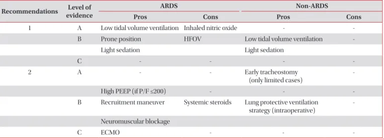
The recommendation and level of evidence of 12 topics were categorized and summarized in Table 1. The details of each topic were as follows.
1. Low tidal volume ventilation
1) Recommendation
- We recommend low tidal volume ventilation can be ap-plied to patients with ARDS to reduce mortality (grade 1A).
2) Key point
- The tidal volume should be maintained less than 6 mL/kg of predicted body weight in patients with ARDS.
- The plateau pressure should be maintained less than 30 cmH2O in patients with ARDS.
According to analyses of the causes of death in patients with ARDS, a majority of patients died of multiple organ
dysfunc-Table 1. Summary of recommendations and level of evidence for mechanically ventilated patients with or without acute respiratory distress syndrome
Recommendations evidenceLevel of ARDS Non-ARDS
Pros Cons Pros Cons
1 A Low tidal volume ventilation Inhaled nitric oxide -
-B Prone position HFOV Low tidal volume ventilation
-Light sedation Light sedation
C - - -
-2 A - - Early tracheostomy
(only limited cases)
-High PEEP (if P/F ≤200) - -
-B Recruitment maneuver Systemic steroids Lung protective ventilation
strategy (intraoperative)
-Neuromuscular blockage
C ECMO - -
-ARDS: acute respiratory distress syndrome; HFOV: high-frequency oscillatory ventilation; PEEP: positive end-expiratory pressure; ECMO: extracorporeal membrane oxygenation.
tion syndromes (MODS) rather than respiratory failure12, and
the mortality rate was reported to be significantly higher when another organ failure in addition to respiratory insufficiency, was involved13. ARDS occurs due to various causes, and many
kinds of treatments are conducted in individual patients based on the cause. Nevertheless, the fact that MODS is the predominant cause of death has raised the hypothesis that mechanical ventilation, which is essentially and commonly administered to all patients with ARDS, can play a major role in initiating and propagating a systemic inflammatory reac-tion. This hypothesis is termed ventilator-induced lung injury (VILI), and studies on VILI and its relation to systemic inflam-matory response syndrome14 and MODS15,16 have been
con-ducted.
The lung of patients with ARDS is characterized by hetero-geneous inflammation, with congestion and atelectasis of dependent alveoli and relatively normal alveoli on the oppos-ing side2. In this condition, if mechanical ventilation is applied
with 10–12 mL/kg of tidal volume, which is a conventional ventilation strategy, and without positive end-expiratory pres-sure (PEEP), the physical stretch injury will occur in relatively normal alveoli because of overexpansion. Also, damage oc-curs from shearing forces, in which the repeated collapse and reopening of the respiration cycle occurs in basal alveoli affected with congestion and atelectasis, and these two inju-ries are the main mechanisms of VILI17. Barotrauma such as
pneumothorax, pneumomediastinum, and subcutaneous emphysema, and volutrauma such as permeability altera-tion, pulmonary edema, and diffuse alveolar injury, occur via stretch injury. Owing to the shearing force on the site of atelectasis, atelectrauma by repeated alveolar collapse and reopening occurs. In the whole process, biotrauma due to the activation and recruitment of inflammatory cells and media-tors occurs. These inflammatory mediamedia-tors are not limited to the lung, and they enter into the systemic circulation through damaged alveoli-capillary membranes. In infectious lung dis-eases, bacteria within alveoli can also move into the systemic circulation, causing a systemic inflammation similar to sepsis. Such systemic inflammation causes MODS, along with organ perfusion deterioration due to reduced cardiac output by increased pressure within the thorax induced by mechanical ventilation, leading to the death of patients15. This is the
patho-logical mechanism of VILI and MODS18.
Lung protective ventilation (LPV) strategy refers to a me-chanical ventilation strategy to minimize VILI by adminis-tering a tidal volume less than the conventional ventilation volume and limiting the plateau pressure to reduce injury to alveoli by increasing end-expiratory lung volume with PEEP19.
In a broad sense, prone position ventilation and recruitment maneuvers, which minimize VILI by reducing heterogeneity, and extracorporeal membrane oxygenation (ECMO), which reduces the risk of lung injury from mechanical ventilation and a high concentration of oxygen, can also be included in
LPV.
After detailed mechanisms of VILI had been studied and reported, an LPV strategy was conceptualized to prevent VILI, and clinical studies were conducted. In 1998, Amato et al.20
first reported that low tidal volume ventilation had a potential clinical effect on patients with ARDS through a randomized clinical trial. In 53 patients, the conventional ventilation strat-egy with a tidal volume of 12 mL/kg, low PEEP, and target-ing a 35–38 mm Hg partial pressure of carbon dioxide was compared with the protective ventilation strategy with a tidal volume of 6 mL/kg, high PEEP, and permissive hypercapnia. As a result, the protective ventilation group had lower 28-day mortality than the conventional ventilation group (38% vs. 71%), the frequency of barotrauma was low, and weaning rate from mechanical ventilation was higher. However, other clini-cal studies reported at almost the same time showed that low tidal volume had no clinical effects on patients with ARDS21-23,
raising controversy over the clinical effect of low tidal volume. In this circumstance, the ARDS Network study, the most re-markable clinical study related to the clinical effect of low tidal volume ventilation, was reported24. The study was conducted
in 10 institutions in the United States for three years on a large scale. A tidal volume of 12 or 6 mL/kg of predicted body weight was applied to 861 patients, and plateau pressures were limited to 50 cmH2O and 30 cmH2O, respectively. The
low tidal volume ventilation group showed significantly lower mortality compared to the conventional ventilation group (31% vs. 39.8%, p=0.007). Regarding the indices including days without breathing assistance, ventilator-free days, days without failure of nonpulmonary organs or systems and blood interleukin-6 concentrations, the low tidal volume ventilation group showed significant improvement, providing strong clinical evidence for the effect of low tidal volume ventilation.
According to the Cochrane review on the clinical trials25,26,
although there was heterogeneity among studies, it was ana-lyzed that 28-day and hospital mortalities were significantly reduced in the low tidal volume ventilation group. Based on the above results of clinical trials and systematic review, the Surviving Sepsis Campaign Guideline which was revised in 2012 recommended low tidal volume ventilation with a high level of evidence27.
Recently, Amato et al.20 reported the result of a multilevel
mediation analysis about the nine clinical trials of ARDS. Ac-cording to the result, driving pressure (∆P=VT/CRS; VT, tidal
volume; CRS, respiratory system compliance) which reflects
functional lung capacity is more correlated with the mortal-ity of ARDS patients than tidal volume, plateau pressure, and PEEP. However, this was the statistical analysis of previous studies, and clinical studies should be performed to confirm the clinical effects of ∆P.
2. High PEEP
1) Recommendation
- We suggest high PEEP can be applied to patients with ARDS, who have PaO2/FIO2 ≤200 mm Hg to reduce mortality
(grade 2B).
2) Key points
- The application of high PEEP does not increase the risk of barotrauma.
- If high PEEP is applied, PaO2/FIO2 at day 1 and three can
be improved compared to the application of low PEEP group. PEEP is an easily applicable intervention and an essential component of the care of critically ill patients who require ven-tilator support. The end-expiratory pressure is elevated above atmospheric pressure to prevent atelectasis and correct the hypoxemia caused by alveolar hypoventilation. Mechanisms that PEEP improves through gas exchange and pulmonary function are increased functional residual capacity, alveolar recruitment, redistribution of extravascular lung water and improved ventilation-perfusion matching28. Also, it may
pre-vent repetitive alveolar collapse. High PEEP has been defined differently in each study. In ALVEOLI study29, it was based on
PEEP table, and in EXPRESS study30, PEEP was raised as
pla-teau pressure reached 28–30 cmH2O. It can be expected that it
may reduce VILI with the above physiological effect. However, side effects include an increase in physiologic dead space, de-creased cardiac output, and inde-creased risk of barotrauma31-33.
A randomized clinical trial that applied high PEEP was con-ducted with 53 patients with early ARDS20. In the group that
was exposed to high PEEP and low tidal volume, mortality on day 28 (38%) was significantly lower than the conventional ventilation group (71%), but there was no significant differ-ence in survival to hospital discharge. In this study, mortality of conventional ventilation group was too high, and the effect of high PEEP was observed on day 3. In a second randomized study, ICU mortality, hospital mortality, and ventilator-free days at day 28 all favored the high PEEP group34.
According to the Cochrane’s review, which meta-analyzed seven randomized control trials20,29,30,34-37 conducted from 1998
to 2009 to compare the effects of the applications of low PEEP and high PEEP, there was heterogeneity that five29,30,35-37 out
of seven included trials had the application of the same tidal volume in both high and low PEEP groups, whereas two stud-ies20,34 had different tidal volumes for the two groups.
There-fore, as a result of analyzing 2,565 patients with ARDS, who were administered the same tidal volume with different PEEP, high PEEP did not contribute to the reduction of mortality in the hospital (relative risk [RR], 0.90; 95% confidence interval [CI], 0.81–1.01). In the group with PaO2/FIO2 ≤200 mm Hg,
high PEEP decreased mortality within the ICU (RR, 0.67; 95% CI, 0.48–0.95). The comparison between the two groups on
barotrauma did not show a significant difference (RR, 0.97; 95% CI, 0.66–1.42). However, PaO2/FIO2 at days 1 and 3 was
improved in the high PEEP application group. In a follow-up study38 of the two large-scale studies30,36 in 2014, the group in
which PaO2/FIO2 increased by more than 25 mm Hg within 2
hours by applying PEEP showing decreased mortality (31% vs. 51%; odds ratio [OR], 0.8; 95% CI, 0.72–0.89). Also, the reduced mortality was observed in patients with increased PaO2/FIO2
regardless of PEEP among ARDS patients with PaO2/FIO2
≤150 mm Hg.
If PEEP recruits collapsing alveoli, atelectrauma of alveoli can be reduced39 whereas alveolar injury may be increased by
raising the intensity delivered to alveoli if they are not recruit-ed40. It is expected that further clinical trials will apply different
PEEP to patients with enough alveoli to be recruited. Based on the above data, high PEEP can be applied to ARDS patients with PaO2/FIO2 ≤200 mm Hg.
3. Prone position
1) Recommendation
- We recommend prone position can be applied to patients with moderate or above ARDS to reduce mortality if it is not contraindicated (grade 1B).
2) Key points
- The prone position should be applied when there is no improvement of oxygenation at an early stage of mechanical ventilation.
- Prone position is recommended at least for 10 hours. - Lung protective strategy should also be applied during prone positioning.
Based on the theory that the use of the prone position would reduce lung injury caused by lung stress and strain exerted on the lungs during artificial ventilation in ARDS41-43
since its first attempt by Bryan in 197444, studies have been
performed to prove its utility by a number of researchers. In a post hoc analysis of Gattinoni et al. in 200145, among PaO
2/
FiO2 <88 mm Hg, Simplified Acute Physiology Score II (SAPS
II) ≥49 and tidal volume >12 mL/kg of predicted body weight groups, the group that used the prone position had a lower 10-day mortality45, and other meta-analyses reported that
the patients using the prone position in the case of PaO2/FiO2
<100 mm Hg had lower mortality46-48, but there were a lot of
controversies over the insistence of the use of the prone posi-tion can decrease mortality in ARDS. However, a large-scale randomized study has recently been conducted by Guerin et al.49, reporting that in the PaO
2/FiO2 <150 mm Hg group at
FiO2 >0.6 and PEEP 5 cmH2O, the group that conducted the
prone position for at least 16 hours per day within 36 hours had a statistically significant decreased 28-day mortality rate (16.0% prone group vs. 32.8% supine group: hazard ratio [HR],
0.39; 95% CI, 0.25–0.63; p<0.001) and 90-day mortality (23.6% prone group vs. 41.0% supine group: HR, 0.44; 95% CI, 0.29– 0.67; p<0.001) compared to the group with the supine posi-tion, despite the mechanical ventilation for 12 or 24 hours49. In
the meta-analysis conducted after the publication of the large-scale study by Guerin et al.49, the use of the prone position was
found to decrease mortality of patients with moderate ARDS50-52.
However, patient’s severity, low tidal ventilation, time of prone position use, and the degree of PEEP were different in each study, showing significant heterogeneity41,50,52. When using
a low tidal volume ventilation concomitantly, the mortality was decreased with a statistical significance (RR, 0.66; 95% CI, 0.5–0.85; p=0.02)49,53-56, but the group without low tidal volume
ventilation did not show decreased mortality (RR, 1.00; 95% CI, 0.88–1.13; p=0.949)56. The time of using the prone position
showed differences concerning mortality. When the prone position was conducted for over 10 hours, a clearly decreased mortality was found (OR, 0.62; 95% CI, 0.48–0.79; p<0.001), and the randomized study by Taccone et al.53 and Mancebo
et al.54, as well as meta-study by Hu et al.51 and Beitler et al.56,
reported that the decrease in mortality was evident when the prone position was conducted for 12 hours or more. In the large-scale randomized study conducted by Guerin et al.49, the
prone position was performed for more than 19 hours49,51,53-55.
However, when the prone position period was less than 12 hours, there was no decrease in mortality (OR, 1.04; 95% CI, 0.80–1.36; p=0.757). No reduction in mortality was shown when analyzing patients with acute lung injury or mild ARDS (OR, 1.02; 95% CI, 0.76–1.36; p=0.920)50. Although there were
not many studies on the degree of PEEP, the meta-analysis published by Hu et al.51 reported that the group maintaining
high PEEP ≥10 cmH2O–13 cmH2O showed a lower 60-day
mortality (RR, 0.82; 95% CI, 0.68–0.99; p=0.04) and 90-day mortality (RR, 0.57; 95% CI, 0.43–0.75; p<0.0001), compared to the group maintaining PEEP <10 cmH2O when conducting
prone position.
In randomized clinical trials and meta-analyses, the use of the prone position was reported to improve hypoxemia. Com-pared to the group using the supine position, the prone posi-tion group showed an increase of PaO2/FiO2 by 25%–36% for
the first 3 days44,45,52, which contributed to the improvement of
perfusion imbalance by reducing the collapse of the depen-dent portion of the lung, as well as edema41,57,58.
In three randomized clinical trials, there was a study on mechanical ventilation period53,55,59, showing no difference in
the mechanical ventilation period between the prone posi-tion and supine posiposi-tion patient groups. In two randomized clinical trials, the result of ICU length of stay showed no differ-ence between the prone position and supine position patient groups53,55.
In the study of Guerin et al.49, patients were ventilated while
in the prone position for more than 19 hours. A meta-study by Lee et al.50 reported that the decrease in mortality in the prone
position for more than 10 hours was statistically significant, and randomized study of Taccone et al.53 and Mancebo et
al.54, and meta-study of Hu et al.51 and Beitler et al.56 showed
that the decrease in mortality was clear when using the prone position for more than 12 hours49,51,53-55. However, if the
dura-tion of the prone posidura-tion is less than 10 hours, there was no decrease in mortality (OR, 1.04; 95% CI, 0.80–1.36; p=0.757)50.
Studies49,50,53-56 showed different indications regarding the
dis-continuation time when conducted in the prone position. In Guerin et al.49, the prone position was discontinued when (1)
there was improvement of hypoxemia (PaO2/FIO2 ≥150 mm
Hg with PEEP ≤10 cmH2O and FIO2 ≤0.6), (2) when PaO2/FiO2
deteriorated by 20% or more compared to the supine position, and (3) when complications occurred due to the prone posi-tion. It is thought that further studies on daily use time and total treatment period for the prone position will be needed.
The complications related to prone position include extuba-tion, endotracheal tube obstrucextuba-tion, selective main bronchus intubation, pneumothorax, cardiac arrest, arrhythmia, loss of venous or arterial access, increased pressure ulcers, pneumo-nia related to mechanical ventilation, and increased use of sedatives41,54,56. Rarely, there was no statistical difference of
op-tic nerve injury, retinal scarring, cardiac arrest, loss of arterial access, and pneumonia related to mechanical ventilation be-tween prone and supine position patient groups50. There are
contraindications for the use of prone position41,49; (1) patients
with intracranial pressure >30 mm Hg or cerebral perfusion pressure <60 mm Hg, (2) patients with massive hemoptysis, (3) patients who have received tracheal surgery or sternotomy within 15 days, (4) patients with head injuries within 15 days, (5) patients who had deep vein thrombosis within 15 days, (6) patients who have received an inserted cardiac pacemaker within 15 days, (7) patients with spine, femur, or pelvis frac-ture, (8) patients with mean arterial pressure ≥65 mm Hg, (9) pregnant women, (10) patients with thoracic duct in precor-dial region, and (11) patients with abdominal open wound. In the randomized study conducted by Guerin et al.49, patients
with ECMO were subjects to be excluded, but in recent stud-ies, the use of prone position after performing EMCO without complications was reported. Especially, regarding the loss of ECMO was difficult in patients who received venous ECMO, some cases reported a successful loss of ECMO using the prone position60-63.
4. Extracorporeal membrane oxygenation
1) Recommendation
- We suggest ECMO as a rescue therapy in patients with ARDS without improvement of hypoxia by LPV strategy (grade 2C).
Since the first application of ECMO in patients with severe hypoxia and respiratory failure in 197264, two randomized
clinical trials65,66 have been performed, both group of ECMO
and control which showed only a high level of mortality and failed to show any difference in mortality. However, the results of a number of prospective observational studies published after the development of ECMO at the end of the 90s67,68
showed the survival rate of ARDS applied by ECMO contin-ued to increase, and in patients with H1N1 influenza-ARDS receiving ECMO, particularly, a high survival rate was found (RR, 0.45; 95% CI, 0.26–0.79; p=0.008)67. On the other hand, in
the third randomized clinical trial conducted in 2009, ECMO showed a statistically significant difference in death or disabil-ity at 6 months in patients with adult influenza-ARDS patients with severe respiratory failure (RR, 0.69; 95% CI, 0.05–0.97; p=0.03), but did not show a significant decrease in mortality regarding the mortality itself at 6 month or before discharge (RR, 0.73; 95% CI, 0.52–1.03; p=0.07), along with many limi-tations in methodology of study69,70. In addition, a recently
conducted meta-analysis mentioned that the effect of the ap-plication of ECMO in patients with severe ARDS cannot be concluded in the current situation because of the effect on the improvement of mortality was not statistically significant (OR, 0.71; 95% CI, 0.34–1.4; p=0.358)71. Therefore, the application of
ECMO in patients with ARDS must be limitedly or selectively performed, especially for severe ARDS patients, after consid-ering financial and ethical issues thoroughly until the ongoing large-scale randomized study result19 comes out. Regarding
this, a cohort analysis68 in 2013 reported that prognosis would
be bad if age, lactate level, and plateau pressure before the ap-plication of ECMO were high. A domestic retrospective obser-vational study72 has shown that survival rate tended to be high
if relatively early ECMO is conducted in young patients. In patients with ARDS, among mechanical ventilation ap-plied by low tidal volume and enough PEEP, ECMO may be tried as a salvage therapy if low tidal volume ventilation cannot be maintained because of persistent hypoxemia or in-tolerable hypercapnia73. However, the cause of ARDS should
be reversible, or lung transplantation should be possible, and especially the period for the application of mechanical ventila-tion should be at least within seven days before considering ECMO for ARDS. Also, patients must not be in irreversible multi-organ failure or end stage cancers at the time of receiv-ing ECMO. Patients should also not be in the condition of irreversible central nervous disorders. Particularly, there may be limits of use in patients with current acute bleeding or a high bleeding tendency because the use of a lot of anticoagu-lants will be needed during ECMO70. Regarding appropriate
patient selection, the application of Respiratory ECMO Sur-vival Prediction Score (RESP-score) which is derived from the recent the Extracorporeal Life Support Organization (ELSO) data analysis may be considered, but it is not possible to be considered as an absolute indication74. Urgent studies should
be conducted to evaluate the possibilities of domestic applica-tion of such prognosis precursors.
The complications related to ECMO can be largely divided into the complications related to patients and related to the device. According to the data published by ELSO70,
complica-tions related to patients were reported to be complicacomplica-tions related to infection identified for culture (18%), followed by bleeding in the catheterization site (15%), and bleeding in the operation site (14%). Regarding the complications related to the device, the blood coagulation in oxygenation device was reported to be most common with 20%. To prevent such com-plications, multi-disciplinary team related to ECMO should be made to conduct continuous education and simulation programs. It is also important to monitor patients, the device, and the whole team constantly. As the long-term prognosis of patients who receive ECMO is not known yet, further studies about this are also needed.
Currently, the technology of ECMO is developing very quickly, and the new membrane oxygenator and catheter, which are not available in Korea, are being developed. Also, the need for experienced centers in which ECMO can be con-ducted skillfully is constantly rising. Although it is true that the application of ECMO is explosively increased through the ex-perience in ARDS accompanied to H1N1 influenza, there are still some problems, including selecting adequately applicable patient groups, optimal catheter composition, and method, appropriate mechanical ventilation and cost-effectiveness during ECMO. Therefore, until results of some well-planned randomized studies come out, the application of ECMO in patients with ARDS may be conducted relatively early only if there is no improvement after applying known LPV strategies such as PEEP, prone position, and alveolar recruitment ma-neuver.
5. Recruitment maneuver
1) Recommendation
- We suggest recruitment maneuver can be applied to pa-tients with ARDS to reduce mortality (grade 2B).
2) Key points
- Recruitment maneuver has an effect on improving hypox-ia, without increasing the risk of barotrauma.
Recruit maneuver (RM) can prevent repeated opening and closing by opening the collapsed alveoli. Also, if the collapsed alveoli are opened by RM, the lung volume with overall venti-lation will be increased to distribute the same amount of tidal volume to more alveoli, resulting in the reduction of alveolar overdistention in patients with ARDS as well. There are some methods of RM, but the one that uses airway pressure is most commonly used. Usually, RM is conducted so that pressure is applied maintain airway pressure at 35–45 cmH2O for 30–40
seconds. In the past, the method to reach the target pressure by exerting a large volume once in the middle of respiration
was used75. Recently, the method where the target pressure is
obtained by gradually increasing PEEP and decreasing pres-sure support is also used76.
The existing RM studies showed a significant improvement of hypoxia after RM77,78. According to a recent meta-analysis79
and the results of our analysis on RM studies, a statistically significant decrease in mortality was observed in RM group compared to the control group. However, because each study is different with the risk of bias, and there is a possibility that other ventilator interventions conducted along with RM af-fected the outcome, it must be careful to interpret the result. There was no significant difference of barotrauma between RM and control groups77-79.
Low blood pressure often occurs during RM, but most cases are recovered after RM. It is thought that it is because the lung is distended to reduce preload and increase afterload of the right ventricle in RM. A temporary hypoxia may occur during RM because alveoli that are already opened are overexpanded by high airway pressure to press adjacent blood vessels, re-ducing the perfusion of alveoli with good ventilation. However, most instances of hypoxia will be improved when the airway pressure is reduced after RM. Furthermore, if collapsed alveoli are opened because of successful RM, overall hypoxia will be improved, and hypoxic vasoconstriction will also be reduced to decrease the afterload of the right ventricle. Due to high airway pressure in RM, barotrauma is concerned. Fortunately, according to existing prospective studies, the occurrence of pneumothorax is reported to be very small77,78.
The groups expected to have good RM will now be dis-cussed. Patients with ARDS in the early “exudative” phase are more likely to react with RM than the ones in the late “fibrotic” phase80. Extrapulmonary ARDS is more likely to react with
RM than pulmonary ARDS81,82. The case in which ARDS is
dif-fused is more likely to succeed in recruitment than the locally diffused case, and the effect of RM drops when baseline PEEP before RM is high82. Severe ARDS rather than moderate ARDS
is more likely to be recruited.
6. Systemic steroids
1) Recommendation
- The use of systemic steroids cannot reduce mortality in patients with ARDS (grade 2B).
2) Key points
- In the case of a low dose of systemic steroid is used in the early stage, it may improve hypoxemia and reduce the period of mechanical ventilation, the length of ICU stay, and mortal-ity.
Because systemic steroids have strong anti-inflammatory and anti-fibrotic effects in patients with ARDS, it has been considered an effective treatment. Based on this idea83-85,
sev-en randomized control trials have besev-en conducted from 1985 to 200786-92. However, these trials reported different results
regarding the effect of the use of steroids for the reduction of mortality in patients with ARDS. In 1998, Meduri et al.89
re-ported that mortality was decreased as a result of administer-ing methylprednisolone 2 mg/kg in patients with unresolved ARDS within seven days after artificial intubation for 32 days (12.5% for corticosteroids vs. 62.5% for placebo, p=0.04). Based on this, ARDS Network conducted the Late Steroid Rescue Study (LaSRS)90. In this study, mortality was compared after
administering methylprednisolone 2 mg/kg in patients with ARDS, which passed 7 days or more after artificial intubation for 25 days. Here, the 60-day mortality (29.2% for cortico-steroids vs. 28.6% for placebo, p=1.0) and 180-day mortality (31.5% for corticosteroids vs. 31.9% for placebo, p=1.0) could not be reduced, but the 60-day mortality of patients whose ar-tificial intubation passed 14 days was reported to be increased (35% for corticosteroids vs. 8% for placebo, p=0.02). A meta-analysis of randomized and cohort studies also showed con-flict results93-99. There were studies that supported that steroids
did not affect the improvement of mortality94,95 whereas other
studies suggested that the use of low dose of steroids (≤meth-ylprednisolone 2 mg/kg) within 14 days after the occurrence of ARDS might show improvement on short-term mortal-ity96,99. However, because the studies included in the analysis
contain cases with different administration doses of steroids, the severity of disease, the cause of lung injury, starting a pe-riod of steroid administration, and application method of the mechanical ventilation, which make it difficult to conclude the effect of the administration of steroids.
In the LaSRS, it was reported that the use of steroids (meth-ylprednisolone 2 mg/kg) in patients with ARDS within seven days or more of the disease period would improve hypox-emia90. Further, Meduri et al.89 (methylprednisolone 2 mg/kg)
in 1998 reported that the use of steroids improved hypoxemia in patients with ARDS (PaO2/FiO2 of 262 for corticosteroids vs.
148 for placebo; p<0.001), and Confalonieri et al.100 reported
that PaO2/FiO2 showed statistically significant improvement
in the group that was administered steroids (hydrocortisone 200 mg) (p=0.0007). In the same study, the use of steroids in patients with ARDS with 7 days or more of a disease period improved respiratory elastance and blood pressure and re-duced the days of application of a respirator for the first 28 days, shock continuation days, and ICU hospitalization pe-riod90. In Meduri et al.91 and Annane et al.92, there were reports
that it reduced the days of application of mechanical ventila-tor90-92 and ICU hospitalization period91.
Weigelt et al.86, who used a high dose of steroids
(methyl-prednisolone 30 mg/kg every 6 hours for 48 hours)86 reported
that infection rate increased in the group using steroids, and a systemic review of Lamontagne et al.97 also reported that the
risk of infection would increase when using a high dose of ste-roids (RR, 1.77; 95% CI, 1.23–2.54). However, Annane et al.92
(hydrocortisone 50 mg every 6 hours) or Meduri et al.91
(meth-ylprednisolone, 2 mg/kg), and the LaSRS reported that the use of low-dose steroid therapy would not increase infection rate89,91,92. However, because the definitions of the secondary
infection related to steroids were different each other, screen-ings were not properly conducted, and the number of patients was small, it is still controversial whether the secondary infec-tion rate will be increased if steroids are used in patients with ARDS.
When steroids are used in patients with ARDS, steroid-related neuromyopathy makes the body unable to move. When a neuromuscular blocking agent is used, the action of a glucocorticoid will be reinforced. If sepsis is involved, it was considered to occur as it promoted degradation of myopro-tein. In the LaSRS, neuromyopathy occurred in nine patients, and all of them occurred in the group using steroids (p=0.001). However, other studies reported that the use of steroids did not increase myopathy with statistical significance (Meduri et al.91: 6.4% for corticosteroids vs. 3.6% for placebo, p=1.0;
Con-falonieri et al.100: 0% for corticosteroids vs. 13% for placebo,
p=0.001)91,100. Since there is a controversy over steroid-related
myopathy because of insufficient studies on it, further studies will be needed. Complications including gastrointestinal tract bleeding, hyperglycemia, other major organ failures (heart, kidney, and liver), arrhythmia, pneumothorax, and psychiatric disorders were reported, but their incidence did not show a statistically significant increase in patients with the steroid administration group90-92,100. Besides, there was a report that
patients in the steroid administration group had a higher fre-quency of using the respirator again after extubating endotra-cheal tube90.
7. Neuromuscular blockage
1) Recommendation
- We suggest using neuromuscular blockade for 48 hours after starting mechanical ventilation in patients with ARDS (grade 2B).
2) Key points
- The use of neuromuscular blockage in patients with ARDS has an effect on improvement of hypoxemia for first 48 hours.
- The use of neuromuscular blockage can reduce barotrau-ma such as pneumothorax in patients with ARDS.
Neuromuscular blockage can maintain transpulmonary pressure appropriately and reduce lung injury related to me-chanical ventilation by reducing asynchrony of ventilator and patients’ respiration in ARDS101. Including prospective studies
related to neuromuscular blockage102,103 and a retrospective
study related to acute respiratory failure104, meta-analyses
showed the effect of neuromuscular blockage on mortality reduction with great effect (OR, 0.78; 95% CI, 0.39–0.90).
Ad-ditionally, an observational study showed that the early use of neuromuscular blockage had an effect on reducing mortality in severe sepsis caused by the respiratory system104. However,
to confirm the daily use of neuromuscular blockage in ARDS, more studies including larger scaled clinical trials are still re-quired105.
Neuromuscular blockage can reduce elastance of the tho-rax to relieve ventilation/perfusion imbalance in ARDS. In a retrospective study, the use of neuromuscular blockage for 48 hours had an effect on improving hypoxemia up to day 5101. In
a meta-analysis, however, the effect that hypoxemia was sig-nificantly improved at 48 hours after beginning the treatment with neuromuscular blockage when compared to the control group106. Although the mechanism improving hypoxemia is
not clear, it is thought that synchrony between patient respira-tion and mechanical ventilarespira-tion will be improved to change lung compliance and gas exchange for the better. Also, the mechanism is thought to reduce barotrauma and atelectasis in expiration by controlling inhaled air volume and pressure in lung inflammatory reaction, consequently resulting in the reduction of lung and systemic inflammations107. Although it
can be expected that the duration of mechanical ventilation will be increased because of weakened neural muscles due to neuromuscular blockage, the meta-analysis showed that it was not different from the control group. However, successful ventilator-free days were longer in the group using neuromus-cular blockage by comparing the risk of death and period of mechanical ventilation106.
Although neuromuscular blockage is known to be related to weakness generated in ICU, the weakness in patients with ARDS who used it for 48 hours did not increase significantly when compared to the control group in the meta-analysis106.
The method to measure weakening of neural muscles after us-ing neuromuscular blockage is possible to decrease sensitivity and specificity of diagnosis based on quadriplegia that may be clinically measured101,102. However, a recent study evaluated
the weakening of muscles based on Medical Research Coun-cil scores108, and if the weakening of muscles is not discovered
is considered, it can be thought that the use of neuromuscular blockage in ARDS for less than 48 hours did not significantly increase weakening of neural muscles. This is different from the long-term muscle weakening that occurred after using neuromuscular relaxant in patients with asthma and sepsis in the past109,110.
When the neuromuscular blockage is used in patients with ARDS for 48 hours, barotrauma such as pneumothorax was significantly reduced compared to control group. In three retrospective studies, comparative risk (RR, 0.43; 95% CI, 0.20– 0.90; p=0.02; I2=0), indicated a lesser degree of barotrauma
when compared to the control group106.
If a patient has a renal or liver function disorder as well as cardiovascular problems, the careful selection of neuromus-cular blockage will be needed. Patients with normal renal and
liver functions prefer pancuronium, and the ones with renal or liver function disorders prefer atracurium or cisatracurium. In cases of the cardiovascular problem, vecuronium is known to be hemodynamically stable with the least amount of side effects111,112.
8. High-frequency oscillatory ventilation
1) Recommendation
- The use of high-frequency oscillatory ventilation (HFOV) should not be recommended as a standard treatment method in adult patients with ARDS (grade 1B).
2) Key points
- HFOV does not improve survival in patients with ARDS. - HFOV may cause side effects such as barotrauma or low blood pressure.
As a result of meta-analysis, the application of HFOV in pa-tients with ARDS had no difference, compared with conven-tional mechanical ventilation, in the 30-day or overall hospital mortality61,113,114. Rather, based on the result of studies that
were terminated because of significantly increased the mortal-ity of patients with ARDS (RR, 1.33; 95% CI, 1.09–1.62; p<0.01), the regular application of HFOV in patients with ARDS is not recommended115,116. When applying a LPV strategy,
particular-ly, it seems there is no additional benefit of HFOV. In a meta-analysis114 and randomized control trials115-118, there was no
difference in the duration of mechanical ventilation between HFOV and conventional mechanical ventilation. Because HFOV maintains very small tidal volume and high mean air-way pressure, the improvement of oxygenation can be expect-ed. In a meta-analysis excluding children113, the improvement
effect of oxygenation (PaO2/FIO2) by HFOV continued from
the first 24 hours to three days, but it was not identified in the case of including children61. Therefore, it is hard to conclude
that HFOV has an effect on the improvement of oxygenation. Also, even with the improvement of oxygenation, the causes of lung injury or ARDS will not be improved, and most pa-tients with ARDS died of multiple organ failure119. Therefore,
temporary improvement of oxygenation seems difficult to be connected to the improvement of survival rate120.
In a meta-analysis, the application of HFOV tended to increase hemodynamic instability such as barotrauma and low blood pressure, but it was not statistically significant. Such side effects showed that higher mean airway pressure in patients who receive HFOV up to 3 days113, which resulted
in decreased venous return by intrathoracic pressure and in-creased right ventricular afterload121,122.
9. Inhaled nitric oxide in adult and child patients with ARDS
1) Recommendation
- The use of inhaled nitric oxide (iNO) should not be recom-mended as a standard treatment method in adult and child patients with ARDS (grade 1A).
2) Key points
- The use of iNO in patients with ARDS may increase the risk of renal injury in adults.
As a result of meta-analyses, the use of iNO in patients with ARDS showed no significant reduction in mortality when compared to the control group, regardless of the severity of hypoxemia123,124. The iNO could be considered when patients
appear to be at great risk of imminent death from hypoxemia despite all other treatments. In meta-analyses, the use of iNO showed insignificant beneficial effects on the duration of me-chanical ventilation and length of stay in the ICU compared to the control group123,124. As a result of meta-analyses, the
improved oxygenation (PaO2/FIO2) was observed in the first
24 hours123,125,126. Although iNO is supposed to improve
oxy-genation by reducing ventilation-perfusion mismatch at first, it will induce vasodilatation of poorly ventilated areas, increas-ing ventilation-perfusion mismatch127. Improved oxygenation
is not associated with increased survival rate because the temporary improvement of oxygenation does not indicate improved lung function, reduction of lung injury, or resolution of the underlying cause of ARDS including coexisting multi-organ damage128.
With the use of iNO, the difference of pulmonary arterial pressure was initially significant at the first day but no longer present on day 2 to 4123. The use of iNO increased the risk of
renal injury among adult patients with ARDS123,129. Nitric
ox-ide (NO) is known as an important regulator renal vascular tone and a modulator of glomerular function. Therefore, the changes in NO production could cause acute renal injury by altering the function of mitochondria, various enzymes, and DNA. Despite insufficient randomized controlled trials or meta-analyses, the combination therapies with prone posi-tioning or HFOV may help in selected groups of patients or as a salvage therapy because they can enhance the effect of iNO than monotherapy alone128.
10. The prevention of ARDS in mechanically ventilated patients
1) Recommendation
- We recommend low tidal volume ventilation can be ap-plied in patients who require mechanical ventilation for dis-eases other than ARDS (grade 1B). To lower the incidence of pulmonary complications including ARDS in intraoperative
patients, LPV strategy may be applied during the operation (grade 2B).
In mechanically ventilated patients without ARDS, the application of low tidal volume can decrease the incidence of ARDS130. In the only randomized control study, the
occur-rence of ARDS increased in the patient group with a tidal vol-ume of 10 mL/kg rather than the group with tidal volvol-ume of 6 mL/kg (predicted body weight) among ICU patients requiring mechanical ventilation for 72 hours or more (RR, 5.1; 95% CI, 1.2–22.6; p=0.01)130. Moreover, the patient group with high
tidal volume had an increased incidence of ARDS, and this study was terminated earlier. The studies on the prevention of ARDS are mostly observational cohort studies131-135, and
meta-analysis is difficult to be conducted because of heterogeneity between studies. According to a systematic review conducted recently, however, in one randomized control study and most observational studies, the relationship between high tidal volume and the occurrence of ARDS136 was suggested.
How-ever, several observational studies showed that fluid imbal-ance135,137 and transfusion131,134,135,137-139 may also contribute to
the occurrence of ARDS.
In patients requiring endotracheal intubation for general anesthesia and mechanical ventilation during surgery, the application of low tidal volume can lower postoperative pul-monary complications140,141. The studies on mechanical
venti-lation during operation are limited in number, and the studies conducted so far are the results of observational studies142,143
which dealt with the change in inflammatory cytokines considered to mediate the occurrence of ARDS rather than directly identifying the ARDS144-146. Both observational cohort
studies which were conducted in patients without acute lung injury under non-thoracic surgery142 and a case-control study
conducted in patients under various surgeries including lung surgery143 did not reveal the relationship between tidal volume
and ARDS. However, the difference of tidal volume between patient groups might not be wide enough to make an ARDS. The tidal volume as the use of high tidal volume may be re-duced because those studies were conducted after the study that the use of low tidal volume brought the reduction of mor-tality in ARDS. Also, there is an opinion that the use of PEEP was not generalized in the group applied by low tidal volume, so the occurrence of atelectasis could not be prevented142,147.
However, in a recent study148, the application of high PEEP
increased the occurrence of hypotension during surgery, compared to low PEEP. Therefore, it is important to apply ap-propriate PEEP during surgery, but it is difficult to suggest its accurate levels. In the observational cohort study conducted in 1,091 patients who received a pneumonectomy within a 10-year period, the reduction of the occurrence of ARDS was found when a LPV strategy was performed during one-lung ventilation (adjusted OR, 0.34; 95% CI, 0.23–0.75; p=0.0002)141.
There was a randomized control study conducted in patients
who received a pneumonectomy, which showed both PaO2/
FiO2 <300 mm Hg or the occurrence of new lung lesions were
decreased in the group of lung protective strategy whereas the occurrence of ARDS did not show a statistical difference because of its small number149. A recent randomized control
study140 conducted in patients who received abdominal
sur-gery showed a similar result; the LPV strategy including low tidal volume and moderate PEEP lowered the occurrence of respiratory complications for 7 days after the abdominal oper-ation, compared to non-lung protective strategy (adjusted RR, 0.49; 95% CI, 0.32–0.74; p<0.001). However, both the groups had very small number of occurrence of ARDS, and there was no statistical significance between the two groups (adjusted RR, 0.21; 95% CI, 0.02–1.71; p=0.14). In this study, the fact that pneumonia and sepsis occurrence, and the main risk factors for ARDS, were decreased in the LPV strategy group suggests the possibility that the use of LPV strategy during the opera-tion under general anesthesia can reduce the occurrence of ARDS.
11. Sedation, analgesia, and delirium in mechanically ventilated patients
1) Recommendation
- We recommend light sedation should be conducted in critically ill patients who receive mechanical ventilation in-cluding ARDS (grade 1B).
2) Key points
- We suggest pain should regularly be evaluated in critically ill patients who receive mechanical ventilation in ICU.
- It is required to have a proper prevention for the occur-rence of delirium caused by the absence of appropriate anal-gesia and sedation or other physical diseases.
Light sedation should be conducted in the mechanically ventilated patients including ARDS (1B). It is known that waking patients up every day or using light sedation will have clinically better results150,151. As a method to evaluate sedation,
it is recommended to use Richmond Agitation-Sedation Scale (RASS) or Sedation-Agitation Scale (SAS)152,153. A variety of
sedatives has been used to induce adequate sedation in pa-tients. Recently, because using non-benzodiazepine sedatives (propofol and dexmedetomidine) is known to cause better clinical outcomes than using benzodiazepines (midazolam and diazepam) in mechanically ventilated patients includ-ing ARDS, it is recommended to conduct sedation with non-benzodiazepine drugs (2B)154.
It is recommended to evaluate the pain during ICU admis-sion regularly for mechanically ventilated patients including ARDS155. Those who can communicate without endotracheal
intubation can directly describe their pain by conducting visual analog scale or numeric rating scale, etc. However,
patients who cannot directly express their pain because of endotracheal intubation or unconsciousness should evaluate the pain through behavioral pain scale156. It is recommended
to use the intravenous infusion of narcotic analgesics as the primary treatment for pain control in ICU157. Fentanyl,
hy-dromorphone, morphine, remifentanil, and methadone can be used as narcotic analgesics, and they should be applied to each patient individually according to pharmacokinetic and pharmacodynamic properties of each drug. Currently, methadone is not available in South Korea. The use of meperi-dine should be avoided because of risks including neurologic toxicity158. Non-narcotic analgesics (IV acetaminophen, a
cyclooxygenase inhibitor, and ketamine) can be administered concomitantly to reduce side effects by narcotic analgesics. For neuropathic pain, the drugs including gabapentin or car-bamazepine should be orally administered in combination with narcotic analgesics. In addition to IV analgesics admin-istration, thoracic epidural analgesia is also effective for rib fracture or abdominal aortic aneurysm surgery154.
Since the pathophysiology and definitive treatment of delir-ium are not evaluated enough, further studies are still needed. According to the studies published so far, however, risk factors that may cause the occurrence of delirium include the seri-ousness of disease at ICU admission, history of alcohol use, and excessive physical binding in ICU159. Delirium is closely
related to increase of various complications such as increased length of stay in ICU or hospital admission, and increased mortality as well as deterioration of cognitive functions159,160.
Therefore, it is recommended to evaluate the occurrence of delirium regularly. Confusion Assessment Methods for the ICU (CAM-ICU) or Intensive Care Delirium Screening Check-list (ICDSC) can be the most reliable evaluation methods to evaluate delirium in ICU161,162. In delirium, prevention rather
than treatment is more important. For the non-pharmacolog-ical method, early rehabilitation treatment such as early am-bulation is recommended163. If delirium occurs, conservative
treatment should be conducted, and excessive use of sedative should be avoided. There is no clear evidence on atypical antipsychotics such as haloperidol. The prevention and treat-ment of sedation, analgesia, and delirium in ICU cannot make individually, but should be connected each other. Also, to give adequate analgesics and sedatives to all mechanically venti-lated patients including ARDS, a variety of multidisciplinary approaches and studies are required.
12. Early tracheostomy in mechanically ventilated patients
1) Recommendation
- We suggest early tracheostomy in patients who receive mechanical ventilation can be performed only in limited cases (grade 2A).
2) Key points
- Early tracheostomy may decrease the hospital length of stay in limited patients and the use of sedative drugs.
- Early tracheostomy may not lower ICU mortality and the incidence of ventilator-associated pneumonia (VAP), or short-en the duration of mechanical vshort-entilation.
The definition of early tracheostomy varies among studies. Generally, the operation conducted before 7–10 days from in-tra-tracheal intubation can be defined as “early tracheostomy.” Otherwise, “late” or “delayed” tracheostomy means that the operation was performed after 10 days of intubation. Three randomized control studies164-166, two meta-analyses167,168, and
one review article169 were examined. According to the results
of recently published large-scale randomized control stud-ies, all studies by Terragni et al.164, Trouillet et al.165, and Young
et al.166 which were large-scale randomized control studies,
did not show a mortality reduction in the early tracheostomy group. Meta-analyses studies by Wang et al.167 and Huang
et al.168 did not also show an effect on short- and long-term
mortality. The randomized control study by Terragni et al.164,
revealed the number of ventilator-free days was significantly greater in the early tracheostomy group (11 days vs. 6 days, p=0.02), but the other randomized control studies and meta-analyses could not prove the statistical significance. All three randomized control studies and two meta-analyses164-168
showed that early tracheostomy did not shorten ICU length of stay and hospital length of stay. Only Terragni et al.164 showed
that the number of ICU discharges was significantly higher in the early tracheostomy group (48% vs. 39%, p=0.03), but the duration of hospital stay was not different. However, the pa-tients with a chronic pulmonary disorder or respiratory tract infection were all excluded in that study, and those were only a small proportion of mechanically ventilated patients. Thus, interpretation of the result requires special caution in clinical practice.
In the study by Terragni et al.164, the early tracheostomy
group showed a lower incidence of VAP, but the study could not prove the statistical significance (14% vs. 21%, p=0.07). In meta-analyses, early tracheostomy had no effect on the reduc-tion VAP incidence. In the study by Trouillet et al.165, reported
early tracheostomy was associated with less need for intrave-nous sedation; the use of midazolam (mean difference, –0.31 mg/kg/day; 95% CI, –0.53 to –0.09 mg/kg/day), propofol (mean difference, –2.87 mg/kg/day; 95% CI, –4.76 to –0.98 mg/kg/ day), and sufentanil (mean difference, –0.48 µg/kg/day; 95% CI, –0.77 to –0.19 µg/kg/day). Young et al.166 proved days
de-manding any sedative was significantly lower in the 30-day survivor subgroup analysis (5 days vs. 8 days; median differ-ence, 2.4; 95% CI, 1.6–3.6 days; p<0.001).
In the recent meta-analyses and large-scale randomized control studies, early tracheostomy could not lower the mor-tality and incidence of VAP. Also, early tracheostomy did not
significantly affect the duration of ICU stay and hospital stay. However, because these studies have a limitation on the sub-jects, and the results cannot be uniformly applied to all critical patients. Generally, tracheostomy should be considered in the following cases: (1) patients at risk of airway obstruction, (2) patients with recurrent weaning failure, (3) patients who need airway hygiene or toileting due to a bed-ridden or prolonged unconscious status. Thus, the optimal time for performing a tracheostomy should be determined after considering the clinical situation of patients, preference of patients, bronchial secretions, causes of respiratory failure, benefits or disadvan-tages of tracheostomy, and others. The basis for performing an early tracheostomy in patients who receive mechanical venti-lation has not yet been established.
Conclusion
The research network initiated by the National Institutes of Health in the United States was recently dispersed while left decades of therapeutic trials for ARDS. Nevertheless, most of their results were negative, recent positive clinical trials of neuromuscular blockage and prone positioning suggested hopeful directions to make an advance in this uncontrollable clinical syndrome, in addition to the traditional lung protec-tive strategy including low tidal volume ventilation, high PEEP, or recruitment maneuver. Moreover, ECMO, a revisited technique described more than decades ago, might be a still optional choice, however, systemic steroids and iNO would be less or no effective treatment based on up-to-date evidence. Besides ARDS, low tidal volume ventilation and light sedation should be considered in most of the patients who receive me-chanical ventilation in the ICU and early tracheostomy could also be attempted in limited patients. Undoubtedly, over the next decade, ambitious research like the “ECMO to rescue Lung injury in severe ARDS (EOLIA)” trial would be conduct-ed, and their results will contribute to revising this brand-new clinical practice guideline for mechanically ventilated patients with or without ARDS.
Summary of Recommendations
1. We recommend low tidal volume ventilation can be ap-plied to patients with ARDS to reduce mortality (grade 1A).
The tidal volume should be maintained less than 6 mL/kg of predicted body weight in patients with ARDS.
The plateau pressure should be maintained less than 30 cmH2O in patients with ARDS.
2. We suggest high PEEP can be applied to patients with ARDS, who have PaO2/FIO2 ≤200 mm Hg to reduce mortality
(grade 2B).
The application of high PEEP does not increase the risk of
barotrauma.
If high PEEP is applied, PaO2/FIO2 at day 1 and three can be
improved compared to the application of low PEEP group. 3. We recommend prone position can be applied to patients with moderate or above ARDS to reduce mortality if it is not contraindicated (grade 1B).
Prone position should be applied when there is no improve-ment of oxygenation at early stage of mechanical ventilation.
Prone position is recommended at least for 10 hours. Lung protective strategy should also be applied during prone positioning.
4. We suggest ECMO as a rescue therapy in patients with ARDS without improvement of hypoxia by lung protective strategy (grade 2C).
5. We suggest recruitment maneuver can be applied to pa-tients with ARDS to reduce mortality (grade 2B).
Recruitment maneuver has an effect on improving hypoxia, without increasing the risk of barotrauma.
6. The use of systemic steroids cannot reduce mortality in patients with ARDS (grade 2B).
In the case of a low dose of systemic steroid is used in the early stage, it may improve hypoxemia and reduce the period of mechanical ventilation, the length of ICU stay, and mortal-ity.
7. We suggest neuromuscular blockade for 48 hours after starting mechanical ventilation in patients with ARDS (grade 2B).
The use of neuromuscular blockage in patients with ARDS has an effect on improvement of hypoxemia for first 48 hours.
The use of neuromuscular blockage can reduce barotrauma such as pneumothorax in patients with ARDS.
8. The use of HFOV should not be recommended as a stan-dard treatment method in adult patients with ARDS (grade 1B).
HFOV does not improve survival in patients with ARDS. HFOV may cause side effects such as barotrauma or low blood pressure.
9. The use of iNO should not be recommended as a stan-dard treatment method in adult and child patients with ARDS (grade 1A).
The use of iNO in patients with ARDS may increase the risk of renal injury in adults.
10. We recommend low tidal volume ventilation can be ap-plied in patients who require mechanical ventilation for dis-eases other than ARDS (grade 1B). To lower the incidence of pulmonary complications including ARDS in intraoperative patients, lung protective ventilation strategy may be applied during the operation (grade 2B).
11. We recommend light sedation should be conducted in critically ill patients who receive mechanical ventilation in-cluding ARDS (grade 1B).
We suggest pain should regularly be evaluated in critically ill patients who receive mechanical ventilation in ICU.