Full Terms & Conditions of access and use can be found at
http://www.tandfonline.com/action/journalInformation?journalCode=iort20
Download by: [Ewha Womans University] Date: 22 September 2016, At: 00:20
ISSN: 1745-3674 (Print) 1745-3682 (Online) Journal homepage: http://www.tandfonline.com/loi/iort20
Comparison of the ACL and ACL graft forces before
and after ACL reconstruction an in-vitro robotic
investigation
Guoan Li, Ramprasad Papannagari, Louis E Defrate, Jae Doo Yoo, Sang Eun
Park & Thomas J Gill
To cite this article: Guoan Li, Ramprasad Papannagari, Louis E Defrate, Jae Doo Yoo, Sang Eun Park & Thomas J Gill (2006) Comparison of the ACL and ACL graft forces before and after ACL reconstruction an in-vitro robotic investigation, Acta Orthopaedica, 77:2, 267-274, DOI: 10.1080/17453670610046019
To link to this article: http://dx.doi.org/10.1080/17453670610046019
© 2006 Informa UK Ltd All rights reserved: reproduction in whole or part not permitted Published online: 26 Aug 2009.
Submit your article to this journal
Article views: 313
View related articles
Comparison of the ACL and ACL graft forces before
and after ACL reconstruction
An in-vitro robotic investigation
Guoan Li1, Ramprasad Papannagari1, Louis E DeFrate1,2, Jae Doo Yoo1,3,
Sang Eun Park1,4 and Thomas J Gill1
1Bioengineering Laboratory, Department of Orthopedic Surgery, Massachusetts General Hospital/Harvard Medical School, Boston, MA, 2Departments of Mechanical Engineering, Massachusetts Institute of Technology Cambridge, MA, USA, 3Orthopedic Surgery, Mokdong
Hospital, Ewha University, 4Orthopaedic Surgery, Dongguk University International Hospital, Seoul, Korea
Correspondence GL: gli1@partners.org Submitted 05-01-11. Accepted 05-05-19
Copyright© Taylor & Francis 2006. ISSN 1745–3674. Printed in Sweden – all rights reserved. DOI 10.1080/17453670610046019
Background Long-term follow-up studies have indi-cated that there is an increased incidence of arthrosis following anterior cruciate ligament (ACL) reconstruc-tion, suggesting that the reconstruction may not repro-duce intact ACL biomechanics. We studied not only the magnitude but also the orientation of the ACL and ACL graft forces.
Methods 10 knee specimens were tested on a robotic testing system with the ACL intact, deficient, and recon-structed (using a bone-patella tendon-bone graft). The magnitude and orientation of the ACL and ACL graft forces were determined under an anterior tibial load of 130 N at full extension, and 15, 30, 60, and 90° of flex-ion. Orientation was described using elevation angle (the angle formed with the tibial plateau in the sagit-tal plane) and deviation angle (the angle formed with respect to the anteroposterior direction in the transverse plane).
Results ACL reconstruction restored anterior tibial translation to within 2.6 mm of that of the intact knee under the 130-N anterior load. Average internal tibial rotation was reduced after ACL reconstruction at all flexion angles. The force vector of the ACL graft was significantly different from the ACL force vector. The average values of the elevation and deviation angles of the ACL graft forces were higher than that of the intact ACL at all flexion angles.
Interpretation Contemporary single bundle ACL reconstruction restores anterior tibial translation under anterior tibial load with different forces (both magni-tude and orientation) in the graft compared to the intact
ACL. Such graft function might alter knee kinematics in other degrees of freedom and could overly constrain the tibial rotation. An anatomic ACL reconstruction should reproduce the magnitude and orientation of the intact ACL force vector, so that the 6-degrees-of-freedom knee kinematics and joint reaction forces can be restored.
■
Surgical reconstructions of the anterior cruciate ligament (ACL) are usually evaluated by mea-suring their ability to restore “anterior stability” under an anterior tibial load. While current ACL reconstruction techniques have been shown to restore the anterior stability, recent vivo and in-vitro studies have shown that ACL reconstructions cannot reproduce knee kinematics in 6 degrees of freedom (DOF) under physiological loading condi-tions (Nordt et al. 1999, Ristanis et al. 2003). For example, it has been reported that ACL reconstruc-tion might alter tibial rotareconstruc-tion in patients (Nordt et al. 1999). Several long-term clinical studies have reported an increased rate of radiographic arthro-sis and anterior pain in ACL-reconstructed knees (Jomha et al. 1999, Lohmander et al. 2004, Fithian et al. 2005), with approximately 10–50% of ACL reconstruction patients having abnormal knee laxity (Fox et al. 2002, Nedeff and Bach 2002, Thornton et al. 2002) and up to 31% of patients having a second operation within 5 years of the first one (Fox et al. 2002).
A review of the literature suggests that ACL reconstruction might not restore the mechanical function of the replaced ACL. Several studies have compared the in-situ forces of the ACL and the ACL graft under anterior tibial loads (Grontvedt et al. 1996, Markolf et al. 1996, Hoher et al. 2001). In these studies, the graft forces were greater than the ACL forces, although the anterior translation of the tibia was similar to that of the intact knee. The mechanical factors explaining the elevated graft forces are not well understood. Recently, an in-vivo ACL kinematics study performed in our laboratory demonstrated that during weight-bearing flexion, the ACL not only elongates, but also twists (Li et al. 2005). Furthermore, the orientation of the ACL varies with flexion. The graft used in contemporary ACL reconstruction might not reproduce the com-plex function of the ACL.
Failure to reproduce the force vector of the ACL may alter knee kinematics in multiple degrees of freedom. We investigated the force vectors of the intact ACL and of a bone-patellar-tendon-bone (BPTB) graft in response to a 130-N anterior tibial load. Both the magnitude and orientation of the force vectors were compared.
Material and methods
Experimental set-up
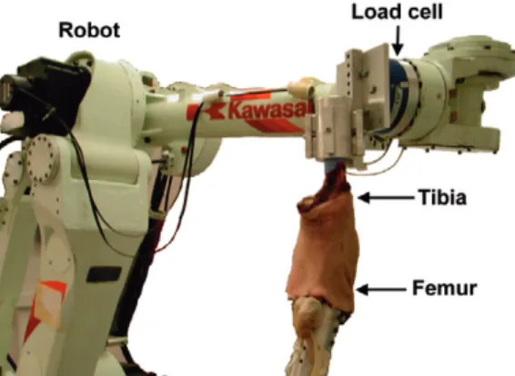
We used 10 fresh frozen cadaveric human knee specimens (age: 52–78 years). Each specimen was thawed overnight at room temperature and then examined using fluoroscopy. In preparation for testing, the specimen was cut approximately 25 cm above and below the knee joint, leaving the soft tis-sues around the knee intact. The specimen was pre-conditioned by manually flexing the knee 10 times before installing it on a robotic testing system (Figure 1). This testing system is composed of a robotic manipulator (Kawasaki Heavy Industry, Japan) and a 6-DOF load cell (JR3 Inc., CA). The testing system can operate in both force and dis-placement control modes and has been described in detail elsewhere (Li et al. 1999, 2004).
After the specimen was installed on the test-ing system, the robot determined the path of least resistance (passive path) in the unloaded knee. The passive path was determined by flexing the knee in
1° increments and finding a position (passive posi-tion) at which all forces and moments applied to the knee in the remaining 5 DOF were minimized. The passive positions formed a passive flexion path of the knee from full extension to 90° of flexion.
The knee was then tested under an anterior tibial load of 130 N at full extension, and 15°, 30°, 60°, and 90° flexion angles on the passive path. At each selected flexion angle, the robot moved the knee in 5 DOF (flexion angle was fixed) until an equilibrium position was reached, where the force transferred through the knee balanced the anterior tibial load. The robot recorded the new equilibrium position of the knee, representing the kinematic response of the knee to the applied load.
Next, the ACL was resected via a small medial arthrotomy with the knee in 30° of flexion. After cutting the ACL, the arthrotomy and skin were closed in layers. The kinematics of the intact knee under the anterior tibial load was replayed at each of the selected flexion angles. The force transmit-ted through the knee joint was measured by the load cell. The difference between this force and the force measured in the intact knee represented the in-situ force in the ACL under the anterior tibial load, using the principle of superposition (Li et al. 1999).
After measuring the ACL forces, the ACL was reconstructed using a 10-mm-wide BPTB graft. The operation mimicked the normal operative procedure using a two-incision technique as previ-ously reported (Gill and Steadman 2002). The tibial tunnel was made anterior to the PCL (7–9 mm) and 7 mm lateral to the medial femoral condyle on
Figure 1. The robotic testing system with a knee specimen installed.
the downslope of the medial tibial spine. A lateral femoral incision was used to place the rear-entry femoral aiming device, to allow femoral tunnel reaming from the outside inward, and to assist in graft fixation using a femoral interference screw. The femoral tunnel was made at the 11 o’clock position of the lateral femoral condyle. After fixing the femoral bone plug with an interference screw, the graft was pre-tensioned with 5 cycles of flex-ion-extension. The tibial bone plug was secured in place with an interference screw at full extension with 90 N of tension applied to the graft. Screws were placed on the anterior surface of the tibial bone plug, adjacent to the cancellous bone plug surface. The arthrotomy was then closed.
The ACL-reconstructed knee was tested using the same protocol as used for the intact knee. After measuring the kinematics of the knee under the anterior tibial load at the selected flexion angles, the ACL graft was released from the specimen. The kinematics of the ACL-reconstructed knee were then replayed at each flexion angle, as the load cell measured the forces transmitted through the knee. Again, using the principle of superposition, the in-situ force of the ACL graft was obtained by calcu-lating the difference between the forces measured before and after releasing the ACL graft.
The force data measured from the load cell con-sisted of three force components. These compo-nents were used to calculate magnitude of the force and two angles representing the orientation of the force: elevation and deviation (Figure 2). The ele-vation angle was defined as the angle between the projection of the force vector in the sagittal plane and the tibial plateau (Figure 2a), and the deviation angle was defined as the angle between the projec-tion of the force vector on the tibial plateau and the sagittal plane (Figure 2b).
Statistics
We used a repeated measures analysis of variance (ANOVA) and the Student-Newman-Keuls (SNK) test to detect statistically significant differences in translation and internal tibial rotation between the intact and ACL-reconstructed knees at each flex-ion angle. Similarly, we used repeated measures ANOVA and SNK test to compare the magnitude of the forces and orientation of the ACL and graft forces. Differences were considered to be statisti-cally significant when p < 0.05. All data are pre-sented using the mean value and 95% confidence interval (CI).
Results
Anterior tibial translation and axial tibial rota-tion
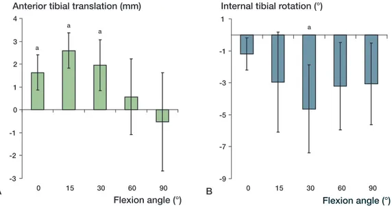
The anterior tibial translation after ACL reconstruc-tion was higher than that measured in the intact knee at all selected flexion angles except 90° (Figure 3a). At full extension, the anterior tibial transla-tion in the ACL-reconstructed knee increased by 1.6 mm (CI: 0.8–2.4 mm) compared to that of the intact knee (p = 0.002). The anterior tibial transla-tion of the reconstructed knee increased until 15° of flexion and decreased thereafter. At 15°, the anterior tibial translation of the ACL-reconstructed knee was 2.6 (CI: 1.8–3.4) mm greater than that of the intact knee (p = 0.0006).
ACL reconstruction reduced the internal tibial rotation of the intact knee at all flexion angles under the anterior tibial load (Figure 3b). At full extension, the ACL reconstruction reduced internal tibial rotation by 1.2° (CI: 0.2–2.2). The internal tibial rotation of the reconstructed knee decreased
Figure 2. Definition of orientation angles of the force. A. The elevation angle is measured in the sagittal plane. B. The deviation angle is measured in the plane of the tibial plateau.
A
further to 30° of flexion, and increased thereafter. At 30° of flexion, internal rotation was reduced by 4.6° (CI: 1.9–7.3) after ACL reconstruction (p = 0.009).
In-situ forces of the ACL and graft
The mean values of the graft forces were greater than those of the corresponding ACL forces at all flexion angles (Figure 4). The ACL force increased with flexion until 15° and decreased thereafter, whereas the graft force increased until 30° of flex-ion and decreased thereafter. The graft force at 15° was 161 N (CI: 142–180), which was statistically significantly greater than the ACL force of 133 N (CI: 146–120) (p = 0.01).
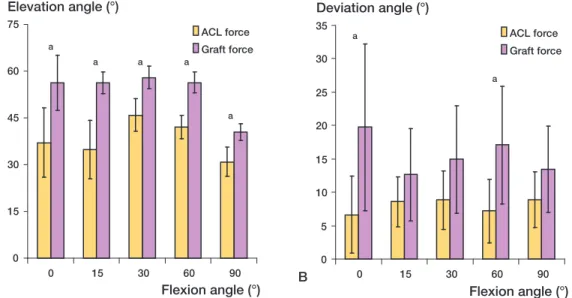
Elevation angles of the ACL and graft forces
There was a statistically significant increase in the elevation angle of the graft forces compared to the corresponding ACL forces at all flexion angles (Figure 5a). At full extension, the elevation angle of the ACL force was 37° (CI: 26–48), while the elevation angle of the graft force was 56° (CI: 47– 65). This represents an increase of approximately 19° (p = 0.02). At 15° of flexion, the mean ACL force elevation angle was 35° (CI: 26–44) and the ACL graft force elevation angle was 56.3° (CI: 53–60) (p = 0.003).
Deviation angles of the ACL and graft forces
The mean deviation angles of the graft forces were consistently greater than those of the ACL forces at all selected flexion angles (Figure 5b). At full extension, the deviation angle of the ACL force was 6.6° (CI: 0.9–12), while that of the ACL graft force was 20° (CI: 7.2–32). This difference was
Figure 3. Change in knee kinematics after ACL reconstruction under the anterior tibial load of 130 N compared to the intact knee: A) change in anterior tibial translation, and B) change in internal tibial rotation. Error bars represent 95% confidence interval. a statistically significant difference.
-3 -2 -1 0 1 2 3 4 0 15 30 60 90 Flexion angle (°) Anterior tibial translation (mm)
a a a -9 -7 -5 -3 -1 1 0 15 30 60 90 Flexion angle (°) Internal tibial rotation (°)
a
A B
Figure 4. In-situ forces of the ACL and graft under an ante-rior tibial load of 130 N. Error bars represent 95% confi-dence interval. a statistically significant difference.
0 40 80 120 160 200 0 15 30 60 90 Flexion angle (°) Force (N) ACL force Graft force a
statistically significant (p = 0.05). At 15° of flex-ion, the ACL force had a deviation angle of 8.6° (CI: 4.9–12) and the graft deviation angle was 13° (CI: 5.7–20°).
Discussion
Experimental studies have demonstrated that the ACL plays an important role in limiting anterior tibial translation (Butler et al. 1980, Fukubayashi et al. 1982). The ACL has been shown to be maximally loaded in the first 30° of flexion under anterior tibial loads and simulated muscle loads (Sakane et al. 1997, Li et al. 1999). The change in ACL force with flexion observed in the current study is consistent with these previous studies.
ACL reconstructions commonly use two types of autogenous graft materials: the BPTB and quadrupled hamstring grafts (Clancy et al. 1982, Brown et al. 1993). There have been many investi-gations into the effect of variations in initial graft tension, graft placement, fixation technique, and graft selection on anterior knee stability after ACL reconstruction (Hefzy and Grood 1986, Kurosaka et al. 1987, Fleming et al. 1992). Previous cadaver studies have demonstrated that ACL reconstruction can restore anterior translation under anterior tibial loads (Markolf et al. 1996, Hoher et al. 2001). In
the current study, after ACL reconstruction, ante-rior tibial translation was restored to within 2.6 mm of the intact knee.
In previous studies, ACL graft forces were measured when the anterior tibial translation was matched to that of the intact knee under an anterior tibial load at 30° of flexion (Markolf et al. 1996, Goss et al. 1997). These graft forces were dramati-cally increased compared to the ACL forces under the anterior tibial load. Our study also found that the mean ACL graft forces were higher than the ACL forces at each flexion angle (significantly higher at 15° of flexion), even though the anterior tibial translation measured in the reconstructed knee was slightly larger than the translation of the intact knee. The difference in force between the ACL and graft measured in this study was not as large as those reported in the literature (Gront-vedt et al. 1996, Markolf et al. 1996, Goss et al. 1997). This may be due to the differences in surgi-cal techniques used in these studies. In our study, we attempted to simulate the procedure used clini-cally.
Even though anterior tibial translation was restored after reconstruction, the average internal tibial rotation was reduced compared to the intact knee. This difference was statistically significant at 30° under the anterior tibial load (Figure 3b). Other studies have also reported that the
ACL-recon-Figure 5. Orientation of the ACL and graft forces under an anterior tibial load of 130 N. A. Elevation angle. B. Deviation angle. Error bars represent 95% confidence interval. a statistically significant difference.
0 15 30 45 60 75 0 15 30 60 90 Flexion angle (°) Elevation angle (°) ACL force Graft force a a a a a 0 5 10 15 20 25 30 35 0 15 30 60 90 Flexion angle (°) Deviation angle (°) ACL force Graft force a a A B
structed knee is externally rotated compared to the contralateral, uninjured limb under dynamic load-ing conditions in vivo (Nordt et al. 1999, Ristanis et al. 2003). The altered tibial rotations observed after ACL reconstruction may have an undesirable effect on patellofemoral joint contact pressures. For example, previous studies have reported that increased external tibial rotation may result in increased patellofemoral contact pressure on the lateral facet of the patellofemoral joint (Li et al. 2004). The effects of altered tibial rotation after ACL reconstruction on knee joint mechanics have not been well described in the literature.
Our findings show that in addition to the magni-tude of the ACL force, the orientation of the ACL graft force may also affect the knee kinematics after ACL reconstruction. Ideally, ACL reconstruction should recreate the force vector of the intact ACL. In contemporary ACL reconstruction, the location of the femoral tunnel was selected based on creat-ing an isometric graft—at the superior region of the ACL footprint on the femur (Musahl and Fu 2003). Although the isometry of a graft is depen-dent on both femoral and tibial graft placement, it has been believed to be less sensitive to changes in location on the tibia (Hefzy and Grood 1986, Hefzy et al. 1989). The effects of tibial tunnel placement on ACL graft forces during physiological loading conditions are, however, unclear.
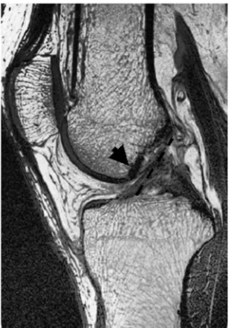
In order to reduce the graft impingement with the femoral condyle notch, the tibial tunnel is usually centered on the posterior half of the ACL footprint, approximately 7–9 mm in front of the PCL (Musahl and Fu 2003). Such an ACL reconstruction proce-dure alters the orientation of the ACL graft com-pared to the intact ACL. As shown in the magnetic resonance image of a normal human knee in Figure 6, the native ACL is hourglass-shaped. This allows the ACL to be attached more anteriorly on the tibial plateau, yet to remain in contact with the intercon-dylar notch during terminal extension. Because the graft does not have this hourglass geometry, it must be placed more posteriorly to prevent notch impingement.
While the current location of the tibial tunnel may help to reduce notch impingement, the result-ing elevation angles of the ACL graft force may be significantly greater than those of the ACL, as demonstrated by our data. The consequence of
the higher elevation angle is that the graft resists anterior tibial loads less efficiently than the ACL. Higher ACL graft forces are therefore required to reproduce the same anterior tibial translation under the anterior tibial load. In our study, we observed a slight increase in anterior tibial translation after ACL reconstruction, with an increased tension in the graft compared to the ACL.
The deviation angle of the graft was also increased compared to the intact ACL, although the differences were statistically significant at only two flexion angles. Since the tibia tunnel is near the insertion of the anteriomedial bundle of the ACL, increased deviation of the ACL graft forces is likely due to the placement of the femoral tunnel. The femoral tunnel is usually positioned in the 10 or 11 o’clock position in the coronal plane. How-ever, the resulting graft orientation may be more lateral and may contribute to an external rotation moment. Thus, the ACL graft resists internal tibial rotation more efficiently, as demonstrated by the reduced internal tibial rotation noted in our study.
The location of the tibiofemoral tunnels also affects the length of the graft. As shown by Heftzy and Grood (1986), moving the tibial tunnel from
Figure 6. MR image of the orientation of the ACL in a human subject in a relaxed, fully extendedposition. The arrow shows ACL contact with the intercondylar notch. The dashed line indicates an ACL graft orientation that is more vertical than that of ACL fibers.
the anterior portion to the posterior portion of the ACL insertion on the tibia may shorten the graft length by over 10 mm. Our previous studies have demonstrated that shortening a graft increases the stiffness of the graft (Li et al. 2003, DeFrate et al. 2004). Consequently, the stiffer graft may experience higher loads during knee motion. The increased graft forces can result in higher tibio-femoral joint contact forces which might alter the biomechanical environment of the joint after ACL reconstruction.
It should be noted that we only evaluated ACL reconstruction at time zero after operation, due to the in-vitro nature of the investigation. Graft healing and post-rehabilitation effects on graft biomechanics have not been addressed. However, our study may provide insight into the mechani-cal function of the ACL graft, and may help in the future development of an optimal ACL reconstruc-tion scheme (using either single or double bundle grafts) that can reproduce the complex function of the ACL. Future studies should quantify ACL bio-mechanics and ACL graft function in vivo.
Author contributions
GL provided the initial concept, contributed to the experi-mental design, the collection and analysis of data and wrote the manuscript. RP and LEDF contributed to the experi-mental design, the collection and analysis of data, and the writing of the manuscript. JDY, SUP and TJG contributed to the experimental design and the collection of data. They performed the surgeries and provided clinical insight into the experimental data.
No competing interests declared.
Brown C H, Jr., Steiner M E, Carson E W. The use of ham-string tendons for anterior cruciate ligament reconstruc-tion. Technique and results. Clin Sports Med 1993; 12 (4): 723-56.
Butler D L, Noyes F R, Grood E S. Ligamentous restraints to anterior-posterior drawer in the human knee. A bio-mechanical study. J Bone Joint Surg (Am) 1980; 62 (2): 259-70.
Clancy W G, Jr., Nelson D A, Reider B, Narechania R G. Anterior cruciate ligament reconstruction using one-third of the patellar ligament, augmented by extra-articular tendon transfers. J Bone Joint Surg (Am) 1982; 64 (3): 352-9.
DeFrate L E. van der Ven A, Gill T J, Li G. The effect of length on the structural properties of an Achilles tendon graft as used in posterior cruciate ligament reconstruction. Am J Sports Med 2004; 32 (4): 993-7.
Fithian D C, Paxton E W, Stone M L, Luetzow W F, Csin-talan R P, Phelan D, Daniel D M. Prospective trial of a treatment algorithm for the management of the anterior cruciate ligament-injured knee. Am J Sports Med 2005; 33 (3): 335-46.
Fleming B, Beynnon B, Howe J, McLeod W, Pope M. Effect of tension and placement of a prosthetic anterior cruci-ate ligament on the anteroposterior laxity of the knee. J Orthop Res 1992; 10 (2): 177-86.
Fox J A, Nedeff D D, Bach Jr, B R, Spindler K P. Anterior cruciate ligament reconstruction with patellar autograft tendon. Clin Orthop 2002; (402): 53-63.
Fukubayashi T, Torzilli P A, Sherman M F, Warren R F. An in vitro biomechanical evaluation of anterior-posterior motion of the knee. Tibial displacement, rotation, and torque. J Bone Joint Surg (Am) 1982; 6 (2): 258-64. Gill T J, Steadman J R. Anterior cruciate ligament
recon-struction the two-incision technique. Orthop Clin North Am 2002; 33 (4): 727-35, vii.
Goss B C, Hull M L, Howell S M. Contact pressure and tension in anterior cruciate ligament grafts subjected to roof impingement during passive extension. J Orthop Res 1997; 15 (2): 263-8.
Grontvedt T, Pena F, Engebretsen L. Accuracy of femoral tunnel placement and resulting graft force using one- or two-incision drill guides. A cadaver study on ten paired knees. Arthroscopy 1996; 12 (2): 187-92.
Hefzy M S, Grood E S. Sensitivity of insertion locations on length patterns of anterior cruciate ligament fibers. J Bio-mech Eng 1986; 108 (1): 73-82.
Hefzy M S, Grood E S, Noyes F R. Factors affecting the region of most isometric femoral attachments. Part II: The anterior cruciate ligament. Am J Sports Med 1989; 17 (2): 208-16.
Hoher J, Kanamori A, Zeminski J, Fu F H, Woo S L. The position of the tibia during graft fixation affects knee kinematics and graft forces for anterior cruciate ligament reconstruction. Am J Sports Med 2001; 29 (6): 771-6. Jomha N M, Borton D C, Clingeleffer A J, Pinczewski L
A. Long term osteoarthritic changes in anterior cruciate ligament reconstructed knees. Clin. Orthop 1999; (358): 188-93.
Kurosaka M, Yoshiya S, Andrish J T. A biomechanical com-parison of different surgical techniques of graft fixation in anterior cruciate ligament reconstruction. Am J Sports Med 1987; 15 (3): 225-9.
Li G, Rudy T W, Sakane M, Kanamori A, Ma C B, Woo S L. The importance of quadriceps and hamstring muscle loading on knee kinematics and in-situ forces in the ACL. J Biomech 1999; 32 (4): 395-400.
Li G, DeFrate L, Suggs J, Gill T. Determination of optimal graft lengths for posterior cruciate ligament reconstruc-tion. A theoretical analysis. J Biomech Eng 2003; 125 (2): 295-9.
Li G, DeFrate L E, Zayontz S, Park S E, Gill T. J. The effect of tibiofemoral joint kinematics on patellofemoral contact pressures under simulated muscle loads. J Orthop Res 2004; 22 (4): 801-6.
Li G, Defrate L E, Rubash H E, Gill T. J. In vivo kinematics of the ACL during weight-bearing knee flexion. J Orthop Res 2005; 23 (2): 340-4.
Lohmander L S, Ostenberg A, Englund M, Roos H. High prevalence of knee osteoarthritis, pain, and functional limitations in female soccer players twelve years after anterior cruciate ligament injury. Arthritis Rheum 2004; 50 (10): 3145-52.
Markolf K L, Burchfield D M, Shapiro M M, Cha C W, Finerman G A, Slauterbeck J L. Biomechanical conse-quences of replacement of the anterior cruciate ligament with a patellar ligament allograft. Part II: forces in the graft compared with forces in the intact ligament. J Bone Joint Surg (Am) 1996; 78 (11): 1728-34.
Musahl V, Fu F H. Fundamentals in ACL reconstruction: The American view. Navigation and robotics in total joint and spine surgery. J B Stiehl, W H Konermann, R G. Haaker. Berlin, Germany, Springer-Verlag 2003: 369-74.
Nedeff D D, Bach B R, Jr. Arthroscopic anterior cruciate ligament reconstruction using patellar tendon autografts. Orthopedics 2002; 25 (3): 343-57; quiz 358-9.
Nordt W E, 3rd, Lotfi P, Plotkin E, Williamson B. The in vivo assessment of tibial motion in the transverse plane in anterior cruciate ligament-reconstructed knees. Am J Sports Med 1999; 27 (5): 611-6.
Ristanis S, Giakas G, Papageorgiou C D, Moraiti T, Ster-giou N, Georgoulis A D. The effects of anterior cruciate ligament reconstruction on tibial rotation during pivot-ing after descendpivot-ing stairs. Knee Surg Sports Traumatol Arthrosc 2003; 11 (6): 360-5.
Sakane M, Fox R J, Woo S L, Livesay G A, Li G, Fu F. H. In situ forces in the anterior cruciate ligament and its bundles in response to anterior tibial loads. J Orthop Res 1997; 15 (2): 285-93.
Thornton G M, Boorman R S, Shrive N G, Frank C B. Medial collateral ligament autografts have increased creep response for at least two years and early immobilization makes this worse. J Orthop Res 2002; 20 (2): 346-52.