*Corresponding authors. Hyoung Doo Shin, Tel: 82-2-705-8615; Fax: 82-2-2026-4299; E-mail: hdshin@sogang.ac.kr; Yong Sung Kim, Tel: 82-42-879-8110; Fax: 82-42-879-8119; E-mail: yongsung@kribb. re.kr
DOI 10.5483/BMBRep.2010.43.12.830
Received 18 June 2010, Accepted 25 October 2010
Keywords: DNA methylation, Epigenetic modification, Gene
ex-pression, Human embryonic stem cell, Retinoic acid
Epigenetic modification of retinoic acid-treated human
embryonic stem cells
Hyun Sub Cheong
1, Han Chul Lee
2, Byung Lae Park
1, Hyemin Kim
3, Mi Jin Jang
3, Yong Mahn Han
3, Seun-Young Kim
2,
Yong Sung Kim
2,* & Hyoung Doo Shin
1,4,*
1Department of Genetic Epidemiology, SNP Genetics, Inc., Seoul 153-803, 2Medical Genomics Research Center, Korea Research Institute of Bioscience and Biotechnology, Daejeon 305-806, 3Department of Biological Sciences and Center for Stem Cell Differentiation, Korean Advanced Institute of Science and Technology, Daejeon 305-701, 4Department of Life Science, Sogang University, Seoul 121-742, Korea
Epigenetic modification of the genome through DNA methyl-ation is the key to maintaining the differentiated state of hu-man embryonic stem cells (hESCs), and it must be reset during differentiation by retinoic acid (RA) treatment. A genome-wide methylation/gene expression assay was performed in order to identify epigenetic modifications of RA-treated hESCs. Between undifferentiated and RA-treated hESCs, 166 differentially me-thylated CpG sites and 2,013 differentially expressed genes were discovered. Combined analysis of methylation and ex-pression data revealed that 19 genes (STAP2, VAMP8, C10orf26,
WFIKKN1, ELF3, C1QTNF6, C10orf10, MRGPRF, ARSE, LSAMP, CENTD3, LDB2, POU5F1, GSPT2, THY1, ZNF574, MSX1, SCMH1, and RARB) were highly correlated with each other.
The results provided in this study will facilitate future inves-tigations into the interplay between DNA methylation and gene expression through further functional and biological studies. [BMB reports 2010; 43(12): 830-835]
INTRODUCTION
Human embryonic stem cells (hESCs) are unique in their abil-ity to maintain pluripotence. This property makes hESCs lead-ing candidates for use in cell therapy and in studies on early human development. Retinoic acid (RA), the most potent natu-ral form of vitamin A, plays an important role in mediating the growth and differentiation of both normal and transformed cells (1, 2). It is essential for many diverse biological functions including growth, vision, reproduction, embryonic develop-ment, differentiation of epithelial tissues, and immune
re-sponses (2). In vitro, RA induces differentiation of hESCs into a number of specific cell types.
Differentiation of a specific cell type involves the establish-ment of a precise epigenetic profile composed of genome- wide epigenetic modifications such as DNA methylation and histone modification. Since epigenetic modifications in gene areas regulate transcriptional activity, the epigenetic profile of the cell reflects the transcriptome, at least partially (3-5).
hESCs have been investigated using multiple techniques, in-cluding gene expression profiling, mitochondrial sequencing, immunocytochemistry, genotyping, functional assays, and DNA methylation assay (6-10). DNA methylation of the ge-nome is the key to maintaining the differentiated state of hESCs (11, 12), and it must be reset during differentiation by RA treatment.
Differences between hESC lines with respect to gene ex-pression profiles have been investigated before (13), and it has also been demonstrated that hESCs have unique DNA methyl-ation profiles compared to other cell types, including embry-onic germ cells, trophoblast stem cells, and several adult stem cell populations (8, 14). Key regulators of development such as Oct4 and NANOG are also controlled by epigenetic mecha-nisms (15, 16). However, a whole-genomic correlation study on DNA methylation and gene expression has not been reported.
The present study utilized DNA methylation and gene ex-pression assays to generate whole-genomic methylation and gene expression profiles for both undifferentiated hESCs and RA-treated hESCs. These results provide valuable information that can be used to identify differentially methylated CpG sites and differentially expressed genes.
RESULTS
We applied a comprehensive DNA methylation profiling ap-proach to assess the epigenetic states of three hESC lines (CHA3-hES, CHA4-hES, and SNUhES3) as well as their epi-genetic modifications after RA treatment. A whole-genome DNA methylation assay method was used to analyze the meth-ylation status of 27,578 CpG sites selected from more than
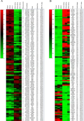
Fig. 1. Heatmaps of differential methylation and gene expression
assays. (A) The 100 most differentially methylated CpG sites in un-differentiated hESCs and RA-treated hESCs. Sample IDs and their average beta values (methylation levels) are shown. Chromosome, coordinate, related gene name, delta beta value, and Diff. P val-ues of each CpG site are also presented. (B) The 100 most differ-entially expressed genes in undifferentiated hESCs and RA-treated hESCs. Sample IDs and their average signal value (expression lev-el) are shown. Gene name, fold change values, and Diff. P val-ues are also presented.
14,000 well-annotated genes. We measured the overall meth-ylation levels after RA treatment. We found that the average methylation level in the RA-treated hESCs (29.5%) was greater than in the undifferentiated hESCs (27.1%). The lower methyl-ation level we obtained for the undifferentiated hESCs was ex-pected since global hypomethylation has been reported often in embryonic stem cells (17).
To discover which CpG sites contribute the most to the epi-genetic modification of hESCs by RA treatment, we compared the DNA methylation patterns between undifferentiated hESCs and RA-treated hESCs. This analysis produced a list of 166 CpG sites from 151 genes that significantly contribute to the separation of the two groups. Among them, the top 100 CpG sites, based on |Δβ|, are shown in Fig. 1A. We then clustered all of the samples based on their relative methylation levels at these 166 CpG sites (data not shown). Three hESC lines were correctly aggregated into the two other major clusters, which comprise undifferentiated hESC lines and RA-treated cells. We also investigated three hESC lines for differential ex-pression of genes upon RA treatment. A total of 9,736 distinct genes (23% of the RefList) passed the expression criteria of a Detection Score ≥0.99. Among them, 2,013 genes were dif-ferentially expressed. We observed that 1,003 genes were up-regulated (>1.5-fold) and 1,010 genes were downup-regulated (<0.66-fold). The extreme differences observed between un-differentiated hESCs and RA-treated hESCs are shown in Fig. 1B. Combined analysis of methylation and expression data re-vealed that 19 genes (STAP2, VAMP8, C10orf26, WFIKKN1,
ELF3, C1QTNF6, C10orf10, MRGPRF, ARSE, LSAMP, CENTD3, LDB2, POU5F1, GSPT2, THY1, ZNF574, MSX1, SCMH1, and RARB) were highly correlated with each other (Pearson
corre-lation coefficient ≥0.8) (Supplementary Table 1).
To validate the methylation status of the highly correlated genes, we selected two genes (CENTD3 and MSX1) and per-formed bisulfate sequencing. Bisulfate sequencing of 400-500 bp including Illumina probe position revealed hypermethy-lation (36.7% and 19.6%) in SNUhES3 cells after RA treatment that was consistent with the genome-wide DNA methylation (Supplementary Fig. 1).
DISCUSSION
Despite their differences in origin, different sample preparation methods, and karyotypes, three hESC lines were correctly ag-gregated into two other major clusters, which comprise un-differentiated hESC lines and RA-treated cells. This suggests that the three hESC lines share a common epigenetic signature, which is likely linked to embryonic stem (ES) cell-specific properties such as self-renewal and pluripotence.
CpGs on the C10orf10, FAM12B, VAMP8, CLDN15, and
FLJ20273 genes were the most hypomethylated, whereas C7orf29, CHFR, GSPT2, HDCMA18P, and MSX1 were the
most hypermethylated after RA treatment. Of these genes, methylation of CHFR is known to be associated with silencing
of CHFR expression in various types of cancer (18), and CHFR is also known as a tumor suppressor (19). This means that cells in which CHFR was epigenetically inactivated constituted dif-ferentiated hESCs.
In order to define the relationship between methylation and expression of genes, we performed gene expression profiling to compare both methylation status and gene expression levels. Among the differentially methylated genes, HOXB5,
INS-IGF2, HOXA5, LCP1, and ANKRD38 were the most highly
upregulated, whereas PRDM14, ZIC2, C9orf135, MIAT, and
SFRP2 were downregulated after RA treatment. Among these
genes, HOXA5 was found to be rapidly induced within mouse ES cells as a result of RA treatment (20). In addition,
knock-down of PRDM14 by siRNA induced expression of early differ-entiation marker genes (21). These previous studies were re-markably consistent with our findings.
Combined analysis revealed that 19 genes were highly cor-related with each other. Among them, we identified seven genes (STAP2, VAMP8, C10orf26, WFIKKN1, ELF3, C1QTNF6 and C10orf10) that were hypomethylated and upregulated upon RA treatment. STAP2 is a signal-transducing adaptor mol-ecule that binds to STAT3 and STAT5, resulting in regulation of integrin-mediated T-cell adhesion through protein degrada-tion of focal adhesion kinase (22). VAMP8 is a member of the vesicle associated membrane protein (VAMP) family and is re-quired for activation-induced degranulation of mature human mast cells (23).
MRGPRF, ARSE, LSAMP, CENTD3, LDB2, POU5F1, GSPT2, THY1, and ZNF574 were identified as hypermethylated and
downregulated (Supplementary Table 1). Among these nine genes, POU5F1 (also known as Oct4) is a transcription factor previously shown to be expressed only in pluripotent cells of the embryo where it promotes differentiation when down-regulated (24-26). RA-induced differentiation of a human em-bryonic carcinoma cell line into neurons is also accompanied by sequential DNA methylation of the promoter regions of
POU5F1 (27). THY1, which plays a critical role in maintaining
the undifferentiated status of ES cells, was also hypermethy-lated and downreguhypermethy-lated (correlation P value = 0.02) (28, 29). The mechanism of THY1 gene inactivation due to hyper-methylation has been previously determined (30, 31).
Three genes (MSX1, SCMH1, and RARB) did not fit in the standard paradigm of extensive methylation being correlated with gene silencing. In previous studies, upregulation of RARB was reported in RA-treated embryonic stem cells and cancer cells (32, 33). In this study, RARB was also hypermethylated and upregulated in RA-treated hESCs (Supplementary Table 1). This methylation, unlike the common epigenetic paradigm, shows positive correlation between the methylation of two up-stream CpG sites and gene expression. Our results indicate that methylation of the upstream CpG sites in these hESC lines was correlated with an increase in RARB expression. The data further suggest that methylation of CpG sites is required for a cell to express high levels of RARB when induced by RA. There is also additional evidence that DNA hypermethylation in specific regions (promoter or genebody) can lead to upregu-lation of transcription (34, 35).
In our combined analysis, CpG sites of 11 genes were lo-cated in the promoter region while others were in the coding region. The relationship between promoter methylation and gene expression is well-known. Recent studies have found that gene-body methylation in differentially expressed genes is a consistent phenomenon throughout the human genome (36- 38). Although we have no functional evidence, the change in DNA methylation levels in the promoter and coding regions of the gene can alter expression levels.
In summary, we presented genome-wide DNA methylation
and gene expression profiles of hESCs upon RA treatment. To our knowledge, this is the first time such research has been reported. The results provided in this study will facilitate inves-tigations into the interplay between DNA methylation and gene expression through further functional and biological studies.
MATERIALS AND METHODS
Human embryonic stem cell culture, RNA, and DNA extraction
Three hESC lines (CHA3-hES, CHA4-hES, and SNUhES3) were analyzed for this study by following Human Subjects Institutional Review Board approved protocols (Supplementary Table 2). The hESCs were maintained on Mitomycin C (Sigma, St. Louis, MO, USA)-treated STO (ATCC CRL-1503) feeders (39). Prior to being treated with retinoic acid (RA), hESCs were transferred onto MatrigelⓇ
(BD Biosciences, Bedford, MA, USA)-coated culture dishes in STO-conditioned medium (CM), as described previously (40). After 2 d of feeder-free culture, 50 μM RA was applied to hESCs for 5 d. When treated with RA under stem-cell conditions, CHA4-hES cells displayed a drastic morphological change as a differentiated state, and Western blot analysis also showed that expression of OCT4 protein was dramatically reduced (Supplementary Fig. 2). Thus, hESCs could be differentiated by treatment with RA for 5 d (26). To extract total RNA from the control and RA-treated hESCs, TRIzol (Invitrogen) was used according to the manu-facturer’s protocol. DNA was extracted from the hESCs using a Qiagen DNeasy kit (Qiagen, Hilden, Germany) in preparation for analysis on bead arrays.
Whole-genome DNA methylation assay
Of the three hESC lines, two cell lines (CHA4-hES and SNUhES3) were run in quadruplicate and one (CHA3-hES) in singlet. One microgram of genomic DNA from each sample was bisulfite converted using a EZ DNA methylation kit (Zymo Research, Orange, CA, USA), and 200 ng of the converted DNA was used for amplification. Amplified DNA was hybri-dized to the HumanMethylation27 BeadChip (Illumina, San Diego, CA, USA), and the arrays were imaged using a Bead-ArrayTM Reader (Illumina). Image processing and intensity data extraction were performed according to Illumina’s instructions. Each methylation signal was used to compute a "Beta" value (β), which is a quantitative measure of DNA methylation rang-ing from 0 for completely unmethylated cytosines to 1 for completely methylated cytosines (8).
Whole-genome gene expression assay
All three cell lines were run in triplicate. RNA isolated from the hESC lines was used for gene expression analysis using the Human-6 Whole-Genome Expression BeadChip (Illumina). Biotin-labeled cRNA was produced by means of a linear am-plification kit (Ambion, Austin, TX, USA) using 300 ng of qual-ity-checked total RNA as an input. Chip hybridizations, wash-ing, Cy3-streptavidin stainwash-ing, and scanning were performed
on a BeadArray™ Reader (Illumina) platform using reagents and by following protocols supplied by the manufacturer. The "Detection Score" was used to determine expression.
Differential DNA methylation/gene expression analysis
Differential DNA methylation and gene expression analysis were performed with the Methylation and Gene Expression Modules in Illumina’s BeadStudio software. The Illumina data were normalized using the background and quantile functions for DNA methylation and gene expression, respectively. We identified CpG sites/genes that were differentially methy-lated/expressed in RA-treated hESCs using the t-test error mod-el implemented in BeadStudio. The methylation/expression difference score (Diff. Score) takes into account background noise and sample variability (41).
In order to identify differentially methylated CpG sites be-tween control hESCs and RA-treated hESCs, we performed a t-test on the difference in mean methylation level between the two groups. We selected sites with a Diff. Score > 20 (P value < 0.01) and with an additional filter of mean |Δβ| > 0.17, the estimated error in β (42). This resulted in a list of 166 sites, the top 100 of which, based on |Δβ|, were chosen to provide a readable list in Fig. 1A. For differential gene expression data analysis, "signal" values below the detection limit were arbitra-rily set to the level of threshold detection in order to avoid nonsense values for expression ratios. Significantly differ-entially expressed genes had a fold change of at least 50% with a Diff. Score > 20 (P value < 0.01). This resulted in a list of 2,013 differentially expressed genes. Among them, the top 100 genes, based on the fold change, are shown in Fig. 1B.
Combined analysis of differentially methylated/expressed data
Differentially methylated CpG sites (n = 166) and differ-entially expressed genes (n = 2,013) were combined based on gene name represented in both data sets. Pearson correlation coefficients were calculated between the expression signal val-ues and methylation β valval-ues. We collected significantly corre-lated sites/genes with a Pearson correlation coefficient >0.8 (P value < 0.05) (Supplementary Table 1).
Bisulfite sequencing
Genomic DNA (1 μg) of SNUhES3 was modified by sodium bi-sulfite using a EZ DNA Methylation kit (ZYMO Research) ac-cording to the manufacturer’s instructions. For amplification of bisulfite-modified DNA, we used the MethPrimer program (43) to design the forward and reverse primer sets of two genes (CENTD3 and MSX1), including Illumina probe position. Bisulfite-modified DNA (1 μl) was amplified in a 20 μl volume containing primers. Primer information is available in Supple-mental Table 3. Samples were heated to 95oC for 12 min and then subjected to 35 cycles of denaturation at 95oC for 45 s,
annealing for 45 s, extension at 72oC for 60 s, and then in-cubation at 72oC for 10 min and cooling to 4oC. The PCR
products were visualized on a 1% agarose gel by ethidium
bromide staining, purified from the gel using a Qiagen Gel Extraction kit, and cloned using pGEM-T Easy Vector (Pro-mega). Ten clones were randomly chosen for sequencing. Complete bisulfite conversion was assured when <0.01% of the cytosines in non-CG dinucleotides in the final sequence were not converted.
Acknowledgements
This research was supported by the Bio R&D program through the National Research Foundation of Korea, funded by the Ministry of Education, Science and Technology (2007-2004101).
REFERENCES
1. Chambon, P. (1996) A decade of molecular biology of ret-inoic acid receptors. Faseb J. 10, 940-954.
2. Soprano, D. R., Teets, B. W. and Soprano, K. J. (2007) Role of retinoic acid in the differentiation of embryonal carcinoma and embryonic stem cells. Vitam. Horm. 75, 69-95.
3. Lewis, J. D., Meehan, R. R., Henzel, W. J., Maurer-Fogy, I., Jeppesen, P., Klein, F. and Bird, A. (1992) Purification, sequence, and cellular localization of a novel chromoso-mal protein that binds to methylated DNA. Cell 69, 905-914.
4. Lee, J. H. and Skalnik, D. G. (2002) CpG-binding protein is a nuclear matrix- and euchromatin-associated protein localized to nuclear speckles containing human trithorax. Identification of nuclear matrix targeting signals. J. Biol.
Chem. 277, 42259-42267.
5. Tate, P. H. and Bird, A. P. (1993) Effects of DNA methyl-ation on DNA-binding proteins and gene expression. Curr.
Opin. Genet. Dev. 3, 226-231.
6. Adewumi, O., Aflatoonian, B., Ahrlund-Richter, L., Amit, M., Andrews, P. W., Beighton, G., Bello, P. A., Benvenisty, N., Berry, L. S., Bevan, S., Blum, B., Brooking, J., Chen, K. G., Choo, A. B., Churchill, G. A., Corbel, M., Damjanov, I., Draper, J. S., Dvorak, P., Emanuelsson, K., Fleck, R. A., Ford, A., Gertow, K., Gertsenstein, M., Gokhale, P. J., Hamilton, R. S., Hampl, A., Healy, L. E., Hovatta, O., Hyllner, J., Imreh, M. P., Itskovitz-Eldor, J., Jackson, J., Johnson, J. L., Jones, M., Kee, K., King, B. L., Knowles, B. B., Lako, M., Lebrin, F., Mallon, B. S., Manning, D., Mayshar, Y., McKay, R. D., Michalska, A. E., Mikkola, M., Mileikovsky, M., Minger, S. L., Moore, H. D., Mummery, C. L., Nagy, A., Nakatsuji, N., O'Brien, C. M., Oh, S. K., Olsson, C., Otonkoski, T., Park, K. Y., Passier, R., Patel, H., Patel, M., Pedersen, R., Pera, M. F., Piekarczyk, M. S., Pera, R. A., Reubinoff, B. E., Robins, A. J., Rossant, J., Rugg-Gunn, P., Schulz, T. C., Semb, H., Sherrer, E. S., Siemen, H., Stacey, G. N., Stojkovic, M., Suemori, H., Szatkiewicz, J., Turetsky, T., Tuuri, T., van den Brink, S., Vintersten, K., Vuoristo, S., Ward, D., Weaver, T. A., Young, L. A. and Zhang, W. (2007) Characterization of human embryonic stem cell lines by the International Stem Cell Initiative. Nat. Biotechnol. 25, 803-816.
7. Andrews, P. W., Benvenisty, N., McKay, R., Pera, M. F., Rossant, J., Semb, H. and Stacey, G. N. (2005) The
International Stem Cell Initiative: toward benchmarks for human embryonic stem cell research. Nat. Biotechnol. 23, 795-797.
8. Bibikova, M., Chudin, E., Wu, B., Zhou, L., Garcia, E. W., Liu, Y., Shin, S., Plaia, T. W., Auerbach, J. M., Arking, D. E., Gonzalez, R., Crook, J., Davidson, B., Schulz, T. C., Robins, A., Khanna, A., Sartipy, P., Hyllner, J., Vanguri, P., Savant-Bhonsale, S., Smith, A. K., Chakravarti, A., Maitra, A., Rao, M., Barker, D. L., Loring, J. F. and Fan, J. B. (2006) Human embryonic stem cells have a unique ep-igenetic signature. Genome Res. 16, 1075-1083.
9. Liu, Y., Shin, S., Zeng, X., Zhan, M., Gonzalez, R., Mueller, F. J., Schwartz, C. M., Xue, H., Li, H., Baker, S. C., Chudin, E., Barker, D. L., McDaniel, T. K., Oeser, S., Loring, J. F., Mattson, M. P. and Rao, M. S. (2006) Genome wide profiling of human embryonic stem cells (hESCs), their derivatives and embryonal carcinoma cells to develop base profiles of U.S. Federal government ap-proved hESC lines. BMC Dev. Biol. 6, 20.
10. Loring, J. F. and Rao, M. S. (2006) Establishing standards for the characterization of human embryonic stem cell lines. Stem Cells 24, 145-150.
11. Brunner, A. L., Johnson, D. S., Kim, S. W., Valouev, A., Reddy, T. E., Neff, N. F., Anton, E., Medina, C., Nguyen, L., Chiao, E., Oyolu, C. B., Schroth, G. P., Absher, D. M., Baker, J. C. and Myers, R. M. (2009) Distinct DNA methyl-ation patterns characterize differentiated human embry-onic stem cells and developing human fetal liver. Genome
Res. 19, 1044-1056.
12. Rodolfa, K., Di Giorgio, F. P. and Sullivan, S. (2007) Defined reprogramming: a vehicle for changing the differ-entiated state. Differentiation 75, 577-579.
13. Abeyta, M. J., Clark, A. T., Rodriguez, R. T., Bodnar, M. S., Pera, R. A. and Firpo, M. T. (2004) Unique gene ex-pression signatures of independently-derived human em-bryonic stem cell lines. Hum. Mol. Genet. 13, 601-608. 14. Shiota, K., Kogo, Y., Ohgane, J., Imamura, T., Urano, A.,
Nishino, K., Tanaka, S. and Hattori, N. (2002) Epigenetic marks by DNA methylation specific to stem, germ and so-matic cells in mice. Genes Cells 7, 961-969.
15. Hattori, N., Nishino, K., Ko, Y. G., Hattori, N., Ohgane, J., Tanaka, S. and Shiota, K. (2004) Epigenetic control of mouse Oct-4 gene expression in embryonic stem cells and trophoblast stem cells. J. Biol. Chem. 279, 17063- 17069.
16. Hattori, N., Imao, Y., Nishino, K., Hattori, N., Ohgane, J., Yagi, S., Tanaka, S. and Shiota, K. (2007) Epigenetic regu-lation of Nanog gene in embryonic stem and trophoblast stem cells. Genes Cells 12, 387-396.
17. Zvetkova, I., Apedaile, A., Ramsahoye, B., Mermoud, J. E., Crompton, L. A., John, R., Feil, R. and Brockdorff, N. (2005) Global hypomethylation of the genome in XX em-bryonic stem cells. Nat. Genet. 37, 1274-1279.
18. Toyota, M., Sasaki, Y., Satoh, A., Ogi, K., Kikuchi, T., Suzuki, H., Mita, H., Tanaka, N., Itoh, F., Issa, J. P., Jair, K. W., Schuebel, K. E., Imai, K. and Tokino, T. (2003) Epigenetic inactivation of CHFR in human tumors. Proc.
Natl. Acad. Sci. U.S.A. 100, 7818-7823.
19. Yu, X., Minter-Dykhouse, K., Malureanu, L., Zhao, W. M., Zhang, D., Merkle, C. J., Ward, I. M., Saya, H., Fang, G.,
van Deursen, J. and Chen, J. (2005) Chfr is required for tu-mor suppression and Aurora A regulation. Nat. Genet. 37, 401-406.
20. Sauter, C. N., McDermid, R. L., Weinberg, A. L., Greco, T. L., Xu, X., Murdoch, F. E. and Fritsch, M. K. (2005) Differentiation of murine embryonic stem cells induces progesterone receptor gene expression. Exp. Cell Res. 311, 251-264.
21. Tsuneyoshi, N., Sumi, T., Onda, H., Nojima, H., Nakatsu-ji, N. and Suemori, H. (2008) PRDM14 suppresses ex-pression of differentiation marker genes in human embry-onic stem cells. Biochem. Biophys. Res. Commun. 367, 899-905.
22. Sekine, Y., Tsuji, S., Ikeda, O., Sugiyma, K., Oritani, K., Shimoda, K., Muromoto, R., Ohbayashi, N., Yoshimura, A. and Matsuda, T. (2007) Signal-transducing adaptor pro-tein-2 regulates integrin-mediated T cell adhesion through protein degradation of focal adhesion kinase. J. Immunol. 179, 2397-2407.
23. Sander, L. E., Frank, S. P., Bolat, S., Blank, U., Galli, T., Bigalke, H., Bischoff, S. C. and Lorentz, A. (2008) Vesicle associated membrane protein (VAMP)-7 and VAMP-8, but not VAMP-2 or VAMP-3, are required for activation-in-duced degranulation of mature human mast cells. Eur. J.
Immunol. 38, 855-863.
24. Niwa, H., Miyazaki, J. and Smith, A. G. (2000) Quantitative expression of Oct-3/4 defines differentiation, dedifferentiation or self-renewal of ES cells. Nat. Genet. 24, 372-376.
25. Pesce, M., Gross, M. K. and Scholer, H. R. (1998) In line with our ancestors: Oct-4 and the mammalian germ.
Bioessays. 20, 722-732.
26. Yeo, S., Jeong, S., Kim, J., Han, J. S., Han, Y. M. and Kang, Y. K. (2007) Characterization of DNA methylation change in stem cell marker genes during differentiation of human embryonic stem cells. Biochem. Biophys. Res. Commun. 359, 536-542.
27. Deb-Rinker, P., Ly, D., Jezierski, A., Sikorska, M. and Walker, P. R. (2005) Sequential DNA methylation of the Nanog and Oct-4 upstream regions in human NT2 cells during neuronal differentiation. J. Biol. Chem. 280, 6257- 6260.
28. Wang, J., Rao, S., Chu, J., Shen, X., Levasseur, D. N., Theunissen, T. W. and Orkin, S. H. (2006) A protein inter-action network for pluripotency of embryonic stem cells.
Nature 444, 364-368.
29. Boyer, L. A., Lee, T. I., Cole, M. F., Johnstone, S. E., Levine, S. S., Zucker, J. P., Guenther, M. G., Kumar, R. M., Murray, H. L., Jenner, R. G., Gifford, D. K., Melton, D. A., Jaenisch, R. and Young, R. A. (2005) Core transcrip-tional regulatory circuitry in human embryonic stem cells.
Cell 122, 947-956.
30. Lung, H. L., Bangarusamy, D. K., Xie, D., Cheung, A. K., Cheng, Y., Kumaran, M. K., Miller, L., Liu, E. T., Guan, X. Y., Sham, J. S., Fang, Y., Li, L., Wang, N., Protopopov, A. I., Zabarovsky, E. R., Tsao, S. W., Stanbridge, E. J. and Lung, M. L. (2005) THY1 is a candidate tumour sup-pressor gene with decreased expression in metastatic na-sopharyngeal carcinoma. Oncogene 24, 6525-6532. 31. Sanders, Y. Y., Pardo, A., Selman, M., Nuovo, G. J.,
Tollefsbol, T. O., Siegal, G. P. and Hagood, J. S. (2008) Thy-1 promoter hypermethylation: a novel epigenetic pathogenic mechanism in pulmonary fibrosis. Am. J.
Respir. Cell Mol. Biol. 39, 610-618.
32. Shen, S., van den Brink, C. E., Kruijer, W. and van der Saag, P. T. (1992) Embryonic stem cells stably transfected with mRAR beta 2-lacZ exhibit specific expression in chi-meric embryos. Int. J. Dev. Biol. 36, 465-476.
33. Qiu, H., Lotan, R., Lippman, S. M. and Xu, X. C. (2000) Lack of correlation between expression of retinoic acid re-ceptor-beta and loss of heterozygosity on chromosome band 3p24 in esophageal cancer. Genes. Chromosomes.
Cancer 28, 196-202.
34. De Larco, J. E., Wuertz, B. R., Yee, D., Rickert, B. L. and Furcht, L. T. (2003) Atypical methylation of the inter-leukin-8 gene correlates strongly with the metastatic po-tential of breast carcinoma cells. Proc. Natl. Acad. Sci.
U.S.A. 100, 13988-13993.
35. Kelavkar, U. P., Harya, N. S., Hutzley, J., Bacich, D. J., Monzon, F. A., Chandran, U., Dhir, R. and O'Keefe, D. S. (2007) DNA methylation paradigm shift: 15-lipoxygenase- 1 upregulation in prostatic intraepithelial neoplasia and prostate cancer by atypical promoter hypermethylation.
Prostaglandins Other Lipid. Mediat. 82, 185-197.
36. Ball, M. P., Li, J. B., Gao, Y., Lee, J. H., LeProust, E. M., Park, I. H., Xie, B., Daley, G. Q. and Church, G. M. (2009) Targeted and genome-scale strategies reveal gene- body methylation signatures in human cells. Nat.
Biote-chnol. 27, 361-368.
37. Hellman, A. and Chess, A. (2007) Gene body-specific
methylation on the active X chromosome. Science 315, 1141-1143.
38. Lee, T. F., Zhai, J. and Meyers, B. C. (2010) Conservation and divergence in eukaryotic DNA methylation. Proc.
Natl. Acad. Sci. U.S.A. 107, 9027-9028.
39. Oh, S. K., Kim, H. S., Ahn, H. J., Seol, H. W., Kim, Y. Y., Park, Y. B., Yoon, C. J., Kim, D. W., Kim, S. H. and Moon, S. Y. (2005) Derivation and characterization of new hu-man embryonic stem cell lines: SNUhES1, SNUhES2, and SNUhES3. Stem. Cells 23, 211-219.
40. Xu, C., Inokuma, M. S., Denham, J., Golds, K., Kundu, P., Gold, J. D. and Carpenter, M. K. (2001) Feeder-free growth of undifferentiated human embryonic stem cells.
Nat. Biotechnol. 19, 971-974.
41. Chudin, E., Kruglyak, S., Baker, S. C., Oeser, S., Barker, D. and McDaniel, T. K. (2006) A model of technical variation of microarray signals. J. Comput. Biol. 13, 996-1003. 42. Bibikova, M., Lin, Z., Zhou, L., Chudin, E., Garcia, E. W.,
Wu, B., Doucet, D., Thomas, N. J., Wang, Y., Vollmer, E., Goldmann, T., Seifart, C., Jiang, W., Barker, D. L., Chee, M. S., Floros, J. and Fan, J. B. (2006) High-throughput DNA methylation profiling using universal bead arrays.
Genome Res. 16, 383-393.
43. Lapointe, J., Li, C., Higgins, J. P., van de Rijn, M., Bair, E., Montgomery, K., Ferrari, M., Egevad, L., Rayford, W., Bergerheim, U., Ekman, P., DeMarzo, A. M., Tibshirani, R., Botstein, D., Brown, P. O., Brooks, J. D. and Pollack, J. R. (2004) Gene expression profiling identifies clinically relevant subtypes of prostate cancer. Proc. Natl. Acad. Sci.