Hes6 Controls Cell Proliferation via Interaction with
cAMP-response Element-binding Protein-binding Protein
in the Promyelocytic Leukemia Nuclear Body
*
□SReceived for publication, September 13, 2007, and in revised form, December 21, 2007 Published, JBC Papers in Press, December 26, 2007, DOI 10.1074/jbc.M707683200 Bokkee Eun‡§, Yool Lee§, Soontaek Hong‡, Jaesang Kim¶, Han-Woong Lee储, Kyungjin Kim§, Woong Sun‡1, and Hyun Kim‡2
From the‡College of Medicine, Brain Korea 21, Korea University, Seoul 136-705,§School of Biological Sciences, Seoul National University, Seoul 151-742,¶Division of Molecular Life Sciences, Ewha Womans University, Seoul 120-750, and储College of Science, Yonsei University, Seoul 120-749, Korea
Hes6 is a basic helix-loop-helix transcription factor that func-tions in the differentiation of pluripotent progenitor cells and during tumorigenesis. However, the molecular mechanism for its function is largely unknown. Here we show that Hes6 is a component of the promyelocytic leukemia nuclear body (PML-NB) complex in the nuclei and that Hes6 inhibits cell proliferation through induction of p21 cyclin-dependent kinase inhibitor. We further show that Hes6 directly interacts with CREB-binding protein (CBP), one of the key compo-nents of PML-NB, via its basic domain. This association is critical for p21 induction through multiple mechanisms, including chromatin remodeling and p53 acetylation. Taken together, these results suggest that the Hes6-CBP complex in PML-NB may influence the proliferation of cells via p53-de-pendent and -indep53-de-pendent pathways.
Transcription factors with the basic helix-loop-helix (bHLH)3motif control critical events during the embryonic development (1), playing instrumental roles in the control of cell fate determination and differentiation. They execute their function by forming heterodimers with ubiquitously expressed E2A proteins with their HLH domain (2– 4). The transcrip-tional activation is subsequently mediated by binding to DNA through their basic domain and/or by recruiting co-activators
that contain histone acetyltransferase activity (3). The Id family molecules that lack the basic motif play the role of negative regulators by interfering with the interaction between bHLH transcription factors and E2A proteins (5). Another class of repressors for bHLH transcription factors consists of the Hes subfamily members, which are vertebrate homologues of Dro-sophilaEnhancer of Split (E/S) genes (6). Hes proteins contain the conserved WRPW tetrapeptide at the C terminus that interacts with co-repressors such as Groucho to execute transcriptional repression. This domain is also involved in the control of protein stability (7, 8). Although most Hes family members are downstream of the Delta-Notch signal-ing and mediate the inhibition of differentiation, Hes6 is known to promote the neuronal differentiation (9, 10). Such unique property of Hes6 has been proposed to be imparted by the suppression of transcriptional repressor activity of other Hes family proteins such as Hes1 (9, 11), and in turn it was suggested that Hes6 is a key molecule for the negative feedback regulation of Hes family-mediated control of cellu-lar differentiation. However, the precise molecucellu-lar mecha-nism of Hes6-mediated control of differentiation has yet to be elucidated.
Mammalian nucleus is a highly organized organelle that is functionally compartmentalized into chromatin territories and nuclear compartments called the nuclear bodies (NBs). There-fore, subnuclear localization of the transcription factors can provide a clue for the role and molecular mechanism of their functions (12, 13). A promyelocytic leukemia nuclear body (PML-NB) is a large protein complex containing transcrip-tional machineries such as PML, RNA polymerase II, CBP, SUMO, Daxx, retinoblastoma protein, and p53 (14). PML-NB is believed to be involved in the variety of cellular processes such as proliferation, apoptosis, and differentiation (15). The first indication that PML-NB is involved in cell growth and differ-entiation was obtained from the observation that the fusion of PML protein and retinoic acid receptor as the result of chromo-somal translocation causes acute promyelocytic leukemia (16, 17). PML is one of the major constituents of PML-NB, and the loss of PML gene disrupts PML-NB formation. Although the precise function is still unknown, PML-NB has been proposed to be the site of the following: 1) excess nuclear protein storage, 2) post-translational modification and degradation of proteins, *This work was supported by Korea Science and Engineering Foundation
Grant M1050000004905J000004900 (to H. K.), Korea Research Foundation Grant KRF-2006-312-C00645 (to W. S.), and in part by the Core Facility Ser-vice of the 21st Century Frontier Brain Research Center Grant M103KV010018-03K2201-01820 (to H. K.). The costs of publication of this article were defrayed in part by the payment of page charges. This article must therefore be hereby marked “advertisement” in accordance with 18 U.S.C. Section 1734 solely to indicate this fact.
□S The on-line version of this article (available at http://www.jbc.org) contains supplemental Figs. S1–S4.
1To whom correspondence may be addressed: Dept. of Anatomy, Korea University College of Medicine, 126-1 Anam-Dong, Sungbuk-Gu, Seoul, Korea. Tel.: 82-2-920-6153-6404; Fax: 82-2-929-5696; E-mail: woongsun@ korea.ac.kr.
2To whom correspondence may be addressed. Tel.: 82-2-920-6153-6404; Fax: 82-2-929-5696; E-mail: kimhyun@korea.ac.kr.
3The abbreviations used are: bHLH, basic helix-loop-helix; PML-NB, promy-elocytic leukemia nuclear body; CBP, CREB-binding protein; CREB, cAMP-response element-binding protein; MEF, mouse embryonic fibroblast; PBS, phosphate-buffered saline; BrdUrd, bromodeoxyuridine; BiFC, bimolecu-lar fluorescence complementation; YFP, yellow fluorescent protein; WT, wild type; HA, hemagglutinin; IR, immunoreactivity.
at Ewha Medical Library on March 22, 2017
http://www.jbc.org/
and 3) specific nuclear events such as transcriptional regulation (18).
One of the key molecules that mediate the function of PML-NB is the tumor suppressor gene p53 (19), a transcription factor that regulates DNA repair, cell cycle arrest, senescence, and apoptosis (20). The cellular functions of p53 are accom-plished, at least in part, by transcriptional activation of down-stream genes such as p21 and BH3-only proteins such as Puma and Noxa (20). The activity of p53 in the PML-NB is regulated by post-translational modifications such as acetylation and phosphorylation (21–23), and co-factors for p53 activation are
often observed in the PML-NB complex. For instance, p53 acetyla-tion is mediated by another PML-NB molecule, CBP (24). PML protein itself also interacts directly with p53 and functions as a co-fac-tor. Consistently, the activity of p53 is greatly impaired in the PML-null primary cells (19).
In this study, we show that Hes6 is associated with PML-NB and modifies the cell cycle by up-regu-lating p21 expression. This function of Hes6 appears to be mediated by a direct interaction with CBP, which acetylates p53 and histones associ-ated with the p21 promoter. Our results suggest a novel role of Hes6 as a regulator of the cell cycle. It is well established that cellular differ-entiation is linked to activation of cyclin-dependent kinase inhibitors such as p21Cip1 (25–27). We thus propose that the differentiation effect of Hes6, as for example seen during neural development, is at least in part mediated by its function in cell cycle regulation.
EXPERIMENTAL PROCEDURES
Cell Culture and Synchroniza-tion—HeLa and ␣T3-1 cells were grown in Dulbecco’s modified Eagle’s medium supplemented with 10% fetal bovine serum (BioWhittaker) and 1% penicillin/streptomycin (Invitrogen). Mouse embryonic fibroblast (MEF) was cultured as described previously (28). For serum starvation, cells were maintained in Dulbecco’s modified Eagle’s medium with 0.2% fetal bovine serum.
Plasmids and Antibodies—Hes6 and its mutants were cloned into pEYFP-C1 (Clontech) and pCMV-Tag2 (Stratagene). Human p21 promoter-luciferase vector containing 2.3 kb of the 5⬘ region of the p21 gene was donated by Dr. H. Lieberman (Columbia University); 0.3-kb p21 promoter region was ampli-FIGURE 1. Localization of HES6 in the PML-NB. YFP::Hes6 (A and B) or HA::Hes6 (C) was expressed in HeLa
(A and C) or␣-T3 cells (B). Inset in C shows a high magnification view of HA::Hes6 speckles. D–O, YFP::Hes6 (green, D–I) or deletion mutants, which⌬ORG (green, J–L) is absent of orange domain and ⌬WRPW (green, M–O) is a tetrapeptide-deleted mutant at the end of C terminus, were transfected to HeLa cells, and co-localization with PML (red) was examined. Nuclei were counterstained with Hoechst 33342 (blue). Schematic representa-tion of mutant constructs and immunoblotting for confirmarepresenta-tion of their expression are shown in P. G–I repre-sent high magnification views (boxed area in F) of Hes6 and PML speckles. Arrows and arrowheads indicate the HES6 and PML-only speckles, respectively.
TABLE 1
Primers for reverse transcription-PCR and chromatin immunoprecipitation assays
Primers Sequence Product Primers Sequence Product
bp bp
Human Hes6 5⬘-AGCTCCTGAACCATCTGCTCG-3⬘ 217 Mouse p21 5⬘-AATCCTGGTGATGTCCGACC-3⬘ 461 5⬘-GCCTGACTCAGTTCAGCCTCA-3⬘ 5⬘-TTGCAGAAGACCAATCTGCG-3⬘
Human p21 Cip1 5⬘-GTGAGCGATGGAACTTCGACTT-3⬘ 330 Mouse p27 5⬘-TATGGAAGAAGCGAGTCAGC-3⬘ 335 5⬘-GGCGTTTGGAGTGGTAGAAATC-3⬘ 5⬘-GCGAAGAAGAATCTTCTGCAG-3⬘
Human p27 5⬘-AGTACGAGTGGCAAGAGGTGG-3⬘ 389 Mouse p53 5⬘-GGAAATTTGTATCCCGAGTATCTG-3⬘ 183 5⬘-TCGAGCTGTTTACGTTTGACG-3⬘ 5⬘-GTCTTCCAGTGTGATGATGGTAA-3⬘ Human p53 5⬘-TGCAGCTGTGGGTTGATTC-3⬘ 375 E-box 5⬘-CGAAGTCAGTTCCTTGTGGA-3⬘ 293
5⬘-TCCGTCCCAGTAGATTACCA-3⬘ 5⬘-ACATCCCGACTCTCGTCAC-3⬘
GAPDHa 5⬘-CCTTCATTGACCTCAACTACAT-3⬘ 339 Proximal p53-binding site 5⬘-CCCGAGGTCAGCTGCGTTAG-3⬘ 265
5⬘-CAAAGTTGTCATGGATGACC-3⬘ 5⬘-GTGATGTGTCTATCCGCTCCC-3⬘
Mouse Hes6 5⬘-CTGCGAGGCCGGGCGCGCGA-3⬘ 444 Distal p53-binding site 5⬘-ATCAGGAACATGTCCCAAC-3⬘ 243 5⬘-TCACCAAGGCCTCCACACAC-3⬘ 5⬘-AGAATCTGACTCCCAGCAC-3⬘
a
GAPDH is glyceraldehyde-3-phosphate dehydrogenase.
at Ewha Medical Library on March 22, 2017
http://www.jbc.org/
fied by PCR and subcloned into pREP4-luciferase vector in which CAT cDNA fragment of pREP4-CAT vector (Invitrogen) was replaced with luciferase gene. By site-directed mutagenesis, E-box consensus sequences on the 0.3-kb p21 promoter were substituted by the sequence GAATCC. Human FLAG-CBP was kindly provided by Dr. S. Baek (Seoul National University, Korea). pFLAG-VN173 and pHA-VC155 vectors encoding N-terminal residues 1–172 (VN173) and C-terminal residues 155–238 (VC155) were provided by Dr. C.-D. Hu (Purdue Uni-versity, IN) (29). CBP was subcloned into pFLAG-VN173 by PCR amplification with primers containing HindIII/NotI sites as follows: 5⬘-AAG CTT ATG GCC GAG AAC TTG CTG GAC G-3⬘ and 5⬘-GCG GCC GCC AAA CCC TCC ACA AAC TTT TC-3⬘. Hes6 was amplified with 5⬘-GAA TTC GCA TGG CTC CGT CCC AGG CGC-3⬘ and 5⬘-CTC GAG CCC AAG GCC TCC ACA CAC TCT G-3⬘ and cloned into EcoRI/XhoI sites of pHA-VC155.
To produce the polyclonal antibody against mouse Hes6, a bacterially expressed glutathione S-transferase-fused Hes6 without bHLH domain was utilized as the antigen. Specificity of the antibody was evaluated by the expression of Hes6 in the cells (supplemental Fig. S1). Antibodies against p21, p27, PML, CBP, Myc, and HA were purchased from Santa Cruz Biotech-nology. FLAG and -actin antibodies were obtained from Sigma. Anti-acetylated histone H3 and anti-acetylated p53 (Lys-373/382) were purchased from Upstate.
Bromodeoxyuridine (BrdUrd) In-corporation and Immunocyto-chemistry—HeLa cells were grown on coverslips to 70% confluency in Dulbecco’s modified Eagle’s medium containing 10% fetal bovine serum. Twenty four hours after transfection, BrdUrd (10M) was applied to the media for 2 h, and the cells were fixed with 4% paraformaldehyde in phosphate-buffered saline (PBS) for 20 min. Following denaturation with 2 N HCl for 20 min, cells were blocked with 3% bovine serum albumin and 0.2% Triton X-100 in PBS, and anti-BrdUrd antibody (1:500) was applied for 1 h. The cells were then rinsed with PBS and incubated with Alexa 568-conjugated secondary antibody (1:500, Molecular Probe) for 30 min. Similar procedure was used for labeling of other proteins except for the denaturation step (PML and p53, 1:500; anti-HA and CBP, 1:1000). Stained cells were observed via fluorescent or confocal microscopy (Zeiss LSM).
Isolation of RNA and RT-PCR Analysis—Total RNAs were iso-lated from the cells using the RNeasy mini kit (Qiagen, Germany) according to the manufacturer’s instructions. For RT-PCR, purified total RNAs (2 g) were reverse-transcribed with reverse transcriptase (Promega) and oligo(dT) primer. An ali-quot of RT products was subjected to PCR amplification with specific primers for the target genes. The PCR primers used in this study are listed in Table 1. The relative ratio of PCR prod-ucts was normalized with glyceraldehyde-3-phosphate dehy-drogenase as an internal control.
Transient Transfection and Luciferase Reporter Assay—For transfection, 1 ⫻ 106HeLa cells were plated onto 100-mm dishes and cultured for 24 h. The plasmids were transfected using the Lipofectamine 2000 (Invitrogen). Twenty four hours after transfection, luciferase activity was assayed (30) in tripli-cate and normalized with the total amount of protein.
Immunoblotting and Immunoprecipitation—To prepare whole cell extracts, cells were washed with PBS and lysed in SDS sample buffer (1% SDS, 100 mMdithiothreitol, 10% glycerol, 50 mMTris-HCl, pH 6.8). After quantification of total protein by BCA assay (Pierce), cell extracts were subjected to SDS-PAGE and transferred onto nitrocellulose membrane (Bio-Rad). After blocking with 5% nonfat milk in TBS-T, the membrane was incubated overnight at 4 °C in primary antibody. Blots were then washed with TBS-T and incubated for 1 h with horserad-ish peroxidase-conjugated secondary antibody (Santa Cruz Biotechnology).
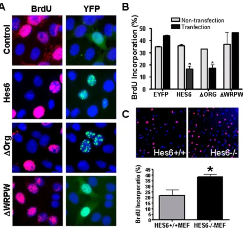
FIGURE 2. Effects of Hes6 on cell proliferation. A, YFP::Hes6 or its mutants were transfected into HeLa cells, and cells in the S-phase were labeled by 2 h of incubation with BrdUrd (BrdU). Following BrdUrd labeling (red), percentages of BrdUrd⫹/YFP⫹(transfection) and BrdUrd⫹/YFP⫺(nontransfection) cells were separately quantified in the same visual field (B). Data⫽ mean ⫾ S.E. n ⫽ 3; *, p ⬍ 0.05 in Student’s t test. C, BrdUrd labeling of Hes6⫹/⫹and Hes6⫺/⫺MEFs demonstrates a significantly higher number of BrdUrd⫹cells in the latter. n⫽ 3; *, p ⬍ 0.05 in Student’s t test.
at Ewha Medical Library on March 22, 2017
http://www.jbc.org/
For immunoprecipitation, extracts from transfected cell or the whole brain (E17.5) lysates were prepared in IP buffer (50 mMHEPES-KOH, pH 7.6, 150 mM NaCl, 10% glycerol, 0.1%
Nonidet P-40, 0.5% deoxycholate, 5 mMEDTA) containing 1⫻
protease inhibitor mixture (CompleteTM, Roche Applied Sci-ence). Cell lysates were precipitated by centrifugation at 15,000⫻ g at 4 °C for 1 h. The supernatant was precleared with protein G-agarose beads and incubated overnight with primary antibody at 4 °C, followed by 2 h of incubation with protein G-agarose. The antibody-conjugated beads were washed with lysis buffer and resuspended in gel loading buffer. After running
the SDS-PAGE and immunoblot-ting, immunoreactive bands were visualized by Amersham Bio-sciences ECL reagents.
Chromatin Immunoprecipita-tion—Hes6-transfected cells were fixed with 1% formaldehyde for 10 min at room temperature, rinsed with PBS containing protease inhib-itor mixtures (Roche Applied Bio-science), suspended in harvest buffer (100 mMTris-HCl, pH 9.4, 10 mMdithiothreitol), incubated for 15
min on ice, and centrifuged for 5 min at 2,000⫻ g. Cells were then washed sequentially with 1 ml of ice-cold PBS, buffer I (0.25% Triton X-100, 10 mM EDTA, 0.5 mM
EGTA, 10 mMHEPES, pH 6.5), and
buffer II (200 mM NaCl, 1 mM
EDTA, 0.5 mM EGTA, 10 mM
HEPES, pH 6.5). Cells were resus-pended in 1 ml of lysis buffer (1% SDS, 10 mM EDTA, 50 mM
Tris-HCl, pH 8.1) containing protease inhibitor mixture (Roche Applied Science). The DNA in the lysate was sheared to 300 –1000 bp in size using a digital sonifier (Branson, model 450). Supernatants were col-lected and diluted with buffer (1% Triton X-100, 2 mMEDTA, 150 mM
NaCl, 20 mM Tris-HCl, pH 8.1),
followed by immunoclearing with 2 l of preimmune serum and single-stranded DNA-protein G-Sepharose (Upstate) for 2 h at 4 °C. Immunoprecipitation was per-formed for 6 h at 4 °C with specific antibodies. After immunoprecipita-tion, 40l of single-stranded DNA-containing protein G-Sepharose was added and incubated for another 2 h. The beads were washed sequentially for 10 min with TSE I (0.1% SDS, 1% Triton X-100, 2 mM
EDTA, 20 mMTris-HCl, pH 8.1, 150
mMNaCl), TSE II (0.1% SDS, 1% Triton X-100, 2 mMEDTA, 20
mMTris-HCl, pH 8.1, 500 mMNaCl), and buffer III (0.25MLiCl,
1% Nonidet P-40, 1% deoxycholate, 1 mMEDTA, 10 mM
Tris-HCl, pH 8.1). Precipitates were then washed twice with TE buffer and eluted three times with 200l of 1% SDS and 0.1M
NaHCO3. Eluates were pooled and heated at 65 °C for 6 h to reverse the formaldehyde-induced cross-linking. DNA frag-ments were purified by phenol/chloroform extraction, followed by ethanol precipitation with glycogen as carrier. For PCR, an aliquot of DNA precipitate was used with 25–35 cycles of amplification with primers (Table 1).
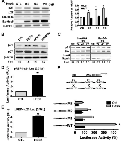
FIGURE 3. Hes6 modifies expression level of p21. A, mRNA levels of p21 and p27 cyclin-dependent kinase inhibitors following transfection of varying amounts of Hes6 expression vector into HeLa cells. Endogenous Hes6 (En-Hes6) and exogenously transfected Hes6 (Ex-Hes6) mRNAs were also examined. Relative levels of transcripts normalized by intensity of glyceraldehyde-3-phosphate dehydrogenase (GAPDH) are shown. n⫽ 3; *, p⬍ 0.05, one-way analysis of variance. CTL, control. B, protein levels of p21 and p27 in HeLa cells transfected with Hes6 and its mutants. Relative intensities of p21 protein bands are indicated at the bottom of images. C, Hes6⫹/⫹and Hes6⫺/⫺MEFs were maintained under serum starvation (SS) conditions for 3 days. Total RNA was isolated from the cells, and the expression of p21, p27, and Hes6 mRNA was assessed by RT-PCR. D and E, transcriptional activation of p21 promoters containing 0.3 kb (D) or 2.3 kb (E) upstream regions by Hes6 overexpression. Luciferase activity was normalized by total protein content of the samples deter-mined by the Bradford assay (Bio-Rad). F, schematic diagram of the p21 promoter and the location of three E-boxes (E1–E3) are shown in the upper panel. The mutations of E-box sequence are shown in gray (upper). Transcriptional activation driven by the wild-type and E-box-mutated 0.3-kb p21 promoters in response to Hes6 co-expression in HeLa cells is shown. Wild-type and mutant E-boxes are illustrated on the left in gray and slashed boxes, respectively (lower). n⫽ 3; *, p ⬍ 0.05 in Student’s t test.
at Ewha Medical Library on March 22, 2017
http://www.jbc.org/
RESULTS
Hes6 Localizes to PML-NB—YFP-tagged Hes6 expression vector was transfected into HeLa cells, and subcellular localiza-tion of YFP::Hes6 fusion protein was examined. YFP::Hes6 sig-nal was predominantly observed as speckles in the nucleus. Speckle-like localization of YFP::Hes6 was also observed in other cell types such as pituitary cell line␣-T3 (Fig. 1B), COS7,
and human embryonic kidney cell HEK293 (supplemental Fig. S2). Such localization pattern is not likely to be due to nonspecific aggre-gation of YFP::Hes6 given that the speckle pattern generated by the fusion protein was reiterated by the HA-tagged Hes6 (Fig. 1C). Because the morphology and distri-bution of Hes6 speckles resembled those of PML-NB, we tested if these Hes6 speckles co-localized with the major PML-NB protein, PML. Immunofluorescence labeling of PML revealed that YFP::Hes6 sig-nals extensively overlapped with PML immunoreactivity (IR) (Fig. 1,
D–I). High magnification observa-tion showed that both PML-IR and YFP::Hes6 signals were co-localized in the same speckles (about 80% of the total speckles), whereas a few speckles exhibited either PML-IR (Fig. 1, G and H, arrows) or YFP::Hes6 signal (Fig. 1, G and H,
arrowhead, both about 10%). We next determined the domain of Hes6 critical for localization in the PML-NB. Hes family proteins contain conserved WRPW tet-rapeptide motif (7, 31). Hes genes also contain the orange domain that is known to stabilize the protein-protein interaction (32). Thus, we tested whether the deletion of these domains modifies the subcellular localization. The orange domain deleted Hes6 (⌬Org) exhibited speckle-like signals that were still co-localized with PML immunore-activity (Fig. 1, J–L). The deletion of WRPW domain (⌬WRPW), on the other hand, resulted in diffuse dis-tribution in the nuclear as well as in the cytoplasmic compartments, albeit weakly in the latter (Fig. 1,
M–O). These results show that WRPW domain is essential for the localization of Hes6 in the PML-NBs.
Hes6 Attenuates the Cell Pro-liferation—PML-NBs are involved in many cellular events, including the control of cell cycle via modification of tumor suppressors such as retinoblastoma protein and p53 (33). We therefore first examined the possible involvement of Hes6 in the regulation of the cell cycle. To assess the proliferation rate, we determined the rate of BrdUrd incorporation following overexpression of the wild-type or mutant Hes6. Approxi-FIGURE 4. p53-dependent and -independent suppression of cell proliferation by Hes6. A,
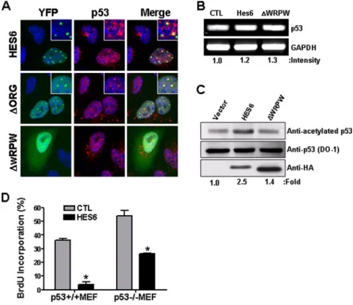
immunofluores-cence images of p53 (red) in HeLa cells transfected with YFP::Hes6 or its mutants. Insets show a high magnifi-cation view. B, levels of p53 mRNA following Hes6 or⌬WRPW mutant overexpression. Relative values of the band intensities after normalization to the level of glyceraldehyde-3-phosphate dehydrogenase (GAPDH) are shown. CTL, control. C, total amounts of p53 protein following Hes6 or⌬WRPW transfection were assessed by Western blot with DO-1 antibody. To assess the level of acetylated p53 level, cell extracts were subjected to immunoprecipitation with anti-acetylated p53 antibody, and the precipitated p53 level was examined with DO-1 antibody. Expression of Hes6 and⌬WRPW was verified by anti-HA staining. Values of acetylated p53 density were normalized to the total amount of p53 protein. D and E, Hes6 was transfected into p53⫹/⫹or p53⫺/⫺MEFs, and cells in the S-phase were quantified after BrdUrd incubation (2 h). BrdUrd⫹cells among Hes6⫹cell (Hes6) and among untransfected cells (CTL) were separately quantified in the same visual field (D). Percentages of BrdUrd incorporated cells in Hes6 transfected versus untransfected cells (% repression) are shown in E. n⫽ 3; *, p ⬍ 0.05 in Student’s t test.
FIGURE 5. Synergistic activation of p21 promoter by p53 and Hes6. Luciferase activity driven by 2.3-kb (A) or 0.3-kb (B) upstream regions to p21 promoters was measured in the presence or absence of Hes6 (500 ng) and different concentrations (ng) of p53 cDNA. n⫽ 3; *, p ⬍ 0.05, in one-way analysis of variance.
at Ewha Medical Library on March 22, 2017
http://www.jbc.org/
mately 40% of the cells were BrdUrd-labeled after a 2-h incuba-tion of actively growing HeLa cells. In contrast, a substantially smaller proportion of cells was labeled in the cell populations expressing wild-type (WT) Hes6 or⌬Org, indi-cating that the cell proliferation is suppressed by the Hes6 or ⌬Org expression (Fig. 2, A and B). Impor-tantly,⌬WRPW failed to show such an anti-proliferating activity, sug-gesting that the localization of Hes6 in PML-NB is a prerequisite for the growth inhibition. Consistent with these observations, we also found that embryonic fibroblasts derived from Hes6⫺/⫺ mouse (MEFs) (10) proliferated substantially faster than WT MEFs and that a signifi-cantly higher level of BrdUrd label-ing was obtained in Hes6⫺/⫺MEFs compared with WT MEFs (Fig. 2C). These results collectively suggest that Hes6 is involved in the control of cell proliferation.
Hes6 Enhances p21 Expression— We next tested whether Hes6 con-trols the cyclin-dependent kinase inhibitor, which acts on G1-S phase to block cell cycle progression. Fol-lowing the expression of exogenous Hes6, we found that the p21 mRNA level increased in a dose-dependent manner, whereas the mRNA level of neither p27 nor endogenous Hes6 was altered (Fig. 3A). Consistently, expression of Hes6 selectively enhanced p21 protein levels but not p27 (Fig. 3B). Importantly, ⌬WRPW only marginally altered the p21 protein level indicating again that localization of Hes6 to PML-NB is required for regulation of p21. To further evaluate the con-tribution of Hes6 to p21 gene expression, we examined the p21 induction in Hes6⫺/⫺MEF. Follow-ing serum starvation, Hes6 mRNA expression was induced in WT MEF (Fig. 3C). Similarly, p21 and p27 mRNA induction was also observed. However, in Hes6⫺/⫺ MEF, although the p27 induction was the same as seen in WT MEF, the
induc-tion of p21 was selectively abrogated, indicating the highly spe-cific nature of the inductive effect of Hes6 on p21. Next, we assessed the effect of Hes6 on p21 promoter activity using a
reporter assay. Overexpression of Hes6 significantly enhanced luciferase activity from reporter plasmids with either 2.3- or 0.3-kb proximal promoter regions of the p21 gene, suggesting
at Ewha Medical Library on March 22, 2017
http://www.jbc.org/
that Hes6 regulates p21 through cis-elements within the 0.3-kb proximal region (Fig. 3, D and E). It is known that several pro-teins of bHLH family are involved in the induction of p21 tran-scription via three E-box (CANNTG) elements found immedi-ately 5⬘ to the core promoter (34, 35). To test whether E-box sequences are necessary to mediate induction of p21 by Hes6, we assayed the activities of 0.3-kb p21 promoter reporters con-taining mutant versions of the E-boxes (Fig. 3F and supplemen-tal Fig. S3). Whereas WT p21 promoter activity increased by ⬃4-fold in response to Hes6 expression, E-box mutation of p21 promoter abrogated the Hes6-induced transcriptional induc-tion. Mutation of E-box also reduced the basal luciferase activ-ity in the absence of Hes6. Considering that HeLa cells do not express Hes6 (data not shown), this result suggests that endog-enous factors other than Hes6 in HeLa cells may contribute to the basal p21 activity.
Anti-proliferating Effect of Hes6 Is Partially Mediated by p53—Considering that p53 is a component of PML-NB and an upstream regulator of p21, we questioned if p53 is involved in the anti-proliferating activity of Hes6. First, we found that p53 was enriched in the Hes6⫹speckles following YFP::Hes6 over-expression (Fig. 4A).⌬Org and ⌬WRPW showed consistent effect on p53 localization with their own tendency to localize to PML-NB; p53 was found in PML-NB in⌬Org⫹cells but not in ⌬WRPW⫹cells. We further assessed whether overexpression of Hes6 altered the expression level of p53, and we found that neither the mRNA nor the protein level was modified by Hes6 overexpression (Fig. 4, B and C). In addition, serum starvation led to an increase in the p53 mRNA level to a similar extent in both Hes6⫹/⫹and Hes6⫺/⫺MEFs (data not shown), suggesting that Hes6 does not contribute to the transcriptional induction of p53. Interestingly, however, we found that acetylation of p53 was increased by Hes6 overexpression. Upon immunoprecipi-tation with an antibody specific for Lys-373/Lys-382 acetylated p53 and subsequent immunoblotting with DO-1, which recog-nizes N terminus of p53, it was shown that acetylation of p53 was increased by 2.5-fold by Hes6 which⌬WRPW failed to do (Fig. 4C).
Finally, we examined whether Hes6 requires the presence of p53 for suppressing cell proliferation (Fig. 4D). Although⬃90% reduction of BrdUrd incorporation was seen as the results of ectopic Hes6 expression in p53⫹/⫹MEFs, the effect of Hes6 overexpression was substantially mitigated in p53⫺/⫺ MEFs showing only 50% reduction. This indicates that anti-prolifer-ating activity of Hes6 is mediated by both p53-dependent and -independent mechanisms.
Hes6 Synergizes with the p53 Activity on p21 Promoter— Given that the function of Hes6 is modulated by p53, we asked whether Hes6 could in turn modify p53 activity using
p21promoter assays (Fig. 5). Overexpression of p53 alone enhanced luciferase activity from the 2.3-kb p21 promoter construct with the maximum of 10-fold induction, whereas Hes6 overexpression alone enhanced luciferase activity by 4-fold. Importantly, co-expression of Hes6 and p53 synergis-tically augmented the induction of luciferase activity up to 30-fold (Fig. 5A). However, the synergistic effect with p53 was not observed with the 0.3-kb p21 promoter that lacks the p53-binding sites (Fig. 5B). These observations are consist-ent with at least two distinct mechanisms that are involved in the anti-proliferating effect of Hes6, one of which is medi-ated by 0.3-kb p21 promoter region independently of p53 and the other mediated by 2.3– 0.3-kb promoter region in a p53-dependent manner.
Hes6 Interacts with CBP—It has been demonstrated previ-ously that PML-NB contains CBP. CBP is known to be an important co-activator of the cell cycle control (36, 37) and cellular differentiation (38 – 40). It has also been reported that CBP is involved in p53-directed transcription via direct acety-lation of p53 (41). Therefore, we asked if CBP was involved in the Hes6-mediated cell proliferation control (Fig. 6). Immuno-fluorescence labeling revealed that YFP::Hes6 speckles were co-localized with CBP in a subset of PML-NBs (Fig. 6A). Consis-tently, the direct protein-protein interaction between Hes6 and CBP was further demonstrated by visualization with bimolecu-lar fluorescence complementation (BiFC) technique (42) (Fig. 6B). CBP and Hes6 were fused to pFLAG-VN and pHA-VC-terminal fragments of enhanced YFP, respectively. Cells singly transfected with either CBP-YN or Hes6-YC failed to produce significant YFP signal, but when CBP-YN and Hes6-YC were co-expressed in HeLa cells, strong BiFC signals were detected as the nuclear speckles. Interestingly, CBP-YN was distributed more or less evenly in the nucleus when transfected alone, and it was recruited into Hes6⫹PML-NBs in co-transfected cells, suggesting that Hes6 is a recruiting factor for CBP into the PML-NB. Similar BiFC signal was detected in living cells (Fig. 6C), implying that CBP and Hes6 interact under physiological conditions. Finally, using anti-Hes6 antibody, we verified that interaction of endogenous Hes6 with CBP in the embryonic mouse brain lysate (Fig. 6D). This Hes6-CBP-PML association appears to be independent of the presence of p53 as Hes6 forms speckles containing CBP in p53⫺/⫺ MEF and p53-deficient H1299 cells (supplemental Fig. S4).
To map the CBP-binding site on Hes6, we constructed trun-cation mutants of Hes6 and co-transfected them with the full-length CBP in HeLa cells (Fig. 6E). Although⌬WRPW was co-immunoprecipitated with CBP, basic domain- and bHLH-truncated mutants (⌬basic and ⌬bHLH, respectively) did not show any evidence of interaction with CBP, suggesting that the
FIGURE 6. Hes6 directly interacts with CBP. A, co-localization of YFP::Hes6 (green) with CBP (red) in HeLa cells revealed by immunofluorescence labeling. B and C, interaction of CBP and Hes6 using BiFC analysis. Interaction of FLAG-CBP-YN and HA-Hes6-YC results in the green fluorescence from the functional complementation of the two halves of the YFP (B). Interaction of CBP and Hes6 proteins in living HeLa cells using BiFC analysis. CBP-YN and Hes6-YC expressed alone as negative controls showing with no detectable BiFC signal (C). D, interaction of CBP with Hes6 proteins from embryonic (E16) brain lysate. E, co-immunoprecipitation of Myc-Hes6 mutants with FLAG-CBP in HeLa cells. Following co-transfection of Hes6 or the Hes6 mutants with FLAG-CBP, cell extracts were immunoprecipitated (IP) with anti-Myc and subjected to Western blot analysis with anti-FLAG antibody. IB, immunoblot. Schematic representation for deletion mutants of Hes6 is shown (left panel). F, localization of YFP::⌬basic (green) with PML-NB (red) in HeLa cells (upper panel). YFP::⌬basic speckles did not co-localize with CBP or p53 (red, middle and lower panels). Insets show large magnification views. G, luciferase activity driven by 2.3 kb upstream to the p21 promoters was measured following the Hes6 mutants and p53 transfection. Similar levels of mutant protein expression were verified by Western blot (data not shown). n⫽ 3; *, p ⬍ 0.05, in one-way analysis of variance.
at Ewha Medical Library on March 22, 2017
http://www.jbc.org/
basic domain is necessary for Hes6-CBP interaction. Accord-ingly, YFP::⌬basic signal was not associated with CBP-IR and p53-IR, although it was seen to be infrequently co-localized with PML-NB (Fig. 6F). Furthermore,⌬basic mutant failed to exhibit transcriptional activation of p21 promoter and synergy with p53 (Fig. 6G). We also found that similar to ⌬basic, ⌬WRPW failed to modify transcriptional activation of p21 pro-moter with or without p53, suggesting that both CBP binding
and PML-NB inclusion of Hes6 are required for the transcriptional acti-vation of the p21 promoter.
Hes6-CBP Complex Promotes the Histone Remodeling and p53 Bind-ing on p21 Promoter—It is known that CBP acetylates histone H3, which affects the transcriptional activation of the target genes. To address this issue, we explored his-tone H3 acetylation patterns of the p21 promoter using chromatin immunoprecipitation assay. Specif-ically, we examined the two p53-binding site (PBX) and three E-boxes in the p21 promoter region and found that histone H3 acetyla-tion on the PBX or E-box were sig-nificantly enhanced by Hes6 over-expression (Fig. 7A). Accordingly, the binding of both the acetylated p53 and Hes6 to the p21 promoter was significantly increased by Hes6 overexpression (Fig. 7, B and C). However, neither ⌬basic nor ⌬WRPW associated with the E-box region, and they failed to increase histone remodeling and p53 bind-ing. In fact, binding of acetylated p53 to the distal PBX or proximal PBX was reduced by the expression of⌬basic or ⌬WRPW. These results suggest that Hes6 may influence the binding of transcription complex containing p53 to the p21 promoter at multiple levels and that the expression of mutant Hes6 inter-feres with in these processes. DISCUSSION
Hes6 Is a Novel Protein in PML-NB Subnuclear Compartments —Tran-scription machinery forms a large complex with DNA, often referred to as the transcriptome, which is typi-cally composed of RNA polymerase, general and specific transcription factors, and transcriptional modula-tors, including mediamodula-tors, co-acti-vators, and co-repressors. PML-NB plays essential roles in controlling cell division and suppression of tumorigenesis (43). PML-NB appears to provide an active zone of transcriptional regulation, because it contains many proteins involved in the transcriptional activation such as PML, RNA polymerase II, CBP, SUMO, Daxx, retinoblastoma pro-tein, and p53 (14). We showed here for the first time that Hes6 is a part of the PML-NB protein complex. In addition, we dem-onstrated that Hes6 interacts with at least one component of FIGURE 7. Histone remodeling and p53 binding are modified by Hes6. A–C, FLAG-Hes6, FLAG-⌬basic, or
FLAG-⌬WRPW were transfected into HeLa cells, and chromatin immunoprecipitation assay was performed with anti-acetylated histone H3 (A), anti-acetylated Lys-373/382 p53 (B), and anti-FLAG (C) antibodies. Recov-ered DNA was employed as template for PCR amplifications using specific primers against distal (dPBX) and proximal (pPBX) p53-binding sites or downstream E-box. Schematic representation of the p21 promoter sequence is shown in the upper panel of A. Arrowheads indicate the positions of primers for PCR amplification. Experiments were repeated three times and representative images are shown. D, hypothetical model for HES6-induced cell cycle arrest. Hes6 interacts with CBP via the basic domain and recruits CBP to the PML-NB complex via WRPW domain. Upon the recruitment of CBP, histone H3 on the proximal p21 promoter and p53 are acetylated, which in turn promotes the binding of p53 onto the p21 promoter. Absence of basic or WRPW domain fails to transactivate the p21 promoter. Although⌬Basic localizes in the PML-NB, it cannot interact with CBP. Conversely,⌬WRPW cannot recruit CBP into PML-NB complex.
at Ewha Medical Library on March 22, 2017
http://www.jbc.org/
the PML-NB, CBP. For the association of Hes6 in the PML-NB, WRPW motif on its C terminus is necessary because the dele-tion of this motif greatly suppressed the Hes6 speckle forma-tion. WRPW domain is recognized as a motif for interaction with transcriptional co-repressors such as Groucho (31) and as a rapid protein degradation signal (7, 8). It therefore appears that Hes6 and PML-NB association is mechanistically related to the WRPW-binding proteins or protein degradation. For instance, NB-included Hes6 may be protected from protein degradation and accumulate to a functionally significant level. However, it is not likely that WRPW domain is sufficient for the PML-NB localization, because we failed to observe Hes1, another Hes family member with the WRPW motif, in the PML-NB when it was overexpressed (data not shown).
p53-dependent and -independent Anti-proliferating Effect of Hes6—Several lines of evidence suggest that PML-NB-medi-ated control of cell proliferation is dependent on p53. PML directly interacts with the DNA binding domain of p53 and controls the transcriptional activity of p53 by enhancing pro-tein stability or promoting post-translational modification such as acetylation (19, 21). Furthermore, transcriptional activation of p21 by p53 is severely compromised in cells lacking PML (19). Interestingly, the PML gene promoter contains the p53-binding site, and PML is a target gene for p53 indicating a pos-itive feedback loop at work between the two genes (44). Because Hes6 is a component of PML-NB and enhances transcription of p21, but not that of p27, it is highly likely that p53 is at least partly involved in the anti-proliferating effect of Hes6. In support of this, we found the following: 1) Hes6 recruits p53 to the PML-NB, although it does not physically interact; 2) Hes6 and p53 increase the p21 promoter activity synergisti-cally; and 3) Hes6 enhances the binding of acetylated p53 to the p21 promoter.
However, Hes6 also appears to be able to activate p21 tran-scription and to suppress cell proliferation independent of p53. We found that the 0.3-kb promoter region of the p21 gene that lacks p53-binding sites was sufficient for the transcriptional activation by Hes6, although it failed to exhibit the synergistic transcriptional activation by Hes6 and p53. Consistently, a sub-stantial reduction of cell proliferation was seen by Hes6 over-expression even in p53⫺/⫺MEF. We also observed that Hes6 can promote p21 promoter activity and interact with the E-box region of p21 promoter in the H1299 cell line, which lacks p53 expression (supplemental Fig. S4). Given that Hes6 does not appear to bind to the known Hes-binding DNA motif for tran-scriptional activation (11), the anti-proliferating effect of Hes6 is possibly mediated by other interacting partners with DNA binding activity. Supporting this idea, we found that the muta-tion in E-box substantially reduces the basal p21 promoter activity in the absence of Hes6. Hes6 has been known to act as an inhibitor for Hes1, which is essential for maintenance of the progenitor pools during development (45– 47). Consistent with such a role, Hes1 has been shown to suppress the p21 promoter activity via E-box elements of the p21 promoter, and co-expres-sion of Hes6 with Hes1 restores p21 promoter activity (48). Induction of proneural bHLH genes such as neurogenin and NeuroD also results in the determination of neural fate in con-junction with cell cycle exit and activation of cyclin-dependent
kinase inhibitors such as p16, p21, and p27 (49 –52). Therefore, Hes6 may influence the p21 promoter activity by suppression of Hes1 or other bHLH transcription factors (51, 53, 54). How-ever, we do not completely rule out the possibility that Hes6 directly interacts with E-box in p21 promoter (55), and we are currently examining the proposed regulatory interaction in greater detail.
CBP Mediates the Anti-proliferating Function of Hes6—As for the mechanism(s) by which p53 is activated by Hes6, we propose that modification (acetylation) of p53 is at least partly responsible. We also observed histone remodeling of p21 pro-moter regions. These two events appear to be mediated by CBP. CBP is an active component for histone acetylation, and CBP can acetylate the C terminus of p53 at lysines 373 and 382, thereby modulating p53 activity and stability (24). Hes6 directly interacts with CBP via its basic domain. Deletion of the basic domain of Hes6 completely abolishes the transcriptional activ-ity on the p21 promoter, and histone remodeling of these regions does not occur, suggesting that CBP is critically involved in the Hes6-mediated modification of p21 promoter activity. Although the basic domain in most bHLH transcrip-tion factors serves as a DNA-binding motif, it has been reported that Hes6 does not bind to DNA (11). Thus, we propose that the absence of transcriptional activity in the ⌬basic mutant is because of the failure to bind to CBP. Despite the failure to promote p21 activity, the⌬basic mutant is still localized to the PML-NB. On the other hand, we observed that ⌬WRPW mutant, which also failed to promote the acetylation of histone H3 on the proximal region of the p21 promoter, could still interact with CBP. Therefore, at least two distinct domains (basic and WRPW) and their distinct protein interactions appear to be necessary for the function of Hes6.
Groucho/TLE proteins interacting with tetrapeptide WRPW of the Hes family has been reported to function in part by recruiting histone deacetylases to repress target genes (56). Our results show to the contrary that native Hes6 containing WRPW motif promoted histone acetylation, and that the elim-ination of WRPW motif abrogated such effect. Because the WRPW motif is necessary for Hes6 to localize in PML-NB, it appears that Hes6 recruits CBP into the PML-NB complex via the WRPW domain of its C terminus. In support of this hypoth-esis, we showed that overexpression of Hes6 augmented PML-NB localization of CBP. It is established that PML leads to co-localization of p53 with CBP and promotes acetylation of p53 at Lys-382 (33). Taken together, Hes6 may be a critical mediator for the PML-CBP-p53 complex formation via basic domain-CBP interaction and WRPW-PML association. A hypothetical scheme for Hes6-mediated anti-proliferation is shown in Fig. 7D.
A Model for Hes6 Involvement in the Development and Tumorigenesis—Hes6 has been known to be an inhibitor for Hes1, a downstream regulatory target of the Delta-Notch path-way (11). Delta-Notch pathpath-way is essential for determination of specific cell fates as well as for maintenance of the progenitor pools during embryonic development (57). Notch signals seem to exert their action by direct transcriptional activation of bHLH transcriptional repressors such as Hes1 and Hes5 (58). Evidence for the mechanistic link between Notch-Hes cascade
at Ewha Medical Library on March 22, 2017
http://www.jbc.org/
and the cell cycle has been described (58, 59). For example, Hes1 knock-out mice have smaller eyes (4, 60), and Hes1 con-tributes to the proliferation of progenitor cells through tran-scriptional repression of cell cycle kinase inhibitors such as p57, p27, and p21 (48, 61, 62). Therefore, depending on the develop-mental context, Notch activation appears to promote cell cycle exit with an up-regulation of the p53 and p21 proteins (63). Furthermore, attenuation of Notch signaling reduces apoptosis in neural progenitor cells indicating that Notch1 may regulate p53-dependent apoptotic cell death (64). In addition, using flu-orescence in situ hybridization analysis in human fibroblast cells, it was demonstrated that PML bodies reside in Notch1 containing genomic loci (65). These observations lead us to speculate that association of Hes6 in the PML-NB and its mod-ulation of p53 activity may feed into the Notch-Hes cascade for the control of cell proliferation and differentiation during development.
The link between p53 and Hes6 also suggests a significant role of Hes6 in tumorigenesis. The expression of Hes6 is up-regulated in a subset of human tumors such as grade II/III astrocytoma and secondary glioblastoma multiformes (66, 67). Interestingly, these tumors are frequently found to have muta-tions in the p53 gene. DNA damage and oncogenic Ras-medi-ated signaling have been shown previously to induce the forma-tion of p53-CBP-PML NBs, which promotes the p53 acetylation and increases p53 stability (19, 33). In this respect, it is possible that increased Hes6 expression may be a result of compensatory feedback signal following the mutation and inac-tivation of p53 in some tumors.
REFERENCES
1. Ross, S. E., Greenberg, M. E., and Stiles, C. D. (2003) Neuron 39, 13–25 2. Iso, T., Kedes, L., and Hamamori, Y. (2003) J. Cell Physiol. 194, 237–255 3. Davis, R. L., and Turner, D. L. (2001) Oncogene 20, 8342– 8357 4. Sasai, Y., Kageyama, R., Tagawa, Y., Shigemoto, R., and Nakanishi, S.
(1992) Genes Dev. 6, 2620 –2634
5. Norton, J. D. (2000) J. Cell Sci. 113, 3897–3905 6. Fisher, A., and Caudy, M. (1998) BioEssays 20, 298 –306
7. Fisher, A. L., Ohsako, S., and Caudy, M. (1996) Mol. Cell. Biol. 16, 2670 –2677
8. Kang, S. A., Seol, J. H., and Kim, J. (2005) Biochem. Biophys. Res. Commun.
332,33–36
9. Gratton, M. O., Torban, E., Jasmin, S. B., Theriault, F. M., German, M. S., and Stifani, S. (2003) Mol. Cell. Biol. 23, 6922– 6935
10. Koyano-Nakagawa, N., Kim, J., Anderson, D., and Kintner, C. (2000) De-velopment(Camb.) 127, 4203– 4216
11. Bae, S., Bessho, Y., Hojo, M., and Kageyama, R. (2000) Development (Camb.) 127, 2933–2943
12. Handwerger, K. E., and Gall, J. G. (2006) Trends Cell Biol. 16, 19 –26 13. Lamond, A. I., and Earnshaw, W. C. (1998) Science 280, 547–553 14. Zhong, S., Salomoni, P., and Pandolfi, P. P. (2000) Nat. Cell Biol. 2,
E85–E90
15. Lin, H. K., Bergmann, S., and Pandolfi, P. P. (2005) Oncogene 24, 5693–5700
16. Pandolfi, P. P., Grignani, F., Alcalay, M., Mencarelli, A., Biondi, A., Lo-Coco, F., Grignani, F., and Pelicci, P. G. (1991) Oncogene 6, 1285–1292 17. de The, H., Lavau, C., Marchio, A., Chomienne, C., Degos, L., and Dejean,
A. (1991) Cell 66, 675– 684
18. Ching, R. W., Dellaire, G., Eskiw, C. H., and Bazett-Jones, D. P. (2005) J. Cell Sci. 118,847– 854
19. Guo, A., Salomoni, P., Luo, J., Shih, A., Zhong, S., Gu, W., and Pandolfi, P. P. (2000) Nat. Cell Biol. 2, 730 –736
20. Vousden, K. H., and Lu, X. (2002) Nat. Rev. Cancer 2, 594 – 604
21. Pearson, M., Carbone, R., Sebastiani, C., Cioce, M., Fagioli, M., Saito, S., Higashimoto, Y., Appella, E., Minucci, S., Pandolfi, P. P., and Pelicci, P. G. (2000) Nature 406, 207–210
22. Hofmann, T. G., Moller, A., Sirma, H., Zentgraf, H., Taya, Y., Droge, W., Will, H., and Schmitz, M. L. (2002) Nat. Cell Biol. 4, 1–10
23. Bode, A. M., and Dong, Z. (2004) Nat. Rev. Cancer 4, 793– 805
24. Ito, A., Lai, C. H., Zhao, X., Saito, S., Hamilton, M. H., Appella, E., and Yao, T. P. (2001) EMBO J. 20, 1331–1340
25. Gill, R. M., Slack, R., Kiess, M., and Hamel, P. A. (1998) Exp. Cell Res. 244, 157–170
26. Ghiani, C. A., Eisen, A. M., Yuan, X., DePinho, R. A., McBain, C. J., and Gallo, V. (1999) Development (Camb.) 126, 1077–1090
27. Coffman, F. D., and Studzinski, G. P. (1999) Exp. Cell Res. 248, 58 –73 28. Abbondanzo, S. J., Gadi, I., and Stewart, C. L. (1993) Methods Enzymol.
225,803– 823
29. Liu, H., Deng, X., Shyu, Y. J., Li, J. J., Taparowsky, E. J., and Hu, C. D. (2006) EMBO J. 25,1058 –1069
30. Wang, L., Oh, D. Y., Bogerd, J., Choi, H. S., Ahn, R. S., Seong, J. Y., and Kwon, H. B. (2001) Endocrinology 142, 4015– 4025
31. Paroush, Z., Finley, R. L., Jr., Kidd, T., Wainwright, S. M., Ingham, P. W., Brent, R., and Ish-Horowicz, D. (1994) Cell 79, 805– 815
32. Dawson, S. R., Turner, D. L., Weintraub, H., and Parkhurst, S. M. (1995) Mol. Cell. Biol. 15,6923– 6931
33. Ferbeyre, G., de Stanchina, E., Querido, E., Baptiste, N., Prives, C., and Lowe, S. W. (2000) Genes Dev. 14, 2015–2027
34. Gartel, A. L., and Tyner, A. L. (1999) Exp. Cell Res. 246, 280 –289 35. Gartel, A. L., and Radhakrishnan, S. K. (2005) Cancer Res. 65, 3980 –3985 36. Iyer, N. G., Ozdag, H., and Caldas, C. (2004) Oncogene 23, 4225– 4231 37. Kiesslich, A., von Mikecz, A., and Hemmerich, P. (2002) J. Struct. Biol.
140,167–179
38. Kawasaki, H., Eckner, R., Yao, T. P., Taira, K., Chiu, R., Livingston, D. M., and Yokoyama, K. K. (1998) Nature 393, 284 –289
39. Roth, J. F., Shikama, N., Henzen, C., Desbaillets, I., Lutz, W., Marino, S., Wittwer, J., Schorle, H., Gassmann, M., and Eckner, R. (2003) EMBO J. 22, 5186 –5196
40. Nguyen, L. A., Pandolfi, P. P., Aikawa, Y., Tagata, Y., Ohki, M., and Kitaba-yashi, I. (2005) Blood 105, 292–300
41. Barlev, N. A., Liu, L., Chehab, N. H., Mansfield, K., Harris, K. G., Hala-zonetis, T. D., and Berger, S. L. (2001) Mol. Cell 8, 1243–1254
42. Hu, C. D., Chinenov, Y., and Kerppola, T. K. (2002) Mol. Cell 9, 789 –798 43. Wang, Z. G., Ruggero, D., Ronchetti, S., Zhong, S., Gaboli, M., Rivi, R., and
Pandolfi, P. P. (1998) Nat. Genet. 20, 266 –272
44. de Stanchina, E., Querido, E., Narita, M., Davuluri, R. V., Pandolfi, P. P., Ferbeyre, G., and Lowe, S. W. (2004) Mol. Cell 13, 523–535
45. Wu, Y., Liu, Y., Levine, E. M., and Rao, M. S. (2003) Dev. Dyn. 226, 675– 689
46. Ishibashi, M., Moriyoshi, K., Sasai, Y., Shiota, K., Nakanishi, S., and Kageyama, R. (1994) EMBO J. 13, 1799 –1805
47. Kageyama, R., Ohtsuka, T., and Tomita, K. (2000) Mol. Cells 10, 1–7 48. Castella, P., Sawai, S., Nakao, K., Wagner, J. A., and Caudy, M. (2000) Mol.
Cell. Biol. 20,6170 – 6183
49. Bertrand, N., Castro, D. S., and Guillemot, F. (2002) Nat. Rev. Neurosci. 3, 517–530
50. Ohnuma, S., Philpott, A., and Harris, W. A. (2001) Curr. Opin. Neurobiol.
11,66 –73
51. Mutoh, H., Naya, F. J., Tsai, M. J., and Leiter, A. B. (1998) Genes Dev. 12, 820 – 830
52. Farah, M. H., Olson, J. M., Sucic, H. B., Hume, R. I., Tapscott, S. J., and Turner, D. L. (2000) Development (Camb.) 127, 693–702
53. Halevy, O., Novitch, B. G., Spicer, D. B., Skapek, S. X., Rhee, J., Hannon, G. J., Beach, D., and Lassar, A. B. (1995) Science 267, 1018 –1021 54. Prabhu, S., Ignatova, A., Park, S. T., and Sun, X. H. (1997) Mol. Cell. Biol.
17,5888 –5896
55. Cossins, J., Vernon, A. E., Zhang, Y., Philpott, A., and Jones, P. H. (2002) Development(Camb.) 129, 2195–2207
56. Chen, G., and Courey, A. J. (2000) Gene (Amst.) 249, 1–16 57. Gaiano, N., and Fishell, G. (2002) Annu. Rev. Neurosci. 25, 471– 490 58. Ohtsuka, T., Ishibashi, M., Gradwohl, G., Nakanishi, S., Guillemot, F., and
at Ewha Medical Library on March 22, 2017
http://www.jbc.org/
Kageyama, R. (1999) EMBO J. 18, 2196 –2207
59. Piscione, T. D., Wu, M. Y., and Quaggin, S. E. (2004) Gene Expr. Patterns
4,707–711
60. Tomita, K., Ishibashi, M., Nakahara, K., Ang, S. L., Nakanishi, S., Guil-lemot, F., and Kageyama, R. (1996) Neuron 16, 723–734
61. Georgia, S., Soliz, R., Li, M., Zhang, P., and Bhushan, A. (2006) Dev. Biol.
298,22–31
62. Murata, K., Hattori, M., Hirai, N., Shinozuka, Y., Hirata, H., Kageyama, R., Sakai, T., and Minato, N. (2005) Mol. Cell. Biol. 25, 4262– 4271 63. Qi, R., An, H., Yu, Y., Zhang, M., Liu, S., Xu, H., Guo, Z., Cheng, T., and
Cao, X. (2003) Cancer Res. 63, 8323– 8329
64. Yang, X., Klein, R., Tian, X., Cheng, H. T., Kopan, R., and Shen, J. (2004) Dev. Biol. 269,81–94
65. Wang, J., Shiels, C., Sasieni, P., Wu, P. J., Islam, S. A., Freemont, P. S., and Sheer, D. (2004) J. Cell Biol. 164, 515–526
66. Somasundaram, K., Reddy, S. P., Vinnakota, K., Britto, R., Subbarayan, M., Nambiar, S., Hebbar, A., Samuel, C., Shetty, M., Sreepathi, H. K., Santosh, V., Hegde, A. S., Hegde, S., Kondaiah, P., and Rao, M. R. (2005) Oncogene
24,7073–7083
67. Kleihues, P., and Ohgaki, H. (1999) Neuro Oncol. 1, 44 –51
at Ewha Medical Library on March 22, 2017
http://www.jbc.org/
Woong Sun and Hyun Kim
Bokkee Eun, Yool Lee, Soontaek Hong, Jaesang Kim, Han-Woong Lee, Kyungjin Kim,
Body
doi: 10.1074/jbc.M707683200 originally published online December 26, 2007 2008, 283:5939-5949.
J. Biol. Chem.
10.1074/jbc.M707683200 Access the most updated version of this article at doi:
Alerts:
When a correction for this article is posted •
When this article is cited •
to choose from all of JBC's e-mail alerts Click here
Supplemental material:
http://www.jbc.org/content/suppl/2007/12/27/M707683200.DC1 http://www.jbc.org/content/283/9/5939.full.html#ref-list-1This article cites 67 references, 26 of which can be accessed free at
at Ewha Medical Library on March 22, 2017
http://www.jbc.org/