Complete resection of a gastric tumor including the adjacent lymph nodes is considered to be the only effec-tive curaeffec-tive treatment modality for patients with gastric cancer (1). In spite of this, local or systemic recurrent le-sions can develop in various locations of the abdomen and pelvis through several routes, therefore, early detec-tion and treatment of recurrence is important for im-proving the quality of life and survival of gastric cancer patients. CT plays an important role in evaluating sus-pected recurrence of tumors, however, early detection of recurrence or detection of small tumors can be diffi-cult identify and correctly diagnose (2, 3). Therefore, we conducted this retrospective review of cases in which recurrence occurred but was not identified on CT
analy-sis to determine the reason the lesions were not identi-fied. The site, shape, and attenuation characteristics of those lesions were reviewed and illustrated.
Patterns of Tumor Recurrence
The main patterns of recurrence after the removal of gastric cancer were as follows:
- Local recurrence: anastomosis or stump, adjacent or-gan, abdominal incision site
- Lymph nodes - Peritoneal seeding
- Hematogeneous metastasis: liver, lung, bone, brain - Unusual metastasis
Local Recurrence
Local recurrence of gastric carcinoma after surgery is defined as histologic evidence of a tumor in the sur-rounding tissues of the resected stomach (4).
Follow up CT Findings of Various Types of Recurrence
after Curative Gastric Surgery
1Hye-Jeong Lee, M.D., Myeong-Jin Kim, M.D., Joon Seok Lim, M.D., Ki Whang Kim, M.D.
1Department of Diagnostic Radiology, Shinchon Severance Hospital,
Yonsei University College of Medicine
Received February 14, 2007 ; Accepted October 4, 2007
Address reprint requests to : Myeong-Jin Kim, M.D., Department of Diagnostic Radiology, Research Institute of Radiological Science. Severance Hospital, Yonsei University College of Medicine, 134, Shinchon-dong, Seodaemun-gu, Seoul 120-752 Korea.
Tel. 82-2-2228-7400 Fax. 82-2-393-3035 E-mail: kimnex@yumc.yonsei.ac.kr
Although the detection of recurred lesions following curative gastrectomy in patients with gastric cancer has increased as the use of computed tomography (CT) has grown, early or small tumor recurrence can be difficult to identify or correctly diagnose using CT. Therefore, in this study, we retrospectively reviewed cases in which tumor recur-rence was missed upon analysis of the follow up CT after gastrectomy. The character-istics of the lesions in various locations of the abdomen and pelvis are illustrated and discussed.
Index words :Stomach neoplasms
Tomography, X-Ray computed Neoplasm recurrence, local Retrospective studies
Anastomosis and gastric stump
Local recurrence of gastric cancer most commonly in-volves the region of the gastric stump or anastomosis (3). Recurrence in this region manifests as an asymmetrical or circumferential thickening of the anastomotic or rem-nant gastric wall (Fig. 1) (1). However, even if thicken-ing of the remnant gastric wall does not occur, the pres-ence of a focal attenuation abnormality at the remnant
stomach or small bowel wall around the anastomosis site should prompt an endoscopic examination (Fig. 2) to differentiate between recurrence and benign conditions, including gastritis. In addition, abnormal distension of the bowel loop around the anastomosis site, including afferent or efferent loops, also suggest the possibility of tumor recurrence (Fig. 3), however this condition may also be caused by an obstruction due to the presence of adhesive bands.
A B
Fig. 1. A 36-year-old woman
under-went total gastrectomy to treat adeno-carcinoma. (A, B) Follow-up CT re-vealed the presence of focal asymmet-ric wall thickening of the anastomosis site (arrows) 12 months after gastrecto-my. The wall thickness measured 1.5 cm. The patient underwent endoscop-ic biopsy, whendoscop-ich revealed that tumor recurrence had occurred at the site of anastomosis.
A B
Fig. 2. A 66-year-old man underwent
subtotal gastrectomy to treat adenocar-cinoma.
A. Follow-up CT 2 years after
gastrec-tomy revealed a suspicious focal wall thickening with low attenuation (ar-row) at the remnant stomach, which was considered to be nonspecific gas-tritis at the time of the CT analysis.
B. After 7 months, a subsequent
fol-low-up CT revealed the progression of mural thickening (arrow) at the rem-nant stomach.
A B C
Fig. 3. A 58-year-old woman underwent subtotal gastrectomy to treat adenocarcinoma. A. There was no abnormality observed on the follow-up CT taken 20 months after gastrectomy.
B. A follow-up CT taken 33 months later, however, revealed the presence subtle wall thickening at anastomosis site; however, this
thickening was not considered to be a recurrent lesion at that time.
C. A follow-up CT taken three months later revealed that the mural thickening (arrows) at the anastomosis site had progressed. An
Abdominal incision site
Recurrences at the abdominal incision site caused by iatrogenic dissemination of cancer cells during the oper-ation are usually seen as a soft tissue attenuoper-ation mass (4). However, differentiation based on a postoperative fi-brosis or granulation may be difficult, therefore, com-parison of a postoperative baseline study with CT taken 3-6 months after surgery can be helpful for determin-ing the correct diagnosis (Fig. 4). In addition, the pres-ence of a painful or growing nodule at the incision site should also be carefully evaluated.
Lymph Nodes
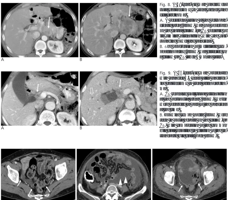
Lymph node metastases comprise the majority of tu-mor recurrences (3), and the most common locations of metastatic lymphadenopathy include the region along the common hepatic artery, celiac axis, the hepatoduo-denal ligament and the periaortic region (4). The pattern of lymphatic recurrence is seen as a conglomerated (4) or infiltrative mass, or as an obliteration of the fat in these regions (Fig. 5, 6). Lymph nodes larger than 10 mm observed in the short axis are usually regarded as abnormal and indicative of metastasis, however, the
A B C
Fig. 4. A 41-year-old man underwent subtotal gastrectomy to treat adenocarcinoma.
A. The baseline CT revealed a fibrotic scar at the incision site (arrow), but no evidence of growth of the mass.
B. A follow-up CT taken 12 months after the gastrectomy revealed the presence of a peripherally enhancing nodule at the
abdomi-nal wall incision site (arrow), however, at the time of the CT no action was taken.
C. A CT scan taken 6 months later revealed that the lesion had grown (arrow).
A B
C D
Fig. 5. An 80-year-old man underwent
total gastrectomy to treat adenocarci-noma.
A, B. Obliteration of the fat
surround-ing the celiac axis (arrow) and SMA root (open arrow) was observed on the follow-up CT taken 15 months after gastrectomy, however at the time that CT was conducted this was consid-ered to be postoperative fibrosis.
C, D. A Follow-up CT taken one year
later revealed that the soft tissue thick-ening around the celiac axis (arrow) and SMA root (open arrow) had grown. After chemotherapy, the soft tissue decreased in size and left a scar.
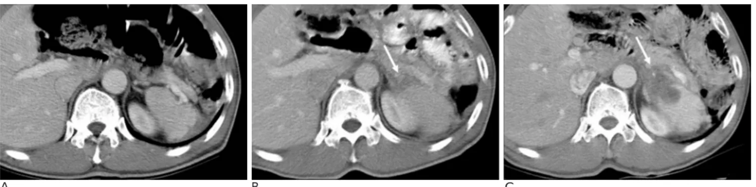
sensitivity and specificity of diagnosis can be poor if it is based solely on size criteria. Therefore the increase in the number and size lymph nodes in comparison to those observed on a previous CT is an important sign of nodal recurrence (Fig. 7). In addition, an area of low at-tenuation within a lymph node is another important sign of recurrence, however, such areas are very small, therefore, occasional analysis using positron emission tomography (PET) may be helpful in the differential di-agnosis of small lymph nodes. Furthermore, metastatic lymph nodes with or without necrosis can mimic nmal structures, such as the snmall bowel or other solid or-gans, therefore careful inspection of the adjacent
sec-tional images is necessary (Fig. 8, 9).
Peritoneal Seeding
Ascites, which is one of the most common findings of peritoneal seeding in cases of stomach cancer, has been reported in up to 74% of cases (5). Ascitic fluid is often loculated and/or septated, and can be seen in non-de-pendent areas of the abdomen, including the greater and lesser sacs, even in when it is not found in dependent ar-eas such as the pelvis (5). Peritoneal implants are soft-tis-sue masses that appear as solitary or multiple nodules and tend to occur at sites that coincide with the natural
A B C
Fig. 6. A 55-year-old man underwent total gastrectomy to treat adenocarcinoma.
A. Follow-up CT did not reveal the presence of any abnormalities in the same location 4 months after surgery.
B. However, a CT scan taken 7 months later revealed new obliteration of fat in the retropancreatic area (arrow), which was
sugges-tive of recurrence.
C. A Follow-up CT taken 10 months later after gastrectomy revealed further progression of the recurrent tumor (arrow).
A B
C D
Fig. 7. A 50-year-old man underwent
subtotal gastrectomy to treat adenocar-cinoma.
A. Follow- up CT conducted 4 months
after the gastrectomy showed no evi-dence of recurrence.
B. Several lymph nodes (5 mm in short
diameter) were seen in the gastros-plenic ligament (arrow) on the next fol-low-up CT, which was taken 2 months later.
C. The lymph nodes were seen as hot
spots (arrow) on the FDG-PET con-ducted at the time.
D. A follow-up CT taken 4 months
lat-er revealed progression of the lymph nodes (arrow).
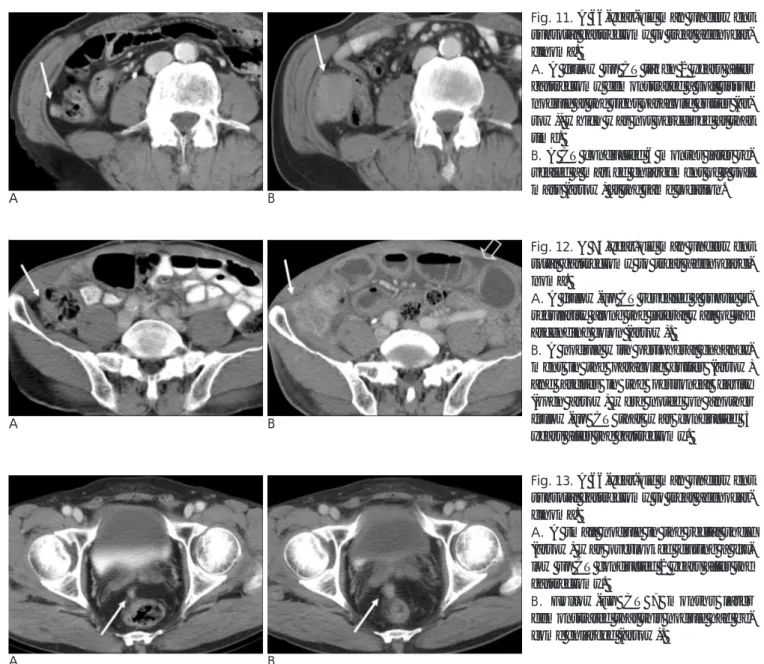
flow of peritoneal fluid. These sites include the superior aspect of the terminal ileum, the medial aspect of the ce-cum, and the superior aspect of the sigmoid colon, the pelvis (especially, cul-de-sac) the paracolic gutter, and the subhepatic and subphrenic spaces on the right. In addition, the peritoneal surfaces of the diaphragm, liver, spleen, and the greater omentum are also common sites of tumor deposition (6). Therefore, careful inspection of these areas is important to ensure that any areas of irreg-ular soft tissue thickening and/or soft tissue nodules
(Fig. 10-13) are identified.
Hematogenous Metastases
Because the venous return from the stomach is drained by the portal vein, the liver is the most common site of hematogeneous metastases (1). Hepatic metas-tases usually appear as areas of hypoattenuation on por-tal venous phase CT. If a dynamic scan is performed, an irregular peripheral rim enhancement around the
hy-A B C
Fig. 10. A 61-year-old woman presented with abdominal distension 3 years after undergoing subtotal gastrectomy to treat signet
ring cell carcinoma.
A. CT depicted a subtle nodular thickening of the peritoneum (arrow) with fluid collection in the pelvic cavity, which was
neglect-ed at the time.
B, C. A follow-up CT taken 3 months later revealed a marked increase in the amount of ascites (arrow heads) and progression of
diffuse peritoneal thickening (arrow).
A B
Fig. 8. A 39-year-old man underwent
total gastrectomy to treat signet ring cell carcinoma.
A. A recurrent lesion at the
retropan-creatic area (arrow) was not observed on the initial follow-up CT because of its similar attenuation with the sur-rounding bowel and pancreas.
B. The progression of metastatic
lym-phadenopathy (arrow) was detected on a follow up CT taken 6 months later.
A B
Fig. 9. A 64-year-old man presented
with jaundice 3.5 years after undergo-ing gastrectomy to treat adenocarcino-ma.
A. CT revealed soft tissue attenuation
at the periportal areas (arrow), which were regarded as part of the pancreas at the time.
B. Growing lymph nodes (arrow) were
seen in porta hepatis on the follow-up CT, with compression of the common bile duct resulting in dilation of the in-trahepatic bile duct (open arrow).
poattenuating center may be seen on the arterial phase and peripheral washout, and central enhancement can be seen on the delayed phase (Fig. 14). Less common sites of hematogenous metastases include the lungs, adrenal glands, kidneys and bones (2). To avoid over-looking metastasis of the lungs and bone, careful inspec-tion of these areas by adjusting the window width is im-portant. Ovarian metastases, which are also known as Krukenberg’s tumors, occur in nearly 10% of the cases of gastric cancer, and are especially common in signet-ring cell type cancer (1), which produces bilateral smoothly enlarged ovaries that are diffusely infiltrated microscopically by tumor cells. Krukenberg’s tumors should be suspected when solid ovarian tumors contain-ing well demarcated intratumoral cystic lesions are ob-served on the CT scan, especially if the walls of such
cysts demonstrate a particularly strong contrast en-hancement (Fig. 15).
Unusual Manifestations of Metastasis
Various forms of other recurrent tumors, including metastatic linitis platisca to the rectum, peribiliary tu-mor spread, portal vein tutu-mor thrombosis, urinary blad-der metastasis and ureter metastasis can also be encoun-tered.
Linitis platisca refers to diffuse proliferation of the connective tissue of a hollow organ. The stomach is the most common primary site of metastatic linitis plastica (7), and the most common finding of metastatic linitis plastica to the rectum was concentric bowel wall thick-ening with a target sign on contrast enhanced CT (Fig.
A B
Fig. 12. A 75-year-old man underwent
total gastrectomy to treat adenocarci-noma.
A. A follow-up CT revealed a subtle
ir-regularity along the lateral wall of the ascending colon (arrow).
B. A nodule with peripheral
enhance-ment in the paracolic gutter (arrow) and ascites in the peritoneal cavity (open arrow) were noted on another follow-up CT that was conducted 3 years after the gastrectomy.
A B
Fig. 13. A 66-year-old man underwent
subtotal gastrectomy to treat adenocar-cinoma.
A. A small nodule in the rectal shelf
(arrow) was overlooked during a fol-low up CT conducted 2 years after the gastrectomy.
B. Follow-up CT 7 months later
demonstrated that this nodule had be-come enlarged (arrow).
A B
Fig. 11. A 66-year-old man underwent
subtotal gastrectomy to treat adenocar-cinoma.
A. A follow up CT taken 2 years after
gastrectomy demonstrated a soft tissue nodule at the right paracolic gutter (ar-row), which was not perceived at that time.
B. A CT conducted 6 months later
re-vealed a marked enlargement of a soft mass (arrow) at the same location.
16) (7).
Peribiliary tumor spread is usually associated with metastatic lymphadenopathy along the common hepatic artery, celiac axis, or hepatoduodenal ligament, and peribilary tumors can be identified based on the pres-ence of irregular thickening of the biliary duct with
proximal biliary duct dilatation (Fig. 17).
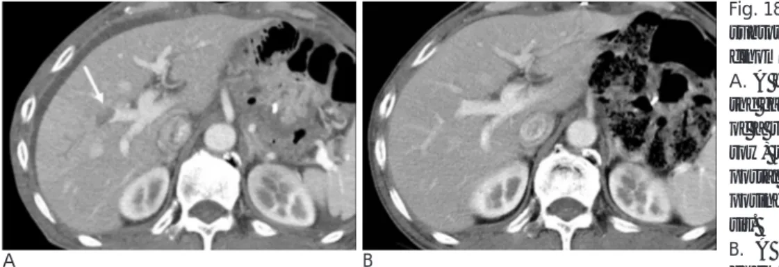
Although rare, portal venous thrombosis may occur in cases of invasive gastric carcinoma, possibly as a result of portal vein invasion by the liver metastatic tumor or direct portal venous invasion from the primary foci of gastric cancer (Fig. 18) (8).
A B C
Fig. 14. A 71-year-old man underwent total gastrectomy to treat adenocarcinoma. A. The initial follow-up CT revealed no abnormal lesions in the liver.
B. Although a newly developed small low attenuated lesion (arrow) was observed on a follow-up CT taken 3 months later, the
le-sion was not observed at that time because of its small size.
C. On the next follow-up CT, a central necrotic mass with an area of peripheral enhancement was observed in the right lobe of the
liver, which is suggestive of metastasis (arrow).
A B C
Fig. 16. A 28-year-old woman presented with constipation 5 years after undergoing total gastrectomy to treat signet ring cell
carci-noma. Follow-up CT (A-C) revealed concentric thickening of the rectal wall with a target sign, which is suggestive of metastatic linitis plastica to the rectum (arrows).
A B
Fig. 15. A 38-year-old woman
under-went total gastrectomy to treat signet ring cell carcinoma.
A. The mass (arrow) was
retrospec-tively observed on the prior follow-up CT; however it was not initially re-garded as a pathologic lesion.
B. A follow-up CT revealed a complex
mass with contrast enhancement of the right ovary, which is suggestive of a Krukenberg tumor (arrow). This pa-tient underwent surgery, and the pathology of the mass was consistent with metastatic signet ring cell carci-noma.
Metastatic tumors of the urinary bladder from distant primary foci are rare and represent only 1.5% of all bladder tumors. Metastatic bladder tumors can be iden-tified by the presence of polypoid lesions that are similar to typical transitional cell carcinoma or focal thickening of the bladder wall (Fig. 19) (9).
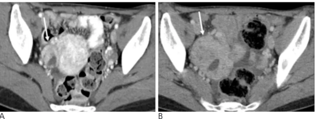
Ureteral metastasis caused by gastric cancer may oc-cur as a result of direct extension, peritoneal deposit, as well as lymphatic, or hematogeneous metastasis. Upon CT analysis, ureteral metastasis presents as concentric or asymmetric wall thickening of the ureter with
ob-structive hydronephrosis (Fig. 20). However, a sclerotic reaction induced by cancer cells, known as malignant retroperitoneal fibrosis, can also invade the periureteral region without direct invasion of the ureter and lead to ureteral obstruction (Fig. 21) (10).
Conclusion
CT scan plays an important role in preoperative stag-ing and postoperative surveillance. Although tumor re-currence indicates a poor prognosis, early identification
A B C
D E F
Fig. 17. A 59-year-old woman presented with jaundice 10 months after undergoing a subtotal gastrectomy to treat adenocarcinoma. A, B. There were no abnormalities observed on the initial follow-up CT conducted after gastrectomy.
C, D. However, on the follow-up CT taken 1 month before the onset of jaundice, slight dilatation of the intrahepatic bile duct (open
arrow) and mild irregular thickening of the proximal common bile duct (arrow) had developed.
E, F. On the next follow-up CT, irregular thickening of the proximal common bile duct (arrow) with obstructive dilatation of the
bil-iary tree (open arrow) was present, which is consistent with peribilbil-iary spread.
A B
Fig. 18. A 70-year-old man underwent
subtotal gastrectomy to treat adenocar-cinoma.
A. A follow-up CT taken 2 years after
the gastrectomy revealed the presence of a right portal vein thrombosis (ar-row) that lacked an anterior segment portal flow. The patient had no predis-posing factors for portal vein thrombo-sis.
B. A follow-up CT conducted after
chemotherapy revealed that the thrombosis had disappeared.
may allow patients to respond better to chemotherapy or radiation therapy.
Following gastrectomy, interpretation of a CT scan may be difficult because alteration of the normal anato-my and postoperative changes may mimic recurrent tu-mors. Moreover, recurrence of gastric cancer can be seen in various forms anywhere in the abdomen and pelvis. Therefore, being thoroughly familiar with the CT findings of recurrence after gastrectomy is important for ensuring accurate diagnosis.
References
1. Gore RM. Gastric cancer. Clinical and pathologic features. Radiol
Clin North Am 1997;35:295-310
2. Kim KA, Park CM, Park SW, Cha SH, Seol HY, Cha IH, et al. CT findings in the abdomen and pelvis after gastric carcinoma resec-tion. AJR Am J Roentgenol 2002;179:1037-1041
3. Kim KW, Choi BI, Han JK, Kim TK, Kim AY, Lee HJ, et al. Postoperative anatomic and pathologic findings at CT following gastrectomy. Radiographics 2002;22:323-336
4. Ha HK, Kim HH, Kim HS, Lee MH, Kim KT, Shinn KS. Local re-currence after surgery for gastric carcinoma: CT findings. AJR Am
J Roentgenol 1993;161:975-977
5. Walkey MM, Friedman AC, Sohotra P, Radecki PD. CT manifesta-tions of peritoneal carcinomatosis. AJR Am J Roentgenol 1988;150: 1035-1041
6. Levitt RG, Koehler RE, Sagel SS, Lee JK. Metastatic disease of the mesentery and omentum. Radiol Clin North Am 1982;20:501-510 7. Ha HK, Jee KR, Yu E, Yu CS, Rha SE, Lee IJ, et al. CT features of
metastatic linitis plastica to the rectum in patients with peritoneal carcinomatosis. AJR Am J Roentgenol 2000;174:463-466
8. Araki T, Suda K, Sekikawa T, Ishii Y, Hihara T, Kachi K. Portal ve-nous tumor thrombosis associated with gastric adenocarcinoma.
Radiology 1990;174:811-814
9. Kim HC, Kim SH, Hwang SI, Lee HJ, Han JK. Isolated bladder metastases from stomach cancer: CT demonstration. Abdom
Imaging 2001;26:333-335
10. Yazdanbod A, Nojavan F, Malekzadeh R. Ureteral metastasis as the first manifestation of asymptomatic gastric cancer. Arch Iran
Med 2005;8:147-149
Fig. 21. A 54-year-old man underwent subtotal gastrectomy
due to adenocarcinoma. A follow-up CT conducted 3 years af-ter gastrectomy revealed the presence of fibrotic soft tissues around the aorta with suspicious left ureteral wall thickening. Decreased perfusion in the Lt. kidney and mild dilatation of the Lt. pelvis were also observed.
A B
Fig. 19. A 50-year-old man underwent
total gastrectomy to treat mucinous adenocarcinoma.
A. A follow up CT conducted 4 years
after the gastrectomy revealed an area of suspicious soft tissue density with small calcification in the anterior wall of the urinary bladder (arrow), which was considered to be a partial volume effect at the time the CT was taken.
B. On the next follow-up CT, which
was conducted 7 months later, a soft tissue nodule with small calcification (arrow) in the same location was clearly detected. The patient underwent surgery and the pathology of the nodule confirmed the presence of metastatic mucinous adenocarcinoma as a result of stomach cancer.
A B
Fig. 20. A 63-year-old man underwent
subtotal gastrectomy due to adenocar-cinoma. A follow-up CT conducted two years after gastrectomy revealed (A) conglomerated paraaortic lym-phadenopathy (arrow head) (B) with left ureteral metastasis (arrow), which resulted in obstructive hydronephro-sis.
대한영상의학회지 2007;57:553-562