Abstract. Osteoclasts (OCLs) are multinucleated cells that
are derived from the monocyte/macrophage hematopoietic lineage in response to receptor activator of NF-κB ligand (RANKL) activation. They are specialized cells responsible for physiological bone resorption and as well as pathologic bone loss. In addition to their unique ability to resorb bone, OCLs also play a potential role in the mobilization of hematopoietic progenitor cells from the bone marrow (BM), particularly under various stress stimuli (e.g. hypoxia, injury or inflammation). We investigated the effect of activated OCLs on the stem cell niche and whether this leads to mobiliz-ation of hematopoietic progenitors. We induced activated OCLs from the RAW264.7 cell line through stimulation with RANKL and we quantified the levels of the stem cell niche component SDF-1 on the osteblasts and CXCR4 on the bone marrow cells (BMCs) by culturing with supernatants from activated OCLs. In addition, we exposed mice to stress by inducing liver injury with CCl4followed by injecting RANKL
to activate OCLs and compared the effect on the mobilization of hematopoietic progenitor cells from the BM. We found that functional OCLs cleaved SDF-1·in the osteoblasts and increased CXCR4 expression in the BMCs. Moreover, under stress in vivo, mobilized hematopoietic progenitor cells were significantly increased after RANKL treatment. These results suggest that OCLs might be involved in alteration of the interaction between SDF-1 and CXCR4 leading to mobilization of hematopoietic progenitor cells from the BM.
Introduction
Osteoclasts (OCLs) are multinucleated cells formed by the fusion of mononuclear progenitors of monocyte/macrophage
lineage. They are specialized resorptive cells of bone, which play a central role in the formation of the skeleton and regulation of its mass (1,2). OCL differentiation is primarily mediated by osteoblast lineage cells and stromal cells through their expression of growth factors and cytokines, including macrophage colony stimulating factor (M-CSF) and receptor activator of NF-κB ligand (RANKL) (3,4). In particular, RANKL acts directly on OCL precursors, via the receptor RANK, to induce differentiation of precursors into multi-nuclear bone resorbing cells (5). On the other hand, osteoblasts, bone forming cells, have an opposite function in skeletal turnover. Osteoblasts secrete bone extracellular matrix proteins which over time become mineralized to form functional and mechanically appropriate bone. Therefore, bone remodeling processes are primarily maintained by OCLs and osteoblasts (4,6). Furthermore, these two types of cells are not only crucial to the axis of bone remodeling, they are also important to the composition of hematopoietic stem cell niche in the bone marrow, and their interactions significantly affect to the mobilization of progenitor cells from the bone marrow (BM) into circulation (7,8).
Osteoblasts reside in the bone surface, including the endo-steum region, which are in close proximity to specialized niches harboring hematopoietic stem and progenitor cells (9,10). In homeostastic condition, these endosteal bone lining osteoblasts maintains stem cell niche by producing hematopoietic growth factors and transferring signals to balance self-renewal versus differentiation rates (11-13). However, in stress situations such as inflammation, injury, hypoxia, or chemotherapy, stress signal induced activated OCLs trigger imbalance of these steady-state homeostasis and induce massive stem cell mobilization (14,15). In stress situation, stress signals increase hepatocyte growth factor (HGF) and stromal cell derived factor-1·(SDF-1·) in the endosteum region. Responding to these signals as well as to RANKL stimulation, bone marrow OCLs precursors differentiate, proliferate and develop into active OCLs. Activated OCLs express their marker enzyme, tartrate resistant acid phosphatase (TRAP) and secrete proteolytic enzymes such as matrix metalloproteinase-9 (MMP-9) and cathepsin K (CTK) that are responsible for the degradation of bone mineral and collagen matrix. Cleavage of SDF-1·, osteopontin, and stem cell factors on the osteoblast cell surface by these
Osteoclast activation by receptor activator of NF-
κκ
B ligand
enhances the mobilization of hematopoietic progenitor
cells from the bone marrow in acute injury
KYUNG-AH CHO1, SUN-YOUNG JOO1, HO-SEONG HAN2, KYUNG-HA RYU3 and SO-YOUN WOO1
1Department of Microbiology, School of Medicine, Ewha Womans University; 2Department of Surgery, Seoul National
University College of Medicine; 3Department of Pediatrics, School of Medicine, Ewha Womans University, Seoul, Korea Received May 3, 2010; Accepted July 5, 2010
DOI: 10.3892/ijmm_00000499
_________________________________________
Correspondence to: Dr So-Youn Woo, Departments of Micro-biology, School of Medicine, Ewha Womans University, 911-1 Mok-6-Dong, Yang-Chun-Gu, Seoul 158-710, Korea
E-mail: soyounwoo@ewha.ac.kr
Key words:osteoclasts, hematopoietic stem cells, CXCR4, SDF-1, and osteoblasts
proteolytic enzymes secreted from activated OCLs lead to weakness of osteoblast-stem cell anchorage and ultimately induce stem cell mobilization into circulation (16,17).
In this study, we focused on activated OCL-induced alteration of the SDF-1·on osteoblasts and CXCR4 on stem cells. Since SDF-1·is an important regulator of osteoblast stem cell-anchorage and CXCR4 is a cognitive receptor for SDF-1·(18,19), we compared the level of expression of SDF-1·and CXCR4 on osteoblasts as well as BMCs as a critical factor to stem cell niche alteration and stem cell mobilization. Next, we induced activation of OCLs by injecting RANKL to mice and compared the mobilized hematopoietic stem cells under liver injury.
Materials and methods
Cell cultures. RAW264.7 mouse monocyte/macrophage cells were purchased from American Type Culture Collection (ATCC, TIB-71™, Rockville, MD). RAW264.7 cells were plated at a density of 2x10⁵cells/well in 6-well plate and
maintained in Dulbecco's modified Eagle's medium (DMEM) containing 10% fetal bovine serum (FBS) and antibiotics (10,000 U/ml penicillin, 10 mg/ml streptomycin and 25 μg/ml amphotericin B in 0.85% NaCl solution). MC3T3-E1 cells (ATCC, CRL-2593™, Rockville, MD) were plated at a density of 105cells/well in 24-well plate and maintained in
Minimum Essential Medium Alpha Medium (·-MEM, Invitrogen, San Diego, CA) supplemented with 10% FBS and antibiotics in a humidified 5% CO2atmosphere at 37˚C.
Isolation of bone marrow cells (BMCs). Six-week-old C57BL/6 female mice were sacrificed by cervical dislocation and lower limbs were removed. The BM was flushed from the medullar cavities of both the femurs and tibias with serum-free RPMI-1640 (Gibco BRL, Carlsbad, CA) medium using a 25-gauge needle, filtrated, and centrifuged at 1,200 rpm for 5 min. Isolated BMCs were incubated in RBC lysis solution (0.15 M NH4Cl, 10 mM NaHCO3, 10 mM EDTA-disodium,
in water) for 2 min and washed twice with phosphate-buffered saline (PBS). Thereafter, the isolated BMCs were cultured under different experimental conditions. All animal studies were approved by the Animal Care and Use Committee in Ewha Medical School (ESM 05-0036) and conforming to international standards.
Osteoclastogenesis. RAW264.7 cells were cultured at a density of 2x10⁵cells/well in 6-well plate in DMEM containing 10%
FBS and antibiotics (10,000 U/ml penicillin, 10 mg/ml strepto-mycin and 25 μg/ml amphotericin B in 0.85% NaCl solutions) in presence of 50 ng/ml recombinant murine receptor activator of NF-κB ligand (RANKL, R&D System, Minneapolis, MN) and 50 ng/ml SDF-1· (R&D System). SDF-1· was replenished daily only for the first 3 days during initial stage of differentiation, while RANKL was treated once every two days for 12 days. Medium was changed every 3 day. Cultures were maintained at 37˚C in a humidified 5% CO2atmosphere.
TRAP staining. Cultured adherent cells were fixed with 10% glutaraldehyde for 15 min at 37˚C. After washing twice with PBS pre-warmed to 37˚C, cells were treated with
TRAP-staining solution (0.2 M of sodium acetate, 0.2 M of acetic acid, 0.3 M of sodium tartrate, 10 mg/ml of phosphate disodium salt, Triton X-100, 0.3 mg/ml Fast Red Violet LB in distilled water) for 10 min at 37˚C. TRAP-positive cells appeared dark red and TRAP-positive multinucleated cells containing three or more nuclei were counted as OCLs. Bone resorption assay. To assess resorption activity of OCLs, RAW264.7 cells were plated onto a carbonated calcium phosphate-coated 24-well plate, OAAS (Oscotec Inc. Cheonan, Korea) in presence or absence of RANKL. After 7 days of culture, the medium was removed, OAAS plate washed with PBS, and the cells incubated with 5% sodium hypochlorite solution for 10 min in order to detach the cells. After washing with PBS for 3 times, plates were dried completely and resorption area observed under a contrast microscope. Reverse-transcription polymerase chain reaction (RT-PCR) analysis. Total RNA was extracted from RAW264.7 cells and differentiated RAW264.7 cells into the OCLs using TRIzol (Invitrogen, Carlsbad, CA) according to the manufacturer's instructions. RT-PCRs for RANK, MMP-9, cathepsin K (CTK) and TRAP were performed as follows: total RNA (1 μg) was transcribed to complementary DNA using the Reverse transcription system (Promega, Madison, WI). PCR was performed by using Taq DNA polymerase (Promega): an initial denaturation step (at 94˚C for 3 min), 30 cycles of PCR (95˚C for 45 sec, 55˚C for 1 min, 68˚C for 1 min) and kept in 4˚C using the Gene Amp PCR system 9700 (Perkin Elmer, Norwalk, CT). The primers for PCR were: 5'-AAA TCA CTC TTT AAG ACC AG-3' and 5'-TTA TTG AAT AGC AGT GAC AG-3' for TRAP (317 bp); 5'-CCT CTC TTG GTG TCC ATA CA-3' and 5'-ATC TCT CTG TAC CCT CTG CA-3' for CTK (490 bp); 5'-CTG TCC AGA CCA AGG GTA CAG CCT-3' and 5'-GTG GTA TAG TGG GAC ACA TAG TGG-3' for MMP9 (383 bp). Glyceraldehyde 3-phosphate dehydrogenase (GAPDH) gene amplified with primer 5'-GTC TTC TCC ACC ATG GAG AAG GCT-3' and 5'-CAT GCC AGT GAG CTT CCC GTT CA-3' (395 bp) served as control for RNA input. PCR products were run on a 1.5% agarose gels in TAE buffer (40 mM Tris-acetate, 2 mM sodium-EDTA), and were visualized by ethidium bromide staining.
Immunoblot. The differentiated RAW264.7 cells were incubated with a lysis buffer (150 mM NaCl, 1% NP-40, 0.5% DOC, 0.1% SDS, 50 mM Tris-Cl (pH 7.5), 2 mM sodium orthovanadate, 20 μg/ml phenylmethylsulfonyl fluoride (PMSF), and 2 μg/ml of aprotinin) for 30 min on ice. The cell lysates were centrifuged at 12,000 x g for 10 min at 4˚C, and the supernatants were collected. Protein concentrations were measured according to the Bradford method (BCA Solution, Sigma). Proteins (10 μg) were subjected to electrophoresis on 10% SDS-PAGE. Proteins were transferred to polyvinylidene fluoride (PVDF) membrane, followed by incubation with blocking solution (5% skim milk in Tris-buffered saline (TBS) containing 0.1% of Tween-20) for 30 min to reduce nonspecific binding. The membranes were incubated with primary antibody against anti-mouse CTK at 1:250 dilution (Santa Cruz Biotechnology Inc., Santa Cruz, CA) for 2.5 h at
room temperature, washed three times and incubated with horseradish peroxidase conjugated goat anti-mouse secondary antibody diluted 1:5,000 in TBST (0.1% Tween-20 in TBS) buffer. Following intensive washing with TBST, the mem-branes were subjected to chemiluminescence detection system (Pierce, Rockford, IL) according to the manufacturer's instructions and exposed to LAS (LAS3000, Fuji, Tokyo, Japan) for acquiring the images.
MMP-9 zymography. After culturing for the indicated period, supernatants were collected from RAW264.7 cells and differentiated OCLs, respectively, and 20 μl of supernatants were loaded on 8% SDS-PAGE gels containing 1 mg/ml gelatin. After running, gels were rinsed in renaturation solution (1 M Tris-HCl, 5 M NaCl, Triton X in distilled H2O) for 1 h
and incubated at 37˚C for 16 h with developing buffer consisting of 1 M of Tris-HCl, 1 M of CaCl2and 10% NaN3.
Then gels were stained with 0.25% Coomassie Blue for 1 h and destained with solutions of 1% acetic acid and 10% isopropyl alcohol. Areas of protease activity appeared as clear bands against dark blue background.
Flow cytometric analysis. Flow cytometric analysis was performed using FACSCalibur and CellQuest software for data collection and analysis (BD). BMCs were analyzed for
cell surface molecule expression after culturing for 4 days in three different supernatants: from OCLs, RAW264.7 cells, and DMEM. Each group of cells were washed with PBS and stained with phycoerythrin (PE)-conjugated anti-CXCR4 monoclonal antibody (BD Pharmingen, San Jose, CA) for 20 min at room temperature. The level of nonspecific staining was assessed by using PE-conjugated rat IgG2b as isotype control. Expression of SDF-1· on osteoblast was detected using human SDF-1·-biotin Fluorokine (R&D, NNS00), which has cross-reactivity with mouse SDF-1·. Biotinylated cytokines reagent was added to wash cell suspension and incubated for 1 h at 4˚C. As a negative staining control, an identical sample of cells was stained with biotinylated negative reagent. After incubation, avidin-conjugated fluorescein isothiocyanate (FITC) reagent were added to each sample without washing. The reaction mixture was incubated for a further 30 min at 4˚C in the dark. The cells were washed twice with wash buffer (saline-protein solution in the kit) to remove unreacted avidin-fluorescein and resuspended with wash buffer for the analysis.
SDF-1·ELISA. After culturing of MC3T3-E1 cells with super-natants from OCLs, RAW264.7, and DMEM as control, the supernatants from three different treatment groups were collected and measured SDF-1·. This assay was performed
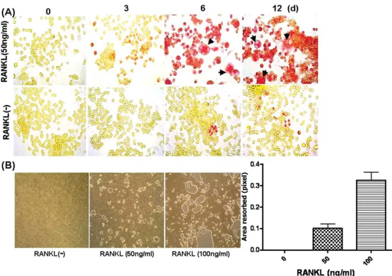
Figure 1. OCLs were differentiated from RAW264.7 cell lines after RANKL stimulation. (A) RAW264.7 cells were plated in 6-well dishes at 2x10⁵cells/well and were grown for 12 day with RANKL (50 ng/ml) or without (control). Cells were fixed and stained for TRAP activity. In presence of RANKL, TRAP-positive OCLs were formed. Arrows indicate multinucleated cells. The original magnification was x100. (B) Functional OCLs which have bone resorbing activity were formed in RAW264.7 cell cultures on a carbonated calcium phosphatase-coated 24-well plate, OAAS, after 7 days in presence of RANKL. The pit-forming activity was dose-dependent on RANKL treatment. The original magnification was x100.
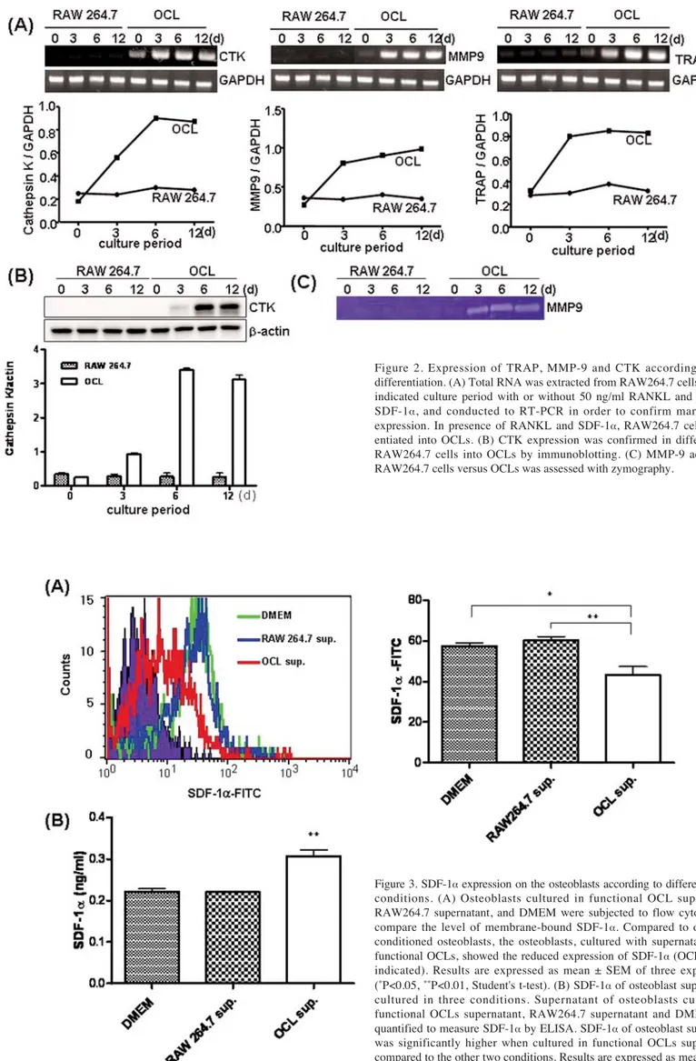
Figure 2. Expression of TRAP, MMP-9 and CTK according to OCL differentiation. (A) Total RNA was extracted from RAW264.7 cells after the indicated culture period with or without 50 ng/ml RANKL and 50 ng/ml SDF-1·, and conducted to RT-PCR in order to confirm marker gene expression. In presence of RANKL and SDF-1·, RAW264.7 cells differ-entiated into OCLs. (B) CTK expression was confirmed in differdiffer-entiated RAW264.7 cells into OCLs by immunoblotting. (C) MMP-9 activity of RAW264.7 cells versus OCLs was assessed with zymography.
Figure 3. SDF-1·expression on the osteoblasts according to different culture conditions. (A) Osteoblasts cultured in functional OCL supernatant, RAW264.7 supernatant, and DMEM were subjected to flow cytometry to compare the level of membrane-bound SDF-1·. Compared to other two conditioned osteoblasts, the osteoblasts, cultured with supernatants from functional OCLs, showed the reduced expression of SDF-1·(OCL sup., as indicated). Results are expressed as mean ± SEM of three experiments (*P<0.05, **P<0.01, Student's t-test). (B) SDF-1·of osteoblast suppernatant cultured in three conditions. Supernatant of osteoblasts cultured in functional OCLs supernatant, RAW264.7 supernatant and DMEM were quantified to measure SDF-1·by ELISA. SDF-1·of osteoblast supernatant was significantly higher when cultured in functional OCLs supernatant, compared to the other two conditions. Results are expressed as mean ± SEM of three experiments (**P<0.01, Student's t-test).
through the quantitative sandwich enzyme immunoassay kit (R&D System) in which a monoclonal antibody specific for SDF-1·has been pre-coated onto a microplate. Standards and samples were pipetted into the wells and any SDF-1·present was bound by the immobilized antibody. After washing away unbound substances, an enzyme-linked polyclonal antibody specific for SDF-1·was added to the wells. Following washing to remove any unbound antibody-enzyme reagent, a substrate solution was added to the wells and color developed in proportion to the amount of mouse SDF-1·bound in the initial step. The color development was stopped and the intensity of the color was measured by ELISA reader (SpectraMax, Molecular Devices, Sunnyvale, CA) and SDF-1· concentrations were calculated by SoftMax Pro program (Molecular Devices).
Cell mobilization in mice. In order to induce liver injury, six-week-old C57BL/6 female mice were injected carbon tetrachloride (CCl4, Sigma) in a 10% solution mixed in
mineral oil intraperitoneally once a day with 10 μl/g of body weight for two consecutive days. After 7 days, mice were sacrificed to collect peripheral blood (PB) in order to perform colony-forming assay. To induce OCLs activation, mouse RANKL (2 μg/injection) was injected subcutaneous over the femur once a day for 4 consecutive days. Mice were sacrificed to collect PB in order to perform colony-forming assay on the following day from the last injection. Mice injected with CCl4as mentioned above, were injected RANKL
sub-cutaneous over the femur the day following the last CCl4
administration. Non-treated six-week-old C57BL/6 female mice were investigated as control of colony-forming assay. For peripheral blood collection, mice were anesthetized by injection of ketamine (0.012 ml/g of body weight, Korea United Pharm, Seoul, Korea) and the chest cavity was opened. A needle was inserted into the ventricle and the blood slowly draw into the syringe. PB was placed in a 14-ml culture tube containing sufficient heparin and added 10 times the volume of RBC lysis solution. After mixing by inverting tube 3-4 times, blood was incubated on ice for 10 min and centrifuged for 7 min at 400 x g. The supernatants were removed and cells were washed 2 times with PBS for colony-forming assay. PB mononuclear cells were seeded in semisolid cultures using MethoCult system (StemCell Technologies, Vancouver, Canada). Colonies were scored 12 days after, according to morphologic criteria provided by the manufacturer.
Results
OCLs formed from RAW264.7 cell lines in response to RANKL. The ability of the RAW264.7 cell lines for OCL differen-tiation was assessed by culture in the presence of RANKL alone. Either TRAP-positive mutinucleated cells (Fig. 1A) or resorption pits (Fig. 1B) were observed in RANKL-treated culture. At least 6 days were needed for complete differ-entiation. In relation to bone resorption activity, it was increased with dose-dependence on RANKL treatment. RAW264.7 cell lines differentiate into OCLs in presence of RANKL plus SDF-1·. To determine OCL differentiation of
RAW264.7 cell lines, we assessed mRNA expression, protein expression and enzyme activity specific to OCLs. To induce the functional OCL differentiation, we treated RANKL during the full culture period, while SDF-1· was treated for 3 consecutive days of the initial stage of the differentiation in order to create a similar condition when OCL precursors activated into functional OCLs. RT-PCR targets included TRAP, MMP-9, and CTK genes, which were highly expressed as OCL differentiated (Fig. 2A). CTK, the major enzyme of OCLs for bone resorbing, was assessed via immunoblotting and confirmed its increased activity according to OCL differentiation (Fig. 2B). For other proteo-lytic enzyme, MMP-9 activity was also evaluated by zymo-graphy, which showed that only RANKL and SDF-1·induced functional OCLs exhibiting MMP-9 activity (Fig. 2C). Activated OCLs reduce the level of SDF-1· on the osteo-blasts. Functional OCLs showed increased expression of both MMP-9 and the major OCLs bone resorbing enzyme CTK (Fig. 2). To evaluate the effect of these enzymes secreted by functional OCLs on osteoblast expressing SDF-1·, we collected supernatants of OCLs after differentiation for 6 days. Osteoblasts were then cultured in these supernatants. Undifferentiated RAW264.7 cultured supernatant was also used and culturing in DMEM was performed as a control. After 5 days of culture, osteoblasts were quantified to compare the level of SDF-1·on the surface by flow cytometry. Compared to culturing condition of osteoblasts with DMEM, osteoblast cultured with supernatant from functional OCLs, showed reduced expression of membrane-bound SDF-1· (Fig. 3A). On the other hand, supernatants of osteoblast cultured in three conditions, as mentioned above, were collected and assayed to detect the SDF-1·by ELISA. SDF-1·of the supernatants was at the highest level when cultured with supernatant from activated OCLs (Fig. 3B). These results indicated that activated OCLs might participate in cleavage of SDF-1·on the osteoblasts.
Activated OCL induces CXCR4 increase on the BMC. CXCR4, a chemokine receptor for SDF-1·, is highly expressed on BMCs and involved in stem cell mobilization from BM to systemic circulation. Therefore, we determined whether activated OCLs induced change of CXCR4 expression on the BMCs. BMCs were cultured in three conditions as in the SDF-1·assay and quantified the level of CXCR4 expression on the cell surface by flow cytometry. CXCR4 on the BMCs, cultured with supernatant from OCLs, highly increased compared with two other conditioned BMCs. In particular, when the BMCs were cultured with supernatant from OCLs which was differentiated for 9 days, the level of CXCR4 expression was significantly increased compared with BMCs cultured in RAW264.7 supernatants (Fig. 4).
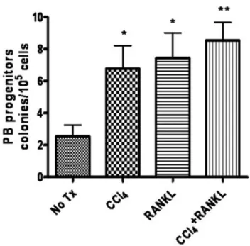
BM progenitor mobilizes under stress situation and RANKL treatment. In order to induce stress situation possibly leading to mobilization of BM progenitors, we injected CCl4to cause
acute liver injury to C57BL/6 mice. By counting mobilized BM progenitors from PB with colony-forming assay, we wished to compare the effect of activation of OCLs via RANKL signaling with liver injury status. Therefore,
treatment with combination of CCl4 and RANKL were
performed in colony-forming assay. Although progenitors mobilized without stimulation as part of homeostasis (Fig. 5A and B), the frequency of progenitors increased approximately 3 times in CCl4, or RANKL treated mice, when compared to
control mice. Moreover, mice treated with combination of CCl4, and RANKL showed additive effect on mobilization
(Fig. 5B).
Discussion
In this study, we identified activated OCL-induced SDF-1· reduction on the osteoblasts and increased CXCR4 expression on the BMCs. In addition, when mice were exposed to stress situation, as inducing liver injury via CCl4injection, the
number of mobilized progenitor cells into PB was significantly increased and such mobilization of progenitor cells could be enhanced significantly by injecting of RANKL, major stimulator of OCLs.
Stem cell niche is composed of various factors, such as SDF-1·, membrane bound stem cell factor (SCF) and osteopontin which were previously shown to have a crucial role in stem cell anchorage, survival and quiescence in the bone marrow. Among them, SDF-1·is the critical factor in osteoblast-BMCs anchorage as a regulator of adhesive interaction. SDF-1·and its cognitive receptor, CXCR4, act as a central axis in stem cell mobilization, including G-CSF induced mobilization, as well as in stem cell homing to the bone marrow (20,21). Thus, we focused on the function of OCLs, alteration of SDF-1·on the osteoblasts and CXCR4 on the BMCs. SDF-1·is required for enhanced secretion of MMP-9 and CTK in the BM, and acted as a OCLs activator, as we treated SDF-1·for the first 3 days during initial stage of differentiation. Functional OCLs secrete proteolytic enzymes, such as MMP-9 and CTK, which allow them to have bone-resorbing activity, but also provide them with SDF-1· degradation. In the mobilizing process, as we observed in this study by ELISA (Fig. 3B), a transient increase of SDF-1· in BM is followed by its proteolytic degradation and the level of CXCR4 expression increase on the surface of stem cells are essential mechanisms (21,22).
In order to confirm the effect of soluble mediators from activated OCLs on osteoblasts and BMCs, supernatant from RAW264.7 cells and from activated OCLs were used. The culture supernatant contains CTK and MMP-9 secreted from OCLs, as we detected (Fig. 2). SDF-1·expression on the surface of osteoblasts cultured with supernatant from
Figure 4. CXCR4 expression on the BMCs according to different culture conditions. (A) Representative result of flow cytometric analysis of CXCR4 expression on BMCs in three different conditions on day 0. (B) CXCR4 expressions on BMCs cultured in OCLs supernatant, RAW264.7 supernatant and DMEM were quantified by flow cytometry and data presented by mean fluorescence intensity (MFI) of BMCs stained with PE-conjugated anti-CXCR4 Ab. CXCR4 expression on BMCs cultured in supernatant of OCLs, which were differentiated for 9 days (d) from RAW264.7 cells, significantly increased compared to BMCs conditioned in RAW264.7 supernatant. Results are expressed as mean ± SEM of three experiments (**P<0.01, Two-way ANOVA).
Figure 5. Acute injury and RANKL injection enhanced the mobilization of hematopoietic progenitor cells. (A) Circulating PB from untreated mice, mice injected with CCl4or RANKL, and in combination of CCl4/RANKL were collected and cultured for colony assay to evaluate the number of mobilized progenitor cells. After 10 days of culture on MethoCult media, colonies were counted. The original magnification was x50. (B) Colony-forming cells of CCl4or RANKL-injected mice were higher in number, compared with those of control mice (No Tx, as indicated). CCl4/RANKL injection group showed the highest number of colony-forming cells. Results are expressed as mean ± SEM of three experiments (*P<0.05, **P<0.01, Student's t-test).
activated OCLs was reduced compared to those with super-natants from undifferentiated RAW264.7 cells or DMEM. These membrane-bound SDF-1· cleavages were also high-lighted through supernatant quantification by ELISA (Fig. 3). SDF-1· of osteoblasts, culturing with supernatant from OCLs, was remarkably high, indicating that high levels of cleaved SDF-1· exists in the supernatant. In contrast, CXCR4 expression on the BMCs increased in culture with OCLs supernatant. Although the mechanism of CXCR4 up-regulation in the mobilizing process has not been precisely determined, it has been reported that stress signals or clinical protocol, such as G-CSF treatment, indirectly induced up-regulation of CXCR4 expression on BMs through mobilizing cytokines activity such as IL-4, IL-6 and IL-8 or as a con-sequence of the potent collapse of SDF-1· concentration within the BMs (19,21). Even though it had limitations of a cell line-based in vitro study, we showed that OCL-mediated CXCR4 regulation on BMCs may exist possibly via secreted enzymes (Fig. 4).
Finally, we confirmed that stress (i.e., liver injury) induced progenitor mobilization in vivo. Under stress situations, including hypoxia, inflammation, chemotherapy and injury, SDF-1, RANKL and HGF in BM are transiently increased and enhance OCLs activation (14,16). As we observed here, mice with damaged liver through CCl4 injection had
significantly higher level of mobilized progenitors, and this outcome was enhancing when performed additional treatment with RANKL, a stimulator of OCL precursors (Fig. 5). Therefore, stress induced the activation of OCLs and such activation of osteoclast seemed to accelerate the mobilization process from the BM, especially more prominent after the treatment with RANKL.
In summary, this study showed that activated OCLs affected the level of expression of SDF-1·on osteoblasts and the CXCR4 expression on BMC via secreted enzymes, e.g., CTK and MMP-9. Moreover, RANKL injection enhanced the mobilization of BM progenitors into the circulation via activation of OCLs in the BM. This means that OCL-osteoblast interaction is a major regulator of turnover for the endosteal stem cell niches. Furthermore, RANKL should be considered together with other mobilizing agents aimed at selective mobilization of progenitor cells for poorly-mobilizing elderly or chemotherapy-treated individuals.
Acknowledgements
This study was supported by a grant of the Korean Health Technology R&D Project, Ministry for Health, Welfare and Family Affairs in Republic of Korea (A084067).
References
1. Boyle WJ, Simonet WS and Lacey DL: Osteoclast differentiation and activation. Nature 423: 337-342, 2003.
2. Teitelbaum SL: Bone resorption by osteoclasts. Science 289: 1504-1508, 2000.
3. Asagiri M and Takayanagi H: The molecular understanding of osteoclast differentiation. Bone 40: 251-264, 2007.
4. Matsuo K and Irie N: Osteoclast-osteoblast communication. Arch Biochem Biophys 473: 201-209, 2008.
5. Udagawa N, Takahashi N, Jimi E, Matsuzaki K, Tsurukai T, Itoh K, Nakagawa N, Yasuda H, Goto M, Tsuda E, Higashio K, Gillespie MT, Martin TJ and Suda T: Osteoblasts/stromal cells stimulate osteoclast activation through expression of osteoclast differentiation factor/RANKL but not macrophage colony-stimulating factor: receptor activator of NF-kappa B ligand. Bone 25: 517-523, 1999.
6. Katagiri T and Takahashi N: Regulatory mechanisms of osteoblast and osteoclast differentiation. Oral Dis 8: 147-159, 2002.
7. Mayack SR and Wagers AJ: Osteolineage niche cells initiate hematopoietic stem cell mobilization. Blood 112: 519-531, 2008.
8. Zhang J, Niu C, Ye L, Huang H, He X, Tong WG, Ross J, Haug J, Johnson T, Feng JQ, Harris S, Wiedemann LM, Mishina Y and Li L: Identification of the haematopoietic stem cell niche and control of the niche size. Nature 425: 836-841, 2003.
9. Lemischka IR and Moore KA: Stem cells: interactive niches. Nature 425: 778-779, 2003.
10. Moore KA and Lemischka IR: Stem cells and their niches. Science 311: 1880-1885, 2006.
11. Calvi LM, Adams GB, Weibrecht KW, Weber JM, Olson DP, Knight MC, Martin RP, Schipani E, Divieti P, Bringhurst FR, Milner LA, Kronenberg HM and Scadden DT: Osteoblastic cells regulate the haematopoietic stem cell niche. Nature 425: 841-846, 2003.
12. Taichman RS, Reilly MJ and Emerson SG: Human osteoblasts support human hematopoietic progenitor cells in vitro bone marrow cultures. Blood 87: 518-524, 1996.
13. Kaplan RN, Psaila B and Lyden D: Niche-to-niche migration of bone-marrow-derived cells. Trends Mol Med 13: 72-81, 2007. 14. Kollet O, Dar A and Lapidot T: The multiple roles of osteoclasts
in host defense: bone remodeling and hematopoietic stem cell mobilization. Annu Rev Immunol 25: 51-69, 2007.
15. Purton LE and Scadden DT: Osteoclasts eat stem cells out of house and home. Nat Med 12: 610-611, 2006.
16. Kollet O, Dar A, Shivtiel S, Kalinkovich A, Lapid K, Sztainberg Y, Tesio M, Samstein RM, Goichberg P, Spiegel A, Elson A and Lapidot T: Osteoclasts degrade endosteal components and promote mobilization of hematopoietic progenitor cells. Nat Med 12: 657-664, 2006.
17. Takayanagi H: Osteoimmunology: shared mechanisms and crosstalk between the immune and bone systems. Nat Rev Immunol 7: 292-304, 2007.
18. Dar A, Kollet O and Lapidot T: Mutual, reciprocal SDF-1/ CXCR4 interactions between hematopoietic and bone marrow stromal cells regulate human stem cell migration and development in NOD/SCID chimeric mice. Exp Hematol 34: 967-975, 2006.
19. Lapidot T and Petit I: Current understanding of stem cell mobilization: the roles of chemokines, proteolytic enzymes, adhesion molecules, cytokines, and stromal cells. Exp Hematol 30: 973-981, 2002.
20. Lapidot T, Dar A and Kollet O: How do stem cells find their way home? Blood 106: 1901-1910, 2005.
21. Petit I, Szyper-Kravitz M, Nagler A, Lahav M, Peled A, Habler L, Ponomaryov T, Taichman RS, Arenzana-Seisdedos F, Fujii N, Sandbank J, Zipori D and Lapidot T: G-CSF induces stem cell mobilization by decreasing bone marrow SDF-1 and up-regulating CXCR4. Nat Immunol 3: 687-694, 2002.
22. Porecha NK, English K, Hangoc G, Broxmeyer HE and Christopherson KW II: Enhanced functional response to CXCL12/SDF-1 through retroviral overexpression of CXCR4 on M07e cells: implications for hematopoietic stem cell trans-plantation. Stem Cells Dev 15: 325-333, 2006.