Endocrinol Metab 2018;33:236-244 https://doi.org/10.3803/EnM.2018.33.2.236 pISSN 2093-596X · eISSN 2093-5978
Original
Article
C-Arm Computed Tomography-Assisted Adrenal Venous
Sampling Improved Right Adrenal Vein Cannulation and
Sampling Quality in Primary Aldosteronism
Chung Hyun Park1,*, Namki Hong1,*, Kichang Han2, Sang-Wook Kang3, Cho Rok Lee3, Sungha Park4, Yumie Rhee1 1Department of Internal Medicine, Endocrine Research Institute, Severance Hospital, Yonsei University College of Medicine; 2Department of Radiology, Research Institute of Radiological Science, Yonsei University College of Medicine; 3Department of Surgery, Severance Hospital, Yonsei University College of Medicine; 4Division of Cardiology, Department of Internal Medicine, Yonsei University College of Medicine, Seoul, Korea
Background: Adrenal venous sampling (AVS) is a gold standard for subtype classification of primary aldosteronism (PA). However, this procedure has a high failure rate because of the anatomical difficulties in accessing the right adrenal vein. We investigated whether C-arm computed tomography-assisted AVS (C-AVS) could improve the success rate of adrenal sampling.
Methods: A total of 156 patients, diagnosed with PA who underwent AVS from May 2004 through April 2017, were included. Based on the medical records, we retrospectively compared the overall, left, and right catheterization success rates of adrenal veins during the periods without C-AVS (2004 to 2010, n=32) and with C-AVS (2011 to 2016, n=124). The primary outcome was adequate bi-lateral sampling defined as a selectivity index (SI) >5.
Results: With C-AVS, the rates of adequate bilateral AVS increased from 40.6% to 88.7% (P<0.001), with substantial decreases in failure rates (43.7% to 0.8%, P<0.001). There were significant increases in adequate sampling rates from right (43.7% to 91.9%,
P<0.001) and left adrenal veins (53.1% to 95.9%, P<0.001) as well as decreases in catheterization failure from right adrenal vein
(9.3% to 0.0%, P<0.001). Net improvement of SI on right side remained significant after adjustment for left side (adjusted SI, 1.1 to 9.0; P=0.038). C-AVS was an independent predictor of adequate bilateral sampling in the multivariate model (odds ratio, 9.01;
P<0.001).
Conclusion: C-AVS improved the overall success rate of AVS, possibly as a result of better catheterization of right adrenal vein.
Keywords: Hyperaldosteronism; Hypertension; Cone-beam computed tomography; Adrenalectomy
INTRODUCTION
Primary aldosteronism (PA) is the most common cause of sec-ondary hypertension characterized by unregulated aldosterone
secretion [1]. Resistant hypertension and hypokalemia are the classic features of hyperaldosteronism. In patients with PA, however, the clinical and biochemical phenotypes are now be-lieved to be more heterogeneous than once thought, varying in
Received: 19 December 2017, Revised: 19 February 2018, Accepted: 8 March 2018
Corresponding author: Yumie Rhee
Department of Internal Medicine, Severance Hospital, Yonsei University College of Medicine, 50-1 Yonsei-ro, Seodaemun-gu, Seoul 03722, Korea
Tel: +82-2-2228-1973, Fax: +82-2-392-5548, E-mail: yumie@yuhs.ac *These authors contributed equally to this work.
Copyright © 2018 Korean Endocrine Society
This is an Open Access article distributed under the terms of the Creative Com-mons Attribution Non-Commercial License (http://creativecomCom-mons.org/ licenses/by-nc/4.0/) which permits unrestricted non-commercial use, distribu-tion, and reproduction in any medium, provided the original work is properly cited.
degree among different subtypes and genotypes [2]. Partly due to the disease heterogeneity, its prevalence tends to change by selected study population and diagnostic methodology, ranging from 3.2% to 12.7% in primary care settings and up to 30% in hypertension units [2]. With the continued rise in the use of al-dosterone-to-renin ratio (ARR) for a screening test, the preva-lence has been found to account for a third of hypertensive pa-tients randomly selected from the general population [1-4].
As the importance of early detection and intervention of PA has become more recognized, the current clinical guidelines recommend that all hypertensive patients at increased risk for PA undergo a screening test, confirmatory tests, and subtype classification [5]. Differentiating between unilateral and bilater-al subtypes is particularly criticbilater-al to opt for surgicbilater-al treatment when feasible. Currently, adrenal venous sampling (AVS) is the gold standard for assessing lateralization of aldosterone secre-tion with the highest sensitivity (95%) and specificity (100%) [5]. Despite its essential role, AVS is still vastly underutilized, performed only in a few major referral centers by an experi-enced interventional radiologist, due to high technical demands. The high failure rates of AVS are attributed mainly to the diffi-culty in accessing the right adrenal vein [6]. C-arm computed tomography (CT) during the right adrenal vein catheterization has been emerged as a novel approach to improve diagnostic outcomes of AVS [7]. C-arm CT-assisted AVS (C-AVS) has been reported to lower the failure rates with minimal complica-tion rates (none in 42 patients, Park et al. [8]; 1 adrenal vein hemorrhage in 19 patients, Plank et al. [7]) [7-9]. We introduced the C-AVS protocol in our hospital in 2011. Before and after this transition were designated as p and post-C-AVS era, re-spectively. In this study, we aim to compare the bilateral and unilateral catheterization success rates between two periods by retrospective analysis.
METHODS
Study designThis is a retrospective study of patients diagnosed with PA from May 2004 through April 2017 at Severance Hospital, Seoul, Korea (Fig. 1). This study was approved by the Institutional Re-view Board of Severance Hospital (no. 4-2017-0838). Informed consent for procedure was obtained from each individual prior to AVS. We identified subjects who underwent AVS before and after 2011, respectively, the transition at which C-AVS was in-troduced into our facility. The two periods were defined as pre- and post-C-AVS era, respectively. Based on the medical records,
we compared the baseline demographics, AVS and CT results, overall, left, and right catheterization success rates between pre- and post-C-AVS era.
Case detection
To determine ARR, we measured plasma aldosterone concentra-tion (PAC) to plasma renin activity ratio using the radioimmu-noassay method. ARR greater than 30 (in ng/dL per ng/mL/hr) with PAC greater than 10 ng/dL were used as the cutoff. Saline infusion test (SIT) was used to confirm the diagnosis of PA. If PAC measured after 2 L intravenous infusion of 0.9% normal saline over 4 hours at the recumbent position was not suppressed to below 5 ng/dL, PA was confirmed [3,10]. For the screening and confirmatory tests, diuretics, β-blockers, angiotensin-con-verting enzyme inhibitors, and angiotensin II receptor blockers were ceased at least 2 weeks; mineralocorticoid receptor antago-nists were stopped for at least 4 weeks. Non-dihydropyridine calcium channel blockers and/or doxazosin were used in the cases with uncontrolled hypertension. All patients who under-went AVS also undertook contrast-enhanced adrenal CT scan to assess the location and size of adrenal nodules, if present.
AVS and C-arm CT protocol
AVS was performed sequentially via transfemoral approach un-der 50 μg/hr cosyntropin infusion initiated 30 minutes before the procedure. The right transfemoral vein was accessed under ultrasound-guidance and a 5-F vascular sheath was inserted. The blood samples were obtained in the order of right adrenal vein, left adrenal vein, and infrarenal inferior vena cava. To catheterize the right adrenal vein, a 5-F Cobra catheter (Cook, Bloomington, IN, USA) was primarily used, and additional sideholes were made at the tip of catheter. When the selection of right adrenal vein was technically challenging, various types of angiographic catheters were used. Under fluoroscopy-guidance, 5-F catheter was positioned into the presumed right adrenal vein and venogram was obtained. Then, C-arm CT was performed with manual injection of about 3 to 4 mL of diluted contrast ma-terial (25% to 50% in 0.9% normal saline). The C-arm CT scans were processed in the workstation (syngoXWP, Siemens, Forchheim, Germany) and displayed as three-dimensional mul-tiplanar reconstruction images. Blood samples were acquired if C-arm CT demonstrated proper catheter position. After sam-pling, venogram was repeated to ensure that the catheter re-mained engaged in the right adrenal vein. When the right adnal vein was not properly catheterized, the catheter tip was re-positioned until the right adrenal gland was opacified on C-arm
CT image. Left AVS was performed with a 5-F angled taper catheter (Terumo, Tokyo, Japan) without the assistance of C-arm CT scan.
Selectivity and lateralization indices
With continuous cosyntropin infusion protocol, the adequacy of each AVS was determined based on the selectivity index (SI) cutoff value of 5: sampling is considered adequate if SI >5 and inadequate if SI ≤5 [5,11]. Overall, the quality of catheteriza-tion was categorized as adequate bilateral sampling if both sides were adequately catheterized, and as adequate unilateral sam-pling if only one side was adequately catheterized, and AVS failure if the sampling failed or fell under the inadequate cate-gory on both sides. The patients with at least unilateral adequate AVS were further assessed for lateralization. In the case of ade-quate bilateral AVS, lateralization index (LI) greater than 4, or between 3 and 4 with contralateral suppression (cortisol
correct-ed aldosterone ratio between the non-dominant adrenal vein and peripheral vein, contralateral suppression index, is less than 1) were used as the lateralization criteria. For adequate unilateral AVS, the presence of contralateral suppression was considered sufficient to determine lateralization [11].
Surgical outcomes for unilateral PA
Among those 61 patients with unilateral PA in post-C-AVS era, 55 patients underwent adrenalectomy. Their clinical and bio-chemical outcomes were evaluated using the follow-up data in-cluding blood pressure, the number of antihypertensive medica-tion, serum potassium, ARR, all measured at 6 to 12 months. The evaluation criteria were modified from the Primary Aldo-steronism Surgical Outcome (PASO) international consensus [12]. Complete clinical success was defined as normal blood pressure without use of antihypertensive medication. Partial clinical success was defined as same blood pressure as before
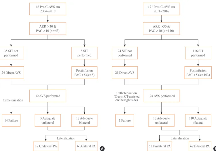
46 Pre-C-AVS era
2004−2010 171 Post-C-AVS era2011−2016
ARR >30 &
PAC >10 (n=43) PAC >10 (n=140)ARR >30 &
32 AVS performed 124 AVS performed
5 Adequate
unilateral 13 Adequate unilateral
12 Unilateral PA 61 Unilateral PA
13 Adequate
bilateral 110 Adequatebilateral
6 Bilateral PA 62 Bilateral PA
14 Failure 1 Failure
8 SIT
performed performed116 SIT
Postinfusion
PAC >5 (n=8) PAC >5 (n=103)Postinfusion
35 SIT not
performed 24 SIT notperformed
24 Direct AVS 21 Direct AVS
A B
Fig. 1. Flowcharts of the study subjects in (A) pre- and (B) post-C-arm CT-assisted AVS (C-AVS) era. ARR, aldosterone-to-renin ratio; PAC, plasma aldosterone concentration; SIT, saline infusion test; AVS, adrenal venous sampling; PA, primary aldosteronism; CT, computed tomography.
Catheterization
Catheterization (C-arm CT-assisted
on the right side)
surgery with reduced number of drugs or lower blood pressure with either the same or reduced number of drugs. Absent clini-cal success indicates unchanged or increased blood pressure with either the same or increased number of drugs. Biochemical success was considered complete if hypokalemia and ARR were normalized. We used the cutoff value of <3.5 mmol/L for hypo-kalemia and >30 ng/dL per ng/mL/hr for positive ARR. Partial success indicates correction of hypokalemia and a raised ARR with ≥50% decrease in baseline PAC. Absent success indicates persistent hypokalemia, or persistently raised ARR, or both.
Statistical analysis
Data were presented as a mean±SD, median (interquartile range [IQR]), or as number (%) as appropriate. Continuous variables were compared using the t test, and categorical vari-ables and catheterization success rates between pre- and post-C-AVS era were analyzed using the chi-square test. Statistical sig-nificance between nonparametric variables was assessed by Mann-Whitney U test. Adjusted right SI was calculated by sub-tracting SI of left adrenal vein from that of the right adrenal vein. Net improvement of adjusted right SI was compared be-tween pre- and post-C-AVS era. Multivariate logistic regression analysis was performed to identify independent predictors of adequate bilateral sampling. Statistical analyses were performed using Stata version 12.0 SE (StataCorp, College Station, TX, USA). A P value less than 0.05 was considered to be statistically significant.
RESULTS
Study populationWe identified 46 and 171 patients who underwent ARR screen-ing test in pre- and post-C-AVS era, respectively, by retrospec-tively reviewing their medical records (Fig. 1). In each group, 43 and 140 patients were tested positive. Among these patients, eight out of 43 and 116 out of 140 patients underwent SIT. In a pre-C-AVS era, 24 out of 35 patients who did not undergo SIT underwent directly to AVS, and so did 21 out of 24 patients in the post-C-AVS era. In total, 32 and 124 cases of AVS were per-formed in pre- and post-C-AVS era groups, respectively, which were included in this analysis to assess selectivity and lateral-ization. In each group, 13 (40.6% of the total AVS-performed population) and 110 patients (88.7%) resulted in adequate bilat-eral catheterization, five (15.6%) and 13 patients (10.5%) re-sulted in unilateral adequate catheterization, and 14 (43.8%) and one patient (0.8%) resulted in bilateral catheterization failure, respectively. Patients in the adequate bilateral and adequate uni-lateral groups were classified as either uniuni-lateral or biuni-lateral PA based on LI and contralateral suppression index. The number of patients with unilateral and bilateral PA in the pre-C-AVS era was 12 (66.6%) and six (33.3%), respectively, and in the post-C-AVS era was 61 (49.6%) and 62 (50.4%).
Baseline characteristics
Baseline characteristics of 156 patients who underwent AVS (32
Table 1. Baseline Characteristics of the Study Subjects in Pre- and Post-C-AVS Era
Variable Pre-C-AVS (n=32) Post-C-AVS (n=124) P value
Age, yr 46.2±10.5 50.0±11.4 0.093
Sex 0.438
Male 14 (43.7) 45 (36.2)
Female 18 (56.2) 79 (63.7)
Body mass index, kg/m2 24.7±3.1 24.8±3.8 0.879
Systolic blood pressure, mm Hg 157±21 146±14 0.001
Diastolic blood pressure, mm Hg 94±14 91±12 0.333
Serum potassium, mmol/L 3.1±0.6 3.7±0.5 <0.001
PRA, ng/mL/hr 0.11 (0.07–0.20) 0.21 (0.10–0.42) 0.006
PAC, ng/dL 24.7 (18.4–55.5) 34.4 (20.5–56.3) 0.329
ARR, ng/dL per ng/mL/hr 389 (157–1,068) 121 (55–428) 0.002
Values are expressed as mean±SD, number (%), or median (interquartile range).
C-AVS, C-arm computed tomography-assisted adrenal venous sampling; PRA, plasma renin activity; PAC, plasma aldosterone concentration; ARR, al-dosterone to renin ratio.
and 124 in pre- and post-C-AVS era, respectively) were summa-rized in Table 1. In subjects before and after C-AVS, the ages were 46.2±10.5 and 50.1±11.4 years, 56.2% and 63.7% were women, and the body mass indexs were 24.7±3.1 and 24.8± 3.8 kg/m2, respectively, without a significant statistical
differ-ence. The patients in pre-C-AVS group had higher systolic blood pressure (157±21 mm Hg vs. 146±14 mm Hg, P= 0.001), lower serum potassium level (3.1±0.6 mmol/L vs. 3.7± 0.5 mmol/L, P<0.001), and higher ARR (389 ng/dL per ng/mL/ hr [IQR, 157 to 1,068] vs. 121 ng/dL per ng/mL/hr [IQR, 55 to 428], P=0.002) at initial visit, whereas diastolic blood pressure and PAC did not differ significantly.
AVS and CT results
As summarized in Table 2, the SIs of both left and right adrenal veins increased significantly (left, 5.4 [IQR, 1.9 to 7.4] to 15.5 [IQR, 9.1 to 24.6]; right, 4.0 [IQR, 1.4 to 18.6] to 26.1 [IQR, 16.2 to 34.1]; P<0.001). The increase was more prominent on the right side, in which C-AVS was performed. The convention-al AVS identified 12 patients (66.6%) with unilaterconvention-al forms of PA and six patients (33.3%) with bilateral PA. With C-AVS, the proportion of bilateral PA tended to increase (50.4%), although it did not reach statistical significance. Changes in the concor-dance rates between AVS and CT findings in two study groups were analyzed as shown in Table 2. The findings were
consid-ered concordant if unilateral lesions were visualized on CT and an excess aldosterone secretion was lateralized to the same side
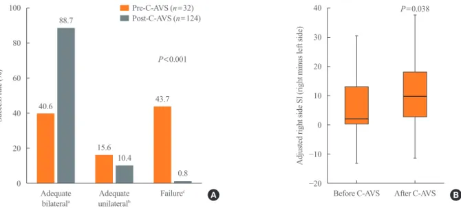
100 80 60 40 20 0 40 30 20 10 0 −10 −20 Success rate (%)
Adjusted right side SI (right minus left side)
Adequate
bilaterala unilateralAdequateb Failure Before C-AVS After C-AVS
c
Fig. 2. Comparison of (A) the rates of adequate bilateral adrenal venous sampling (AVS) and (B) selectivity indices in pre- and post-C-arm computed tomography-assisted AVS (C-AVS) era. Adjusted right side selectivity index (SI) was calculated by subtracting left SI from right SI. The middle line of the box indicates the median. The lower and the upper end of the whiskers indicate the minimum and the maximum observations blow the upper fence (1.5 interquartile range above the 75th percentile), respectively. aAdequate bilateral indicates adequate
sampling in both sides; bAdequate unilateral indicates adequate sampling in any one side with catheterization failure or inadequate sampling
on the other side; cFailure indicates catheterization failure or inadequate sampling on both sides.
A B 40.6 15.6 43.7 88.7 10.4 0.8 Pre-C-AVS (n=32) Post-C-AVS (n=124) P<0.001 P=0.038
Table 2. AVS and CT Results of the Study Subjects in Pre- and Post-C-AVS Era
Variable Pre-C-AVS (n=32) Post-C-AVS (n=124) P value
Median selectivity index
Left 5.4 (1.9–7.4) 15.5 (9.1–24.6) <0.001 Right 4.0 (1.4–18.6) 26.1 (16.2–34.1) <0.001 Subtype 0.176 Unilateral PAa 12 (66.6) 61 (49.5) Bilateral PAb 6 (33.3) 62 (50.4) AVS-CT concordance 0.934 Concordantc 12 (60.0) 75 (60.9) Discordantd 8 (40.0) 48 (39.0)
Values are expressed as median (interquartile range) or number (%). AVS, adrenal venous sampling; CT, computed tomography; AVS, C-arm computed tomography-assisted AVS; PA, primary aldosteronism.
aUnilateral PA includes aldosterone-producing adenomas and unilateral
adrenal hyperplasia; bBilateral PA includes bilateral adrenal hyperplasia
or bilateral idiopathic hyperaldosteronism; cConcordant findings
in-clude unilateral lesions on CT and ipsilateral lateralization on AVS, and
bilateral lesions on CT and no lateralization on AVS; dDiscordant
find-ings include unilateral lesions on CT and contralateral or no lateraliza-tion on AVS.
on AVS as well as if bilateral lesions were visualized on CT and no lateralization was confirmed on AVS. The conflicting results such as CT unilateral and AVS contralateral or no lateralization were considered discordant. The concordance rates between two periods were comparable (60% vs. 60.9%, P=0.934). Ad-renal nodule sizes also did not differ significantly between two groups (1.5±5.5 cm vs. 1.6±13.3 cm, P=0.558). For each C-arm CT-assisted procedure, the total procedural time, total fluo-roscopy time, and total radiation dose were 45, 7 minutes, and 160 to 170 mGy, respectively. The additional procedure time for the C-arm CT was 5 to 10 minutes, and the additional radiation exposure was 90 to 100 mGy (60% to 70%) (data not shown).
Changes in the overall success rates and adjusted right side SI of AVS
As shown in Fig. 2A, with the assistance of C-arm CT, the rate of adequate bilateral sampling increased significantly from 40.6% to 88.7%, with a marked reduction in adequate unilateral sampling and overall failure (15.6% to 10.4% and 43.7% to 0.8%, respectively; P<0.001 for all). The catheterization suc-cess rates of each adrenal vein are summarized in Supplemental Table S1. The results showed a marked increase in the success rate of adequate sampling from a right adrenal vein (43.7% to 91.9%, P<0.001) and left adrenal vein (53.1% to 95.9%,
P<0.001) as well as a decrease in inadequate sampling from
both adrenal veins. The catheterization failure previously
asso-ciated with a right adrenal vein (9.3%) was reduced to 0.0% (P<0.001). Considering the possible training effect originated from the temporally sequenced design of our study, we demon-strated the presence of a net improvement in the right side cath-eterization with C-AVS (Fig. 2B). The adjusted right side SIs, calculated by subtracting left side SI from right side SI, were compared before and after C-AVS. The median value of the post-C-AVS adjusted right side SI increased significantly (1.1 to 9.0, P=0.038), indicating that the significant improvement of sampling quality in right adrenal vein, with the left side im-provement taken into account, could not be entirely attributable to the training effect.
Independent association of C-AVS with adequate bilateral sampling
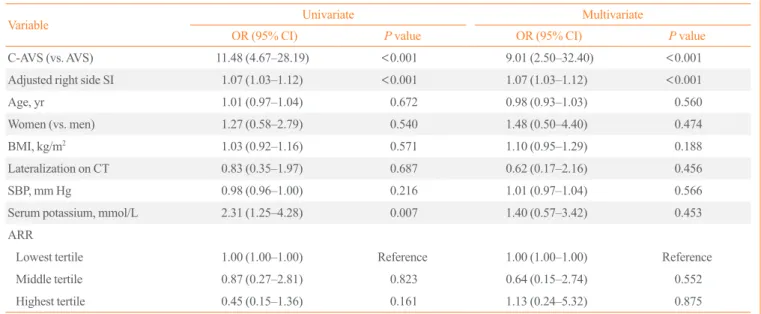
Adoption of C-AVS was found to be associated with adequate bilateral sampling (odds ratio [OR], 11.48; 95% confidence in-terval [CI], 4.67 to 28.19; P<0.001) (Table 3). This association remained robust after adjustment for potential confounders in the multivariate model (OR, 9.01; 95% CI, 2.50 to 32.40;
P<0.001). The adjusted right side SI was also an independent
predictor of adequate bilateral sampling (OR, 1.07; 95% CI, 1.03 to 1.12; P<0.001) along with C-AVS, indicating that im-provement of success rate in the post-C-AVS era is at least part-ly mediated by improvement of sampling adequacy in right side adrenal vein.
Table 3. Independent Association of C-AVS with Adequate Bilateral Sampling
Variable Univariate Multivariate
OR (95% CI) P value OR (95% CI) P value
C-AVS (vs. AVS) 11.48 (4.67–28.19) <0.001 9.01 (2.50–32.40) <0.001
Adjusted right side SI 1.07 (1.03–1.12) <0.001 1.07 (1.03–1.12) <0.001
Age, yr 1.01 (0.97–1.04) 0.672 0.98 (0.93–1.03) 0.560
Women (vs. men) 1.27 (0.58–2.79) 0.540 1.48 (0.50–4.40) 0.474
BMI, kg/m2 1.03 (0.92–1.16) 0.571 1.10 (0.95–1.29) 0.188
Lateralization on CT 0.83 (0.35–1.97) 0.687 0.62 (0.17–2.16) 0.456
SBP, mm Hg 0.98 (0.96–1.00) 0.216 1.01 (0.97–1.04) 0.566
Serum potassium, mmol/L 2.31 (1.25–4.28) 0.007 1.40 (0.57–3.42) 0.453
ARR
Lowest tertile 1.00 (1.00–1.00) Reference 1.00 (1.00–1.00) Reference
Middle tertile 0.87 (0.27–2.81) 0.823 0.64 (0.15–2.74) 0.552
Highest tertile 0.45 (0.15–1.36) 0.161 1.13 (0.24–5.32) 0.875
C-AVS, C-arm CT-assisted AVS; OR, odds ratio; CI, confidence interval; AVS, adrenal venous sampling; SI, selectivity index; BMI, body mass index; CT, computed tomography; SBP, systolic blood pressure; ARR, aldosterone-to-renin ratio.
Surgical results of unilateral primary aldosteronism
The clinical and biochemical outcomes of adrenalectomy for patients with unilateral PA are shown in Supplemental Fig. S1. Among those 61 patients with unilateral PA in post-C-AVS era, 55 patients underwent adrenalectomy. Based on the criteria modified from the PASO international consensus, the clinical and biochemical remission rates were assessed as complete, partial, and absent. Among 41 patients with available follow-up data, 39% achieved complete success, and 61% achieved at least partial remission. In terms of biochemical remission, near-ly all patients achieved complete success (29 of 31 patients). These results suggest that the C-arm CT-assisted AVS was con-siderably effective in subtype diagnosis of PA.
DISCUSSION
Although AVS plays a crucial role in subtype classification of PA, it is not as widely used as demanded due to technical diffi-culties. The poor success rate of the right adrenal vein, ranging from 55% to 98%, is of the main concern to overcome [9]. The conventional AVS relies primarily on venographic findings to identify the adrenal veins. In case of a right adrenal vein, the venographic patterns are often nonspecific, and renal capsular vein and accessory hepatic vein are commonly mistaken as a right adrenal vein. These erroneous catheterizations can be im-mediately corrected by contrast-enhanced CT images on site [8,13,14]. In this study, we found that the success rate of right adrenal vein catheterization was markedly improved with the use of intraprocedural C-arm CT. Adoption of C-AVS and ad-justed right side SI were positively associated with adequate bi-lateral sampling, and these associations were independent of possible confounders.
To define adequate sampling under adrenocorticotropic hor-mone (ACTH) stimulation, we strictly adhered to the current guidelines using SI cutoff value of 5 [5,11]. We found a marked improvement in the success rates of overall (88.7%) and right AVS (91.9%), which are comparable to those reported in other studies (overall AVS, 87% to 95.7%; right AVS, 95% to 100%) [8,9,13,15]. These studies, however, used different CT tech-niques including dyna-CT, C-arm CT, and angio-CT and differ-ent protocols with or without ACTH stimulation varying in se-lectivity criteria. In our study, the success rate of right AVS would have been 94.3% (vs. currently reported 91.9% in the right AVS) if SI cutoff value of 3 was chosen instead of 5. In our study, we also showed improved results on left AVS as repre-sented by increased SI in the post-C-AVS era. Even when the
possible training effect was corrected by measuring the differ-ence between right and left SI, as denoted by the term adjusted right side SI, the rise in the success rate of right AVS was evi-dent. The adjusted right side SI was a strong independent pre-dictor of adequate bilateral sampling. Therefore, as suggested by previous studies, with C-AVS, we were able to achieve better sampling outcome overall, substantially reducing the technical failure rate of right AVS in a large cohort from a single institu-tion using unified protocol.
Sequential sampling with continuous ACTH infusion is a widely used protocol by which stress-induced fluctuations in al-dosterone release can be avoided, and alal-dosterone secretion from aldosterone-producing adenoma (APA) as well as the cor-tisol gradient between the adrenal vein and inferior vena cava can be maximized to increase lateralization and selectivity indi-ces, respectively [11,16]. In a previous study conducted to dem-onstrate the role of C-arm CT in improving success rates of AVS, the effect of ACTH stimulation was also measured [14]. Selective sampling was considered successful if SI >5 with ACTH stimulation and SI >3 without ACTH stimulation. The success rates increased from 19% (6/32) to 91% after ACTH stimulation [15]. In this study, we found that C-AVS further im-proved the diagnostic success rate even in the setting of continu-ous ACTH infusion during AVS, indicating the additive impact of C-AVS on sampling accuracy.
The concordance rate between AVS and CT was 60%, consis-tent with the rates reported by others [17,18]. Interestingly, this value was not affected by C-AVS (60% to 60.9%, P=0.934), suggesting that CT-guided diagnosis alone would be unreliable in subtype differentiation, similar to the previous reports [3]. Also, our results showed that the proportion of unilateral PA had tended to decrease in the post-C-AVS era (66.6% to 49.5%,
P=0.276), a trend also found in a previous study with
temporal-ly sequenced study design (70.7% to 54.5%, P=0.19) [14]. It is conceivable that increased number of patients screened with ARR might have led to improved detection of milder forms of the disease. Given the prevalence of APA and bilateral adrenal hyperplasia found in a recent, large prospective cohort (27% and 65% of the patients with PA, respectively), our finding may also indicate that the reduction in disproportionality between subtypes might have been driven by the better diagnostic accu-racy of C-AVS [3]. It should be considered; however, that a cer-tain degree of disproportionality is likely to remain as our study was conducted in a tertiary referral center with a population of predominantly ARR positive patients.
were not randomized but were temporally sequenced, rendering it a major limitation to this study. The success rate of left adre-nal venous catheterization was unexpectedly low (53.1%) in pre-C-AVS era. This may be explained largely by the lack of experience, where there were only 32 cases of AVS performed until 2011. As demonstrated by the German Conn’s Registry, up to 45% of inadequate bilateral sampling rate was reported among centers when adequate sampling was defined as SI ≥5.0 as in our study [19]. Centers with experience of less than 20 procedures had low adequate sampling rate around 31%, even for left adrenal vein [19]. However, as we have experienced a significant improvement in the success rates of AVS with the utilization of C-arm CT, they also emphasized the increase in success rates, with the introduction of such measures as intraop-erative CT, rapid cortisol assay during AVS, and defined stan-dard operation procedure protocol between 2008 and 2009. Al-though training effect could have possibly contributed to the improved overall success rates of AVS, we found that the net improvement of SI in right adrenal vein over left adrenal vein by C-AVS independently contributed to the advancement of success rate, suggesting the preferential enhancement in right adrenal vein sampling adequacy by adopting C-AVS. Another limitation is the issue of additional radiation exposure during C-arm CT imaging. However, the total radiation dose in our study was lower than previously reported radiation dose in similar C-arm CT studies (160 to 170 mGy vs. 600 to 670 mGy) [14]. Differences in collimation or manufacturers of CT scanners could be one explanation for lower total radiation dose in our study. Despite the additional exposure, C-AVS would decrease the amount of radiation overall by limiting the number of tech-nical failures, in which the procedure would have to be repeat-ed. Lastly, some of the patients bypassed SIT and directly went on to AVS, yet the majority of them being in the pre-C-AVS era which relatively lacked an established protocol shared within the facility. SIT would also not have been performed if the pa-tients had underlying conditions or were reluctant to undergo the examination.
In conclusion, the adoption of the C-AVS improved overall success rates of AVS, partly as a result of better localization of right adrenal vein. Given the clinical impact of accurate subtyp-ing in the treatment of PA, C-AVS might have potential to im-prove not only the diagnostic but also the therapeutic outcomes of PA. Proper indications for applying C-AVS and its correla-tion with treatment outcomes need to be assessed in further studies.
CONFLICTS OF INTEREST
No potential conflict of interest relevant to this article was re-ported.
ACKNOWLEDGMENTS
We thank Sung Il Park, a dedicated former radiologist, for es-tablishing the equipment and protocol of C-AVS.
AUTHOR CONTRIBUTIONS
Conception or design: C.H.P., N.H., Y.R. Acquisition, analysis, or interpretation of data: C.H.P., N.H., K.H., S.W.K., C.R.L., S.P., Y.R. Drafting the work or revising: C.H.P., N.H., Y.R. Final ap-proval of the manuscript: C.H.P., N.H., K.H., S.W.K., C.R.L., S.P., Y.R.
ORCID
Yumie Rhee https://orcid.org/0000-0003-4227-5638
REFERENCES
1. Piaditis G, Markou A, Papanastasiou L, Androulakis II, Kaltsas G. Progress in aldosteronism: a review of the preva-lence of primary aldosteronism in pre-hypertension and hy-pertension. Eur J Endocrinol 2015;172:R191-203.
2. Kayser SC, Dekkers T, Groenewoud HJ, van der Wilt GJ, Carel Bakx J, van der Wel MC, et al. Study heterogeneity and estimation of prevalence of primary aldosteronism: a systematic review and meta-regression analysis. J Clin En-docrinol Metab 2016;101:2826-35.
3. Monticone S, Burrello J, Tizzani D, Bertello C, Viola A, Buffolo F, et al Prevalence and clinical manifestations of primary aldosteronism encountered in primary care practice. J Am Coll Cardiol 2017;69:1811-20.
4. Olivieri O, Ciacciarelli A, Signorelli D, Pizzolo F, Guarini P, Pavan C, et al. Aldosterone to renin ratio in a primary care setting: the Bussolengo study. J Clin Endocrinol Metab 2004;89:4221-6.
5. Funder JW, Carey RM, Mantero F, Murad MH, Reincke M, Shibata H, et al. The management of primary aldosteronism: case detection, diagnosis, and treatment. An Endocrine So-ciety clinical practice guideline. J Clin Endocrinol Metab 2016;101:1889-916.
6. Daunt N. Adrenal vein sampling: how to make it quick, easy, and successful. Radiographics 2005;25 Suppl 1:S143-58.
7. Plank C, Wolf F, Langenberger H, Loewe C, Schoder M, Lammer J. Adrenal venous sampling using Dyna-CT: a practical guide. Eur J Radiol 2012;81:2304-7.
8. Park SI, Rhee Y, Lim JS, Park S, Kang SW, Lee MS, et al. Right adrenal venography findings correlated with C-arm CT for selection during C-arm CT-assisted adrenal vein sampling in primary aldosteronism. Cardiovasc Intervent Radiol 2014;37:1469-75.
9. Onozawa S, Murata S, Tajima H, Yamaguchi H, Mine T, Ishizaki A, et al. Evaluation of right adrenal vein cannula-tion by computed tomography angiography in 140 consecu-tive patients undergoing adrenal venous sampling. Eur J En-docrinol 2014;170:601-8.
10. Mulatero P, Milan A, Fallo F, Regolisti G, Pizzolo F, Fardel-la C, et al. Comparison of confirmatory tests for the diagno-sis of primary aldosteronism. J Clin Endocrinol Metab 2006;91:2618-23.
11. Monticone S, Viola A, Rossato D, Veglio F, Reincke M, Go-mez-Sanchez C, et al. Adrenal vein sampling in primary al-dosteronism: towards a standardised protocol. Lancet Dia-betes Endocrinol 2015;3:296-303.
12. Williams TA, Lenders JWM, Mulatero P, Burrello J, Rotten-kolber M, Adolf C, et al. Outcomes after adrenalectomy for unilateral primary aldosteronism: an international consensus on outcome measures and analysis of remission rates in an international cohort. Lancet Diabetes Endocrinol 2017;5:
689-99.
13. Chang CC, Lee BC, Liu KL, Chang YC, Wu VC, Huang
KH. Non-stimulated adrenal venous sampling using Dyna computed tomography in patients with primary aldosteron-ism. Sci Rep 2016;6:37143.
14. Chang CC, Lee BC, Chang YC, Wu VC, Huang KH, Liu
KL, et al. Comparison of C-arm computed tomography and on-site quick cortisol assay for adrenal venous sampling: a retrospective study of 178 patients. Eur Radiol 2017;27: 5006-14.
15. Georgiades CS, Hong K, Geschwind JF, Liddell R, Syed L, Kharlip J, et al. Adjunctive use of C-arm CT may eliminate technical failure in adrenal vein sampling. J Vasc Interv Ra-diol 2007;18:1102-5.
16. Monticone S, Satoh F, Giacchetti G, Viola A, Morimoto R, Kudo M, et al. Effect of adrenocorticotropic hormone stimu-lation during adrenal vein sampling in primary aldosteron-ism. Hypertension 2012;59:840-6.
17. Young WF, Stanson AW, Thompson GB, Grant CS, Farley
DR, van Heerden JA. Role for adrenal venous sampling in primary aldosteronism. Surgery 2004;136:1227-35.
18. Nwariaku FE, Miller BS, Auchus R, Holt S, Watumull L,
Dolmatch B, et al. Primary hyperaldosteronism: effect of adrenal vein sampling on surgical outcome. Arch Surg 2006; 141:497-502.
19. Vonend O, Ockenfels N, Gao X, Allolio B, Lang K, Mai K, et al. Adrenal venous sampling: evaluation of the German Conn’s registry. Hypertension 2011;57:990-5.