Research Article
Association between Serum Matrix Metalloproteinase- (MMP-) 3
Levels and Systemic Lupus Erythematosus: A Meta-analysis
Jiwon M. Lee
,
1Andreas Kronbichler
,
2Se Jin Park,
3Seong Heon Kim
,
4Kyoung Hee Han
,
5Hee Gyung Kang
,
6Il Soo Ha,
6Hae Il Cheong
,
6Ki Hwan Kim,
7Gaeun Kim,
8Dong Soo Kim,
9Hyun Wook Chae,
9Chul Ho Lee,
9Keum Hwa Lee,
9and Jae Il Shin
9,10,111Department of Pediatrics, Chungnam National University Hospital, Daejeon, Republic of Korea
2Department of Internal Medicine IV (Nephrology and Hypertension), Medical University Innsbruck, Innsbruck, Austria 3Department of Pediatrics, Ajou University Hospital, Ajou University School of Medicine, Suwon, Republic of Korea 4Department of Pediatrics, Pusan National University Children’s Hospital, Yangsan, Republic of Korea
5Department of Pediatrics, Jeju National University School of Medicine, Jeju, Republic of Korea 6Department of Pediatrics, Seoul National University Children’s Hospital, Seoul, Republic of Korea
7Department of Pediatrics, Incheon St. Mary’s Hospital, The Catholic University of Korea, Seoul, Republic of Korea 8Keimyung University College of Nursing, Daegu, Republic of Korea
9Department of Pediatrics, Yonsei University College of Medicine, Seoul, Republic of Korea 10Department of Pediatric Nephrology, Severance Children’s Hospital, Seoul, Republic of Korea 11Institute of Kidney Disease Research, Yonsei University College of Medicine, Seoul, Republic of Korea Correspondence should be addressed to Jae Il Shin; [email protected]
Received 5 February 2019; Accepted 4 June 2019; Published 18 July 2019 Academic Editor: Massimiliano Castellazzi
Copyright © 2019 Jiwon M. Lee et al. This is an open access article distributed under the Creative Commons Attribution License, which permits unrestricted use, distribution, and reproduction in any medium, provided the original work is properly cited. Introduction. Matrix metalloproteinase (MMP) is an emerging disease marker in rheumatic diseases. This is a meta-analysis aimed at systematically reviewing association between serum MMP-3 levels and systematic lupus erythematosus (SLE) activity, which sought to raise interest in MMP-3 as a putative biomarker. Methods. We conducted a meta-analysis of serum MMP-3 levels in patients with SLE and controls. We performed a PubMed search, EMBASE search, and forward search of the retrieved articles published until Oct. 1, 2018. In addition to this, we included data from a case-control study on a national pediatric SLE cohort, in which serum MMP-3 levels were measured in 11 SLE patients and 9 controls (unpublished). Subgroup analyses based on gender and disease activity were performed. Results. A total of 662 cases and 771 controls including 651 patients and 762 controls from 11 publications were studied. We observed significantly higher MMP-3 levels in SLE patients compared to healthy controls (P < 0 001, Hedges’ g: 2.104, 95% CI 1.426-2.782). In subgroup analyses, we found a significant elevation of MMP-3 in the patients with nephritis compared to those without (P = 0 006, Hedges’ g: 0.611, 95% CI 0.611-1.704). This finding was consistent between patients with persistent proteinuria and those without (P = 0 023, Hedges’ g: 1.535, 95% CI 0.207-2.862). Meta-analysis showed no association between MMP-3 levels and gender or anti-double strand DNA antibody titer. Conclusions. Our meta-analysis demonstrated significantly higher MMP-3 levels in SLE patients than in controls and in patients with renal involvement than in those without.
1. Introduction
Systemic lupus erythematosus (SLE) is a multisystemic autoimmune disease [1]. Although the pathogenesis of SLE remains yet to be elucidated, studies have reported its
association with dysregulation of matrix metalloproteinases (MMPs) [2, 3]. MMPs, a family of enzymes, were discovered for the ability to degrade extracellular matrix (ECM) and basement membrane components [4]. Since they have important roles in wound healing through processes
implicated in tissue remodeling [4, 5], an imbalance between MMPs and their endogenous inhibitors, such as tissue inhib-itors of metalloproteinases (TIMPs), may lead to tissue destruction and associated inflammatory diseases [4, 5].
A body of literature investigated MMP as a potential bio-marker in various rheumatic diseases, namely, rheumatoid arthritis, Kawasaki disease, giant cell arteritis, Takayasu arteritis, and anti-neutrophil cytoplasmic antibody (ANCA) -associatedvasculitis [6–10]. In addition, our group has pre-viously studied the expression profiles of all known MMPs and TIMPs in children with IgA vasculitis (former Henoch-Schönlein purpura (HSP)) [11]. Increased levels of MMPs and TIMPs in children with IgA vasculitis were observed [11]. In patients with SLE, MMPs including MMP-2, 3, 9, and 13 are proposed to correlate with SLE activity [2, 3, 12, 13]. However, conflicting data on serum MMP-3 levels and its correlation with SLE [2, 3, 12] prompted us to further investigate its role.
We performed this meta-analysis to review serum MMP-3 levels in patients with SLE compared to those in healthy controls and determine the correlation of MMP-3 levels with disease activity of SLE.
2. Methods
2.1. Search Strategy and Data Extraction. We performed a PubMed, EMBASE, and Google Scholar search to identify eligible articles. Furthermore, a forward search of the retrieved articles was performed, and“Google Scholar” was assessed to screen for nonindexed publications. The last search in EMBASE and PubMed was performed on Oct. 1, 2018. The search terms included the following: systemic lupus erythema-tosus OR“SLE” OR “lupus” OR “lupus nephritis” AND matrix metalloproteinase 3 OR “matrix metalloproteinase-3” OR “MMP 3” OR “MMP-3” OR “Stromelysin 1” OR “Stromelysin-1”. The detailed search strategy is as follows.
PubMed and MEDLINE search strategy (last search performed on Oct. 1, 2018):
#1 “systemic lupus erythematosus” [All Fields] or [Mesh]
#2 “lupus nephritis” [All Fields] #3 “lupus” [All Fields]
#4 #1 OR #2 OR #3
#5 “matrix metalloproteinase 3” [All Fields] or [Mesh] #6 “matrix metalloprotease-3” [All Fields]
#7 “MMP 3” [All Fields] OR “MMP-3” [All Fields] #8 “Stromelysin 1” [All Fields] OR “Stromelysin-1” [All
Fields]
#9 #5 OR #6 OR #7 OR #8 #10 #4 AND #9
We examined and screened the articlesfirstly by titles, followed by abstracts, and eventually by assessing and
read-ing the respective full texts. The detailed process of reviewread-ing the articles is presented in Figure 1.
2.2. Eligibility Criteria. We included cross-sectional or longi-tudinal studies which measured MMP-3 levels in the sera of patients with SLE and compared them with controls. We excluded studies that measured MMP-3 in the jointfluid or kidney tissues. Animal studies were also excluded. The deci-sion to include or exclude was made independently by two authors (Lee JM and Shin JI), and any disagreements were settled by discussion.
2.3. Quality Assessment. The meta-analysis followed the Pre-ferred Reporting Items for Systematic Reviews and Meta-Analysis (PRISMA) statement (Supplementary Table S1). We used the Newcastle-Ottawa Scale (NOS) [14] to score the quality of the studies, recommended by the Cochrane Collaboration [14]. The scoring was performed independently by two researchers (Shin JI and Lee JM). The NOS ranges from 0 to 9 stars; a study can be awarded a maximum of one star for each numbered item within the Selection and Exposure categories. A maximum of two stars can be given for comparability. If more than 6 stars were given, the study is assumed to have a high quality (Supplementary Table S2).
2.4. Unpublished Data from Pediatric SLE Cohort. In order to reinforce the power of the meta-analysis, we included our data from a case-control study, which we performed earlier on the national pediatric SLE cohort (KPS) (data not pub-lished). We were able to quantify serum MMP-3 levels in 11 children with SLE and 9 healthy controls. Detailed information with regard to this cross-sectional study is provided as Supplementary Materials and in Supplementary Table S3.
2.5. Statistical Analysis and Evaluation of Heterogeneity and Publication Bias. We calculated Hedges’ g, and correspond-ing 95% confidence intervals (CIs) were used to compare serum MMP-3 levels. All meta-analyses were performed using random and fixed effects models, but only random effects models were used because true differences among the studies were expected due to heterogeneity.
We assessed the heterogeneity of the studies by using the CochranQ test, and a P value of < 0.05 was considered signif-icant. The inconsistency across the studies was also measured byI2metric, as a measure of the percentage of total variation across the studies because of heterogeneity.I2values of<25, 25-75, and>75% were considered to represent low, moder-ate, and high levels of heterogeneity, respectively. Publication bias of each article was estimated by inspecting a funnel plot and using the Egger test. All analyses were conducted using Comprehensive Meta-Analysis v.2.0 (Biostat, Englewood, NJ, USA).
3. Results
3.1. Study Selection and Characteristics. We were able to identify 202 articles using electronic and manual researches. After reviewing titles and abstracts, 31 studies were selected
for full-text reading. Of them, 17 were excluded due to duplicates, irrelevance, or inappropriateness. Of the remain-ing 14 studies, 3 were excluded (2 lacked numericalfigures, and 1 did not report MMP-3 levels in controls) to finally include 11 eligible articles (Figure 1) [2, 3, 12, 15–22]. Here, we included results from a national pediatric SLE cohort (KPS) involving 11 SLE and 9 healthy controls.
The respective characteristics of included studies are summarized in Table 1. The PRISMA checklist for meta-analyses is shown in Supplementary Table S1. Study quality assessed by using the Newcastle-Ottawa scale (NOS) scored 6 in two studies, 7 in three studies, and 8 in four studies (range: 1 (very poor) to 9 (very high); Supplementary Table S2).
3.2. Meta-analysis of MMP-3 Levels in SLE Patients Compared to Controls. A meta-analysis on SLE patients and healthy controls was performed. Extracting data from 12 studies (11 published articles and KPS data), there were 662 patients with SLE and 771 controls. The results revealed that MMP-3 levels were significantly higher in the SLE group than in the control group (P < 0 001, Hedges’ g: 2.104, 95% CI 1.426-2.782) (Table 2 and Figure 2). We then performed the same analysis excluding our KPS data to confirm that the results were not affected by including pediatric data. The results consistently showed that MMP-3 levels were significantly higher in SLE patients than in controls
(P = 0 001, Hedges’ g: 1.963, 95% CI 1.276-2.650) (Table 2
and Figure 3).
77 articles reviewed by abstract screening
44 articles were excluded 11 were not about MMP-3
8 were genetic studies 7 were reviews 4 were not related to SLE
4 were animal studies 4 were about pathophysiology 3 were immunohistochemistry studies
2 were about treatment 1 was about clinical manifestations
1 was a case report 1 was a duplicate 202 articles reviewed by title screening
125 articles were excluded 110 were duplicates 7 were not related to SLE 2 were not about MMP-3 2 were genetic studies 1 was about treatment 1 was an animal study
1 was a review 1 was a meeting report
1 did not report the MMP-3 levels in the controls
31 articles were selected for full-text search
17 articles were excluded 5 were reviews 4 were not about MMP-3 4 were abstracts of congress
2 were not about lupus 1 was an introduction only
1 was a meeting report
11 eligible articles were included in the meta-analysis related to SLE and MMP-3 published until
Oct. 1, 2018 2 lacked numerical figures
Table 1: Characteristics of all studies included in the meta-analysis. Auth or (year) Study groups N Sex Age Subgroup analysis within SLE MMP-3 in SLE MMP-3 in HC ELISA kit used M/F Mean (SD or range) Mean (SD or range) Mean (SD or range) Jin et al. (2013) [15] SLE 31 0/31 48.7 (12.7) — 25.2 (23.4) 10.2 (4.4) Quantikine; R&D Systems Inc., UK HC 150 0/150 45.8 (11.3) Zhu et al. (2015) SLE 60 3/57 33.3 (10.8) — 21.4 (7.3) 14.4 (8.4) Quantikine; R&D Systems Inc., UK HC 60 3/57 33.4 (10.9) De Leeuw et al. (2006) [16] SLE 72 9/63 41.0 (12.0) — 19 (14.1) 8.0 (4.4) BioSource, Europe S.A., Belgium HC 36 3/33 41.0 (12.0) Ribbens et al. (2002) [17] Cutaneous lupus 7 0/7 34 (15-57 ) Cutaneous lupus 11.5 (4.4) (female data) 9.2 (2.8) BioSource, Europe S.A., Belgium Renal lupus 7 2/5 30 (15-54 ) Renal lupus HC 96 50/46 44 (25-64 ) Zucker et al. (1999) [12] SLE 73 N/A N/A — 416.0 (252.0) 125.0 (93.0) In-house method using human Ab (Mac078, Celltech) HC 39 N/A N/A Ic hikawa et al. (1998) [18] SLE 21 3/18 38.2 (13.5) — 239.1 (199.6) 63 (64.1) Fuji Chemical Industries Ltd., Toyama, Japan HC 20 0/20 56.8 (4.6) Kot ajima et al. (1998) SLE 124 8/116 N/A With or without dsDNA 193.0 (171.5) 61.8 (33.9) In-house method using human Ab (NB1RGB) HC 117 67/50 N/A Akiyam a et al. (1997) [19] SLE 13 5/7 35.6 (3.5) — 155.7 (23.4) 61.7 (3.15) Fuji Chemical Industries Ltd., Toyama, Japan HC 154 N/A N/A Shi ngu et al. (1995) [20] SLE 67 6/61 46.5 (15.6) — 117.3 (107.4) 42.1 (29.2) Fuji Chemical Industries Ltd., Toyama, Japan HC 170 50/120 N/A Zucker et al. (1994) [21] SLE 17 3/14 36.2 (N/A) — 258.4 (124.3) 50.0 (124.3) In-house method using human Ab (Mac078, Celltech) HC 53 30/23 42.0 (N/A) Gheit a et al. (2015) [22] SLE 42 0/42 33.2 (11.6) With or without nephritis 80.9 (45.8) 10.01 (2.6) N/A HC 30 0/30 N/A KP S study (unpublished) SLE 11 3/8 14.5 (11.8-18) With vs. without nephritis 195.3 (50.3) 26.4 (19.3) Ab Frontier, Seoul, Korea HC 9 2/7 12.2 (10.0-15.0) With vs. without dsDNA Abbrev iations used :dsDN A: do uble-strand ed DNA antibod ies; F: female; HC: health y cont rols; M: male; MMP -3: ma trix met allopro teinase-3; N :number ;N/A: not ava ilable; SD: st andard deviation; SLE: sy stemic lupus er ythemat osus. MMP -3 le vels were all mea sured in ng/m L.
Table 2: Summary of the results of meta-analysis. Group-wise No. of stud ies No. of subjects Meta-analysis Heterogeneity Egger ’s bias Hedges ’g 95% CI P value I 2(%) Tau 2 P value P value Total SLE vs. HC (including KPS data) 12 SLE 662 HC 771 2.104 1.426 2.782 < 0.001 96.046 1.308 < 0.001 0.02 SLE vs. HC (excluding KPS data) 11 SLE 651 HC 762 1.963 1.276 2.650 < 0.001 96.217 1.256 < 0.001 0.02 Sex Ma le vs. female SLE 2 Male 6 Female 26 0.360 -0.491 1.212 0.407 0.000 0.000 0.369 — Re nal manifestation SLE with vs. without nephritis 3 With 53 Wi thout 39 0.639 0.221 1.057 0.003 0.000 0.000 0.841 — SLE with vs. without proteinuria 2 With 57 Wi thout 82 1.535 0.207 2.862 0.023 79.694 0.751 0.023 — Disease activity SLE (+) vs. (-) anti-dsDNA 2 (+) 57 (-) 82 0.094 -1.352 1.540 0.898 83.360 0.924 0.014 — Abbrev iations used: dsDNA : doub le-stranded DNA antibod ies; HC: healt hy cont rols; SLE: syst emic lup us erythe mat osus. P values we re all two-ta iled.
3.3. Meta-analysis of MMP-3 Levels in Subgroups by Gender. In subgroup analyses, we firstly compared serum MMP-3 levels in male vs. female SLE patients. Data were extracted from two studies; Ichikawa et al. [18] and KPS (unpublished). The results revealed no significant difference (P = 0 407, Hedges’ g: 0.360, 95% CI -0.491-1.212) (Figure 4).
3.4. Meta-analysis of MMP-3 Levels in SLE Patients with Renal Involvement and Those Without. We compared serum MMP-3 levels in SLE patients with active nephritis (n = 53) and those without (n = 39). Data were extracted from three studies. Kotajima et al. [2] defined active nephritis according to the SLEDAI score, while Gheita et al. [22] and KPS (unpublished) defined it as biopsy-proven nephritis. The meta-analysis showed that MMP-3 levels were significantly
higher in the lupus nephritis group than in the nonnephritis group (P = 0 003, Hedges’ g: 0.639, 95% CI 0.221-1.057) (Table 2 and Figure 5).
In addition, subgroup meta-analysis involving two studies [2, 18] was performed on patients with proteinuria
(n = 57) and those without (n = 82). Proteinuria was defined
as >0.5 gm/24 hours according to the SLEDAI score. The results revealed that serum MMP-3 levels were significantly higher in patients with overt proteinuria than in those with-out (P = 0 028, Hedges’ g: 1.583, 95% CI 0.167-3.000) (Table 2 and Figure 6).
3.5. Meta-analysis of MMP-3 Levels of SLE Patients with Positive Anti-dsDNA Titer. Further meta-analyses were con-ducted on SLE patients in subgroups based on abnormal
Model Study name Statistics for each study Hedges' g and 95% CI
Jin, 2013 Zhu., 2010 De Leeuw, 2006 Ribbens, 2002 Zucker, 1999 Ichikawa, 1998 Kotajima, 1998 Akiyama, 1997 Shingu, 1995 Zucker, 1994 Gheita, 2015 KPS (unpublished) Fixed Random Hedges' g Standard error Lower limit Upper limit
Variance Z value P value 0.697 0.200 0.040 0.305 1.089 3.488 0.000 0.884 0.190 0.036 0.511 1.257 4.648 0.000 0.925 0.212 0.045 0.509 1.340 4.357 0.000 0.714 0.327 0.107 0.074 1.354 2.186 0.029 1.369 0.217 0.047 0.944 1.795 6.304 0.000 1.153 0.332 0.110 0.503 1.803 3.475 0.001 1.044 0.137 0.019 0.775 1.312 7.620 0.000 13.364 0.786 0.617 11.824 14.904 17.008 0.000 1.208 0.154 0.024 0.906 1.510 7.836 0.000 1.658 0.309 0.096 1.052 2.264 5.362 0.000 1.998 0.289 0.084 1.432 2.565 6.909 0.000 4.080 0.776 0.601 2.560 5.600 5.261 0.000 1.208 0.065 0.004 1.081 1.336 18.550 0.000 2.104 0.346 0.120 1.426 2.782 6.082 0.000 −4.00 −2.00 0.00 2.00 4.00 Decreasing MMP-3 Increasing MMP-3
Figure 2: Forest plot of random effects meta-analysis of MMP-3 levels in SLE patients compared with healthy controls.
Model Study name Statistics for each study Hedges' g and 95% CI
Jin, 2013 Zhu., 2010 De Leeuw, 2006 Ribbens, 2002 Zucker, 1999 Ichikawa, 1998 Kotajima, 1998 Akiyama, 1997 Shingu, 1995 Zucker, 1994 Gheita, 2015 Fixed Random Hedges' g Standard error Lower limit Upper limit
Variance Z value P value 0.697 0.200 0.040 0.305 1.089 3.488 0.000 0.884 0.190 0.036 0.511 1.257 4.648 0.000 0.925 0.212 0.045 0.509 1.340 4.357 0.000 0.714 0.327 0.107 0.074 1.354 2.186 0.029 1.369 0.217 0.047 0.944 1.795 6.304 0.000 1.153 0.332 0.110 0.503 1.803 3.475 0.001 1.044 0.137 0.019 0.775 1.312 7.620 0.000 13.364 0.786 0.617 11.824 14.904 17.008 0.000 1.208 0.154 0.024 0.906 1.510 7.836 0.000 1.658 0.309 0.096 1.052 2.264 5.362 0.000 1.998 0.289 0.084 1.432 2.565 6.909 0.000 1.188 0.065 0.004 1.060 1.316 18.172 0.000 1.963 0.351 0.123 1.276 2.650 5.598 0.000 −4.00 −2.00 0.00 2.00 4.00 Decreasing MMP-3 Increasing MMP-3
Figure 3: Forest plot of random effects meta-analysis of MMP-3 levels in SLE patients compared with healthy controls (excluding pediatric data from KPS).
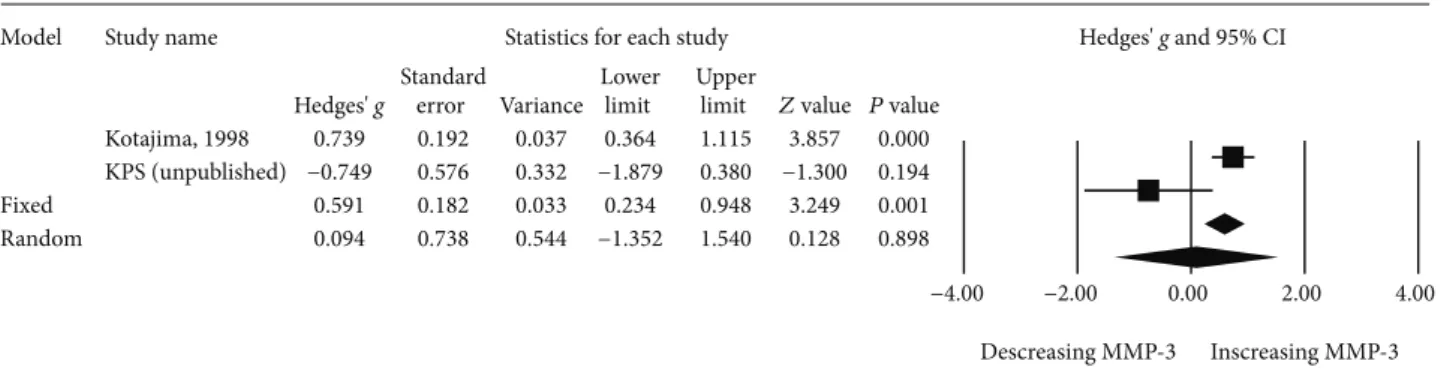
anti-double strand DNA antibody (anti-dsDNA Ab) titer at the time of sample collection. The results involving two studies—Kotajima et al. [2] and KPS (unpublished)—-demonstrated no significant difference in MMP-3 levels between SLE patients with abnormally increased anti-dsDNA Ab titer and those without (P = 0 898, Hedges’ g: 0.094, 95% CI -1.325-1.540) (Table 2 and Figure 7).
3.6. Assessment of Heterogeneity and Publication Bias. We assessed statistical heterogeneity between the included stud-ies (Table 2). In the meta-analysis of serum MMP-3 levels comparing SLE patients with healthy controls and subgroup analysis of proteinuria, the I2 test showed a value > 50%, indicating substantial heterogeneity. Random effects models were used for meta-analyses. Although the funnel plot showed symmetry (Figure 8), Egger’s regression analysis indicated possibility of publication bias (Table 2).
4. Discussion
Due to a remitting-relapsing disease course of most patients with SLE, biomarkers reflecting disease activity are desirable. One of the candidate biomarkers is the MMP family. MMP-3, also known as Stromelysin-1, degrades tissue proteins including collagen types II, III, IV, IX, and X, proteoglycans, fibronectin, laminin, and elastin [12]. It can also activate other MMPs, such as MMP-9, which is suggested to be involved in the pathogenesis of SLE [13]. A recently pub-lished meta-analysis involving 12 studies, however, showed that circulating MMP-9 levels did not differ between SLE patients and healthy controls [23].
In this meta-analysis, serum MMP-3 levels were reviewed in 662 SLE patients and 771 controls. There were 621 patients and 762 controls extracted from 11 publications and 11 patients and 9 controls from a pediatric lupus cohort, KPS.
Model Study name Statistics for each study Hedges' g and 95% CI
Ichikawa, 1998 KPS (unpublished) Fixed Random Hedges' g Standard error Lower limit Upper limit
Variance Z value P value 0.745 0.610 0.372 −0.450 1.940 1.222 0.222 −0.036 0.619 0.383 −1.250 1.177 −0.059 0.953 0.360 0.434 0.189 −0.491 1.212 0.829 0.407 0.360 0.434 0.189 −0.491 1.212 0.829 0.407 −4.00 −2.00 0.00 2.00 4.00 Decreasing MMP-3 Increasing MMP-3
Figure 4: Forest plot of random effects meta-analysis of MMP-3 levels in SLE patients; male vs. female.
Model Study name Statistics for each study Hedges' g and 95% CI
Gheita, 2015 Kotajima, 1998 KPS (unpublished) Fixed Random Hedges' g Standard error Lower limit Upper limit
Variance Z value P value 0.758 0.314 0.099 0.143 1.373 2.415 0.016 0.489 0.337 0.114 −0.172 1.150 1.449 0.147 0.678 0.572 0.327 −0.443 1.800 1.186 0.236 0.639 0.213 0.045 0.221 1.057 2.998 0.003 0.639 0.213 0.045 0.221 1.057 2.998 0.003 −4.00 −2.00 0.00 2.00 4.00 Decreasing MMP-3 Increasing MMP-3
Figure 5: Forest plot of random effects meta-analysis of MMP-3 levels in SLE patients; with vs. without nephritis.
Model Study name Statistics for each study Hedges' g and 95% CI
Ichikawa, 1998 Kotajima, 1998 Fixed
Random
Hedges' g Standard Lower Upper error Variance limit limit Z value P value 2.333 0.587 0.345 1.182 3.483 3.974 0.000 0.960 0.195 0.038 0.578 1.342 4.925 0.000 1.096 0.185 0.034 0.734 1.459 5.927 0.000 1.535 0.677 0.459 0.207 2.862 2.266 0.023 −4.00 −2.00 0.00 2.00 4.00 Descreasing MMP-3 Inscreasing MMP-3
The results showedfirstly that serum MMP-3 levels were sig-nificantly higher in patients with SLE than in healthy controls and secondly that serum MMP-3 levels were significantly elevated in patients with renal involvement than in those without, both for active lupus nephritis and persistent pro-teinuria. Previous studies suggested a correlation of serum MMP-3 levels and hematologic indices, such as white blood cells (WBC) and platelet counts [22]. However, in our meta-analysis, subgroup comparisons were available only for renal manifestations, sex, and serum dsDNA anti-body titer due to paucity of quantifiable data. Subgroup comparison by sex and serum anti-dsDNA antibody titer showed no significant difference in the serum MMP-3 levels. With regard to MMP-3, several studies have reported elevation of circulatory MMP-3 levels in SLE patients [2, 3, 12, 17, 22]. Our meta-analysis results were in agreement with these studies. However, the correlation of serum MMP-3 elevation and disease activity of SLE had been inconsistent [2, 3, 12, 22]. Precisely, Kotajima et al. reported that increased levels of serum MMP-3 in SLE are related to clinical features relevant to lupus nephritis [2]. They found that serum MMP-3 levels were significantly higher in SLE patients with active clinical presentation such as persistent proteinuria, malar rash, and laboratory parame-ters, such as cellular casts, anti-dsDNA antibodies, decreased complement C3 and C4 levels, circulating immune com-plexes, and hypoalbuminemia [2]. Similarly, Gheita et al. found that serum levels of MMP-3 correlated with the sys-temic lupus erythematosus disease activity index (SLEDAI)
and Systemic Lupus International Collaborating Clinics/-damage index (SLICC/DI) scores [22]. However, Zucker et al. found an increase in serum concentrations of MMP-3 in SLE but reported no correlation with disease activity [12]. Moreover, Zhu et al. [3] reported that serum MMP-2, MMP-3, and MMP-13 levels in SLE patients were signi fi-cantly higher than those in controls but found no overall cor-relation between serum levels of the three MMPs and disease activity scores. Our data supported the relationship between serum MMP-3 levels and renal involvement of SLE, implicat-ing its correlation with disease activity. With regard to renal involvement, a few studies investigated its association with serum MMP levels. In a study by Gheita et al., the serum MMP-3 levels correlated with class of lupus nephritis, show-ing the highest levels in patients with class IV nephritis [22]. Thiyagarajan et al. speculated in an animal study that MMPs may represent some component of membrane disintegration in progressive nephritis [24]. Our results and previous works suggested that serum MMP-3 levels may reflect the presence and possibly histological severity of lupus nephritis in patients with SLE.
There are several limitations in this study. First, the mean values of MMP-3 serum levels in SLE patients were relatively high in those studies published in more remote years (before the year 2000) and significantly lower in those studies per-formed after 2000. We speculate that different ELISA kits may have made a general comparability of results impossible. This issue led to different nonreproducible results in the past (biomarker biology), but we have only included studies with respective control cohorts and observed similar regulation in most studies. Still, this is a major limitation in this study. Second, this meta-analysis had small sample sizes, lowering the power of the study. In those meta-analyses involving two studies, the conclusions drawn may be subject to bias because they are affected by the small sample size of clinical studies. In order to alleviate this, we used a random effects model in this study. However, we speculate that such a limitation should raise attention and subsequently increase publications in this subject. This is one of the reasons for performing this work. Third, the data included in this meta-analysis are extracted from heterogeneous groups. The patients had different demographics, such as age, sex, and ethnicity, and varying clinical manifestations which may have affected the results. In particular, this meta-analysis included data from one pediatric cohort (KPS) and
−20 0 1 2 3 4 5 6 7 8 −10 0 Hedges' g P recisio n (1/S td Er r)
Funnel plot of precision by Hedges' g
10 20
Figure 8: Funnel plot of standard error in meta-analysis of MMP-3 levels in SLE patients compared with healthy controls.
Model Study name Statistics for each study Hedges' g and 95% CI
Kotajima, 1998 KPS (unpublished) Fixed Random Hedges' g Standard error Lower limit Upper limit
Variance Z value P value 0.739 0.192 0.037 0.364 1.115 3.857 0.000 −0.749 0.576 0.332 −1.879 0.380 −1.300 0.194 0.591 0.182 0.033 0.234 0.948 3.249 0.001 0.094 0.738 0.544 −1.352 1.540 0.128 0.898 −4.00 −2.00 0.00 2.00 4.00 Descreasing MMP-3 Inscreasing MMP-3
11 studies on adult patients which may have increased het-erogeneity of the data. Lastly, there remains a possibility of existing literature that was not accessible and the presence of publication bias.
Although the results require cautious interpretation, we speculate that this meta-analysis may provide some evidence-based results regarding a controversial issue, based on current publications. In the future, meta-analysis using individual patient data and propensity scoring would make a more powerful study.
Firstly, the results of the present study revealed that serum MMP-3 levels were significantly elevated in SLE patients, which is in accordance with previous reports [2, 3, 12, 22]. Secondly, the results showed that MMP-3 was significantly elevated in patients with renal involvement, both in histologically proven lupus nephritis and mere pro-teinuria. Although the correlation of MMP-3 and lupus activity requires further verification, it is yet tempting to speculate that elevated MMP-3 at initial diagnosis of SLE may require more close follow-ups.
5. Conclusions
The present meta-analysis showed that serum MMP-3 levels were significantly higher in patients with SLE than in con-trols and in patients with renal involvement than in those without. Although our meta-analysis suggested that MMP-3 likely correlate with disease activity, further studies in a larger scale are warranted to elucidate the role of MMP-3 as a putative biomarker of SLE.
Data Availability
The raw data supporting this meta-analysis are from previ-ously reported studies and datasets, which have been cited and included as supplementary material. The processed data are included within the article and Supplementary Materials. The full processed data in detail are also available from the corresponding author upon request.
Conflicts of Interest
The authors declare that they have no conflicts of interest.
Authors’ Contributions
Jiwon M. Lee, Andreas Kronbichler, Se Jin Park, and Seong Heon Kim contributed equally to the work.
Acknowledgments
This study was supported by the Chungnam National University Hospital Research Fund, 2017 (to J.M.L.).
Supplementary Materials
Supplementary Table S1: PRISMA 2009 Checklist. Supplementary Table S2: the Newcastle-Ottawa Scale (NOS). Supplementary Table S3: comparison of investigated biomarkers in pediatric SLE patients and controls (KPS data).
Supplementary Material on pediatric SLE (KPS) data. (Supplementary Materials)
References
[1] H. T. Cook and M. Botto,“Mechanisms of disease: the comple-ment system and the pathogenesis of systemic lupus erythema-tosus,” Nature Clinical Practice. Rheumatology, vol. 2, no. 6, pp. 330–337, 2006.
[2] L. Kotajima, S. Aotsuka, M. Fujimani et al.,“Increased levels of matrix metalloproteinase-3 in sera from patients with active lupus nephritis,” Clinical and Experimental Rheumatology, vol. 16, no. 4, pp. 409–415, 1998.
[3] Q. Q. Zhu, T. T. Li, R. Chen et al.,“Elevated serum levels of MMP-2, MMP-3, and MMP-13 in Chinese patients with systemic lupus erythematosus,” Scandinavian Journal of Rheumatology, vol. 39, no. 5, pp. 439–441, 2010.
[4] H. Nagase, R. Visse, and G. Murphy,“Structure and function of matrix metalloproteinases and TIMPs,” Cardiovascular Research, vol. 69, no. 3, pp. 562–573, 2006.
[5] W. C. Parks, C. L. Wilson, and Y. S. Lopez-Boado,“Matrix metalloproteinases as modulators of inflammation and innate immunity,” Nature Reviews Immunology, vol. 4, no. 8, pp. 617–629, 2004.
[6] I. Tchetverikov, L. R. Lard, J. DeGroot et al.,“Matrix metallo-proteinases-3, -8, -9 as markers of disease activity and joint damage progression in early rheumatoid arthritis,” Annals of the Rheumatic Diseases, vol. 62, no. 11, pp. 1094–1099, 2003. [7] H. Senzaki, S. Masutani, J. Kobayashi et al.,“Circulating matrix
metalloproteinases and their inhibitors in patients with Kawasaki disease,” Circulation, vol. 104, no. 8, pp. 860–863, 2001.
[8] M. Segarra, A. Garcia-Martinez, M. Sanchez et al.,“Gelatinase expression and proteolytic activity in giant-cell arteritis,” Annals of the Rheumatic Diseases, vol. 66, no. 11, pp. 1429– 1435, 2007.
[9] A. Matsuyama, N. Sakai, M. Ishigami et al.,“Matrix metallo-proteinases as novel disease markers in Takayasu arteritis,” Circulation, vol. 108, no. 12, pp. 1469–1473, 2003.
[10] P. A. Monach, R. L. Warner, G. Tomasson et al.,“Serum pro-teins reflecting inflammation, injury and repair as biomarkers of disease activity in ANCA-associated vasculitis,” Annals of the Rheumatic Diseases, vol. 72, no. 8, pp. 1342–1350, 2013. [11] J. I. Shin, K. S. Song, H. Kim et al.,“The gene expression profile
of matrix metalloproteinases and their inhibitors in children with Henoch–Schönlein purpura,” British Journal of Derma-tology, vol. 164, no. 6, pp. 1348–1355, 2011.
[12] S. Zucker, N. Mian, M. Drews et al., “Increased serum stromelysin-1 levels in systemic lupus erythematosus: lack of correlation with disease activity,” The Journal of Rheumatol-ogy, vol. 26, no. 1, pp. 78–80, 1999.
[13] A. Lesiak, J. Narbutt, A. Sysa-Jedrzejowska, J. Lukamowicz, D. McCauliffe, and A. Wózniacka, “Effect of chloroquine phosphate treatment on serum MMP-9 and TIMP-1 levels in patients with systemic lupus erythematosus,” Lupus, vol. 19, no. 6, pp. 683–688, 2010.
[14] J. P. Higgins and S. Green, Cochrane Collaboration: Cochrane Handbook for Systematic Reviews of Interventions, Wiley-Blackwell, 2011.
[15] T. Jin, K. Almehed, Y. Zhu, H. Carlsten, and H. Forsblad-d’Elia, “Soluble E-cadherin in systemic lupus erythematosus,”
The Journal of Rheumatology, vol. 40, no. 10, pp. 1677–1682, 2013.
[16] K. de Leeuw, B. Freire, A. J. Smit, H. Bootsma, C. G. Kallenberg, and M. Bijl, “Traditional and non-traditional risk factors contribute to the development of accelerated atherosclerosis in patients with systemic lupus erythematosus,” Lupus, vol. 15, no. 10, pp. 675–682, 2006.
[17] C. Ribbens, M. Martin y Porras, N. Franchimont et al., “Increased matrix metalloproteinase-3 serum levels in rheumatic diseases: relationship with synovitis and steroid treatment,” Annals of the Rheumatic Diseases, vol. 61, no. 2, pp. 161–166, 2002.
[18] Y. Ichikawa, C. Yamada, T. Horiki, Y. Hoshina, and M. Uchiyama,“Serum matrix metalloproteinase-3 and fibrin degradation product levels correlate with clinical disease activity in rheumatoid arthritis,” Clinical and Experimental Rheumatology, vol. 16, no. 5, pp. 533–540, 1998.
[19] K. Akiyama, K. Shikata, H. Sugimoto et al.,“Changes in serum concentrations of matrix metalloproteinases, tissue inhibitors of metalloproteinases and type IV collagen in patients with various types of glomerulonephritis,” Research Communica-tions in Molecular Pathology and Pharmacology, vol. 95, no. 2, pp. 115–128, 1997.
[20] M. Shingu, K. Obata, I. Ezaki et al.,“Stromelysin-1 (MMP-3) level in the sera from patients with rheumatoid arthritis and other connective tissue diseases–clinical significances in early onset rheumatoid arthritis,” Ryūmachi, vol. 35, no. 1, pp. 15–24, 1995.
[21] S. Zucker, R. M. Lysik, M. H. Zarrabi et al.,“Elevated plasma stromelysin levels in arthritis,” The Journal of Rheumatology, vol. 21, no. 12, pp. 2329–2333, 1994.
[22] T. A. Gheita, D. M. Abdel Rehim, S. A. Kenawy, and H. A. Gheita, “Clinical significance of matrix metalloproteinase-3 in systemic lupus erythematosus patients: a potential bio-marker for disease activity and damage,” Acta Reumatológica Portuguesa, vol. 40, no. 2, pp. 145–149, 2015.
[23] Y.-M. Mao, S. Wang, C. N. Zhao et al.,“Circulating matrix metalloproteinase-9 levels in patients with systemic lupus ery-thematosus: a meta-analysis,” Current Pharmaceutical Design, vol. 24, no. 16, pp. 1780–1787, 2018.
[24] D. Thiyagarajan, S. Fismen, N. Seredkina et al.,“Silencing of renal DNaseI in murine lupus nephritis imposes exposure of large chromatin fragments and activation of Toll like receptors and the Clec4e,” PLoS One, vol. 7, no. 3, article e34080, 2012.
Stem Cells
International
Hindawi www.hindawi.com Volume 2018 Hindawi www.hindawi.com Volume 2018Endocrinology
International Journal ofHindawi www.hindawi.com Volume 2018 Hindawi www.hindawi.com Volume 2018
Disease Markers
Hindawi www.hindawi.com Volume 2018 BioMed Research InternationalOncology
Journal of Hindawi www.hindawi.com Volume 2013 Hindawi www.hindawi.com Volume 2018 Oxidative Medicine and Cellular Longevity Hindawiwww.hindawi.com Volume 2018
PPAR Research
Hindawi Publishing Corporation
http://www.hindawi.com Volume 2013 Hindawi www.hindawi.com
The Scientific
World Journal
Volume 2018 Immunology Research Hindawi www.hindawi.com Volume 2018 Journal ofObesity
Journal of Hindawi www.hindawi.com Volume 2018 Hindawi www.hindawi.com Volume 2018 Computational and Mathematical Methods in Medicine Hindawi www.hindawi.com Volume 2018Behavioural
Neurology
Ophthalmology
Journal of Hindawi www.hindawi.com Volume 2018Diabetes Research
Journal ofHindawi
www.hindawi.com Volume 2018
Hindawi
www.hindawi.com Volume 2018
Research and Treatment
AIDS
Hindawi
www.hindawi.com Volume 2018
Gastroenterology Research and Practice
Hindawi www.hindawi.com Volume 2018