i
웅성 백서 비골의 분절성 골 결손에서
15deoxyΔ
12,14
prostaglandin J₂
의
국소적용이 골 재생에 미치는 영향
연세대학교 대학원
치 의 학 과
홍 주 영
ii
웅성 백서 비골의 분절성 골 결손에서
15deoxyΔ
12,14
prostaglandin J₂
의
국소적용이 골 재생에 미치는 영향
지도교수 김 형 준
이 논문을 석사 학위 논문으로 제출함
2013 년 12 월
연세대학교 대학원
치의학과
홍 주 영
iii
홍주영의 석사 학위논문을 인준함
심사위원 김 형 준 인
심사위원 정 원 윤 인
심사위원 박 원 서 인
연세대학교 대학원
2013 년 12 월
iv
감사의 글
연세대학교 치과대학 구강악안면외과학 석사 과정은 치과의사로 살아가는데 기준을 잡아준 감사한 시간 이였습니다. 감탄하고 정진하며 분주하게 공부할 수 있는 기회를 주시고, 조언을 아끼지 않으신 김형준 교수님께 진심으로 존경하며 감사 드립니다. 바쁘신 와중에도 심사를 맡아주시고 오류를 잡아주신 정원윤 교수님, 박원서 교수님 깊이 감사를 드립니다. 시약에 대한 지식을 조언 해 주신 김기림 선생님, 오른팔처럼 든든한 파트너인 권현진 선생님, 마음 따뜻한 나혜영, 백지웅, 곽은정 선생님 덕분에 이 논문이 더 풍성하게 진행 될 수 있었습니다. 감사 드립니다. 조언과 지지를 아끼지 않았던 김혜선 선생님, 구호산나 원장님 감사합니다. 사랑으로 응원해준 가족과 하나님 아버지께 진심으로 감사 드리며 항상 정진하는 진실한 치과의사로 살아가겠습니다.2013 년 12 월
홍 주 영
v
차 례
그림차례 ... vi
표 차례 ... viii
국문 요약 ... ix
I. 서 론 ... 1
II. 연구 대상 및 방법 ... 6
1. 연구 대상 ... 6
2. 연구 방법 ... 6
III. 결 과 ... 12
1. 육안 소견 ... 12
2. 방사선학적 소견 ... 16
4. 통계 분석 ... 27
3. 조직형태학적 소견 ... 29
IV. 고 찰 ... 37
V. 결 론 ... 42
VI. 참고 문헌 ... 43
영문 요약
vi
그 림 차 례
Figure 1. Pathways for the synthesis of prostanoids ... 3 Figure 2. Scheme of the Michael addition ... 4 Figure 3. Experiment design ... 7 Figure 4. Procedure of 15-deoxy-Δ12,14-prostaglandin J₂applied absorbable collagen sponge grafting on SD-rat fibula 7mm defect site. ... 9
Figure 5. Region of interest (ROI) ... 11
Figure 6. Control group at 4week ... 13
Figure 7. Experimental group at 4week ... 13
Figure 8. Control group at 8week. ... 15
Figure 9. Experimental group at 8week. ... 15
Figure 10. Radiographic findings at 4 week control group on Left site. ... 17
Figure 11. Radiographic findings at 4 week experimental group on Right site ... 18
Figure 12. Radiographic findings at 8 week control group on Left site ... 19
Figure 13. Radiographic findings at 8 week experimental group on Right site ... 20
Figure 14. Micro CT : longitudinal & cross sectional view at 4 week Rat5 experimental site ... 21
Figure 15. Micro CT : longitudinal & cross sectional view at 4 week Rat9 experimental site ... 22
Figure 16. Micro CT : longitudinal & cross sectional view at 4 week Rat9 control site ... 22
Figure 17. Micro CT : longitudinal & cross sectional view at 8 week Rat2 experimental site ... 23
Figure 18. Micro CT : longitudinal & cross sectional view at 8 week Rat4 experimental site ... 24
vii
Figure 19. Micro CT : longitudinal & cross sectional view at 8 week Rat4
control site ... 24
Figure 20. 3D Reconstruction of the 4 week experiment site ... 25
Figure 21. 3D Reconstruction of the 4week control site ... 25
Figure 22. 3D Reconstruction of the 8 week experiment site ... 26
Figure 23. Graph of the statistical analysis ... 28
Figure 24. Histologic findings of the 4week experiment site (Rat 5). ... 30
Figure 25. Histologic findings of the 4week experiment site (Rat 8). ... 31
Figure 26. Histologic findings of the 4week experiment site (Rat 9). ... 32
Figure 27. Histologic findings of the 4week control site (Rat 9) ... 33
Figure 28. Histologic findings of the 8week experiment site (Rat 2). ... 34
Figure 29. Histologic findings of the 8week experiment site (Rat 4). ... 35
viii
표 차 례
Table 1. Study design. ... 7 Table 2. Statistical analysis of the 4 week experiment and control group
... 27 Table 3. Statistical analysis of the 8 week experiment and control group
ix
국문 요약
웅성 백서 비골의 분절성 골 결손에서
15
deoxyΔ
12,14prostaglandin J₂의 국소적용이 골 재생에
미치는 영향
< 지도교수: 김 형 준 >
연세대학교 대학원 치의학과
홍 주 영
구강악안면부 골 결손 발생 시 재건과 재생을 위한 다양한 이식재의 연구가 진행되고 있다. 15deoxyΔ12,14prostaglandin J₂(15dPGJ₂) 가 유방암 전이 시 발생하는 골 파괴를 감소 및 회복시키고, 자궁적출 후 발생하는 골다공증을 억제한다는 연구가 2012 년 연세대학교 응용 생명 과학부에서 발표 되었고, 골 파괴 억제 뿐 아니라 골 재생의 가능성까지 제시 된 점에 착안하여 본 연구에서는 15dPGJ₂의 골 형성 능력을 평가하는 목적으로 실험을 진행하였다. 웅성 백서 비골의 7mm 임계크기 골 결손을 형성 한 후 우측 골 결손부에 1mg/500 ㎕ 농도의 15deoxyΔ12,14 prostaglandin J₂ 100 ㎕ 을 적신 absorbable collagen sponge 을 이식 하였고, 대조군인 좌측 비골 결손부에 100 ㎕ saline 을 적신 absorbable collagen sponge 을 이식x
하였다. 근육의 층별봉합을 시행하여 이식편을 고정 하였고 실험 동물의 움직임은 제한하지 않았다. 4 주 희생군 9 마리와 8 주 희생군 8 마리를 실험 및 희생 하였다.
육안소견 관찰, standard x-ray 와 micro CT 를 이용한 방사선학적 소견 관찰, data viewer 를 이용한 3D-reconstruction, 조직학적 평가 및 절단연 부위의 Bone fraction volume(BV/TV), Trabecular thickness (TbTh), Trabecular number (TbN), Trabecular separation (TbSp), Structure model index (SMI) 값의 통계 분석을 시행 하였다. 일부 시편의 결손부위에서 신생골 형성이 관찰되었고, 4 주의 BV/TV, TbN과 8 주의 BV/TV, TbN, TbSp, SMI값이 통계적으로 유의하며 대조군과 차이를 보였다. 육안소견이나 x-ray 소견에서 현저한 골 형성은 관찰되지 않았으나 micro CT 분석과 3D 영상에서 결손부의 연속적인 신생골 형성이 일부 시편들에서 확인 되었다. 조직학적 소견에서는 신생골 및 석회화된 기질들의 생성과 신생혈관형성을 일부 시편에서 관찰 할 수 있었다. 15d-PGJ₂의 골 결손부 이식 시 골 형성 능력은 우수하다고 보기에는 부족한 부분들이 있고 기전의 확인을 위한 추가적인 연구들이 필요 하다고 판단된다. 그러나 핵 수용체 단위에서 조골세포와 파골세포 형성을 자극 및 조절하는 능력이 있어 추가적인 실험을 통해 농도나 carrier 가 효과적으로 적용된다면 더 나은 골 이식 대체제가 될 수 있을 것으로 생각된다. 핵심 단어 : 15d-PGJ₂, 골 재생, 골 형성, 골 이식재
1
I.
서 론
악 안면부의 골절이나 종양으로 인한 광범위 골 결손 부위 골 재생 연구가 많은 관심을 받고 있다. 이식 후 거부반응이 가장 적고 골 형성 능력을 보이는 자가골 이식이 많이 사용되고 있으나 이식 후 흡수량이 많고 공여부의 한계가 있다. 자가골 이식에 많이 사용되는 자가 장골의 경우 이식 후 치유과정에서 40%이상의 흡수를 보인다 (Sbordone et al., 2009). 미세혈관 문합술을 동반한 자가골 이식도 좋은 결과를 보이지만 현저한 이환률을 보이고 있어, 대체 재건술에 대한 신기술 연구가 활발히 진행되고 있다(Adams et al., 2013). 최근에는 분자 생물학의 발달로 골 유도 인자 Bone morphogenic protein(BMPs)이 많이 사용되고 있다(Rubens Moreno de Freitas et al., 2013). rhBMP2(recombinant human BMP2)/ACS (absorbable collagen sponge) 조합의 임플란트는 2007 년 미국 FDA 에서 상악동 거상술 과 치조능 증대술 시 사용 하도록 승인 받았다(Triplett RG et al., 2009). 하지만 임상에서 골 이식재의 이식 후 골절부의 뼈가 잘 유합 되지 않고 마치 관절처럼 움직이거나 세균 감염증으로 가관절 상태가 되어 재 수술을 하는 경우들도 보고되고 있으며 하악골의 재건이나 구개열의 회복을 위한 이식 수술에서 rhBMP 만으로는 재수술이 필요 하거나 이식 실패가 나타나는 결과들도 보고된다(E.J.Woo., 2012).2012 년 Kim 등은 Peroxisome proliferator activated receptor gamma(PPARγ)의 내인성 ligand 인 15-deoxy-Δ12,14-prostaglandin J₂
(15d-PGJ₂)가 유방암 전이 시 발생하는 골 파괴를 감소 및 회복시키고, 자궁적출 후 발생하는 골다공증을 억제한다는 연구결과를 발표하였다. 이
2
연구에서 유선 암종 세포를 주입 한 15dPGJ2 를 매주 3 번씩 피하 주사
하여 6 주간 실험하였을 때 positive control 인 bisphosphonate 계열의 Zoledronic acid(ZA) 보다는 낮지만 정상 상태와 유사한 BV(bone volume) /TV(tissue volume)(%)를 보이며 골 흡수 억제 및 골 형성 능력을 보였다. 특정 농도에서 골 파괴를 억제 한다고 알려진 bisphosphonate 의 경우 파골세포의 활성을 제한 할 뿐만 아니라 조골세포의 동화작용에 영향을 미쳐 골 재생을 촉진 할 수 있다는 결과들이 보고 된다(Kim Ki-Rim et al., 2012). Im 등은 MG-63 세포를 이용한 in vitro 실험에서, 특정 농도의 bisphosphonate 를 적용 했을 때 조골세포가 증식 하고, 골수강 내의 중간엽세포가 조골세포로 분화 유도 함을 보였다. 또한 RT-PCR 검사를 통해 bisphosphonate 가 BMP-2, Type 1 collagen, osteocalcin 의 발현을 증가시킴을 보였다(Im., 2004). 저자는 이에 착안하여 15d-PGJ2 역시
조골세포와 파골세포에 특정 기전으로 작용하여 골의 재생과 항상성 향상에 영향을 줄 수 있다는 가설을 설정하고 검증하고자 본 연구를 진행 하였다.
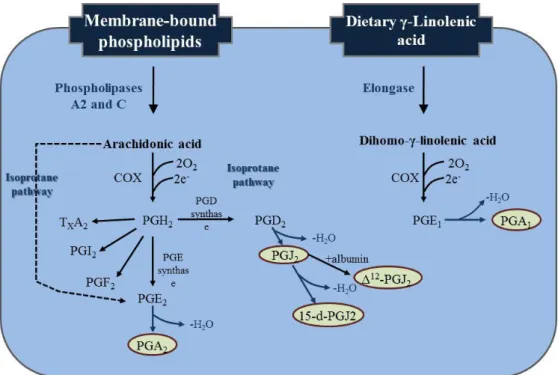
15d-PGJ2 가 속하는 Prostanoids 는 cyclooxygenase biosynthetic
pathway 의 생산물로 다양한 구조와 생물학적 역할들을 하는 지방질 매개체 family 이다. 이 중에 cyclopentone structure 의 prostaglandin(cyPG)은 다양한 기전에 작용하는 친전자적 지방질로, 핵과 세포막 수용체의 활성화에 작용하고 단백질의 cysteine residue 에 바로 작용하여 단백질 기능까지 조절 할 수 있다(Beatriz Diez-Dacal et al., 2010).
cyPG 는 parent PG 의 비효소적 탈수 과정에 의해 생성된다. PGD2 의
탈수 과정에 의해 발생한 prostaglandin J 계열은 계속적인 탈수 과정에 의해 15d
-
PGJ2와 Δ12-
PGJ2를 생산한다(Shibata T et al., 2002).3
Figure 1. Pathways for the synthesis of prostanoids. COX enzymes acting on
polyunsaturated fatty acids generate PG. The PG common precursor PGH2 generated from arachidonic acid can subsequently be transformed by synthases into the various PG and TXA2. It should be taken into account that nonenzymatic transformation of polyunsaturated fatty acids can also give rise to PG through the isoprostane pathway. In addition, nonenzymatic dehydration of some PG leads to the formation of reactive cyPG. 15-d-PGJ2 is synthesized by the nonenzymatic dehydration course (Beatriz Diez-Dacal et al, 2010).
CyPG 의 cyclopentane ring 에는 α,β-불포화 탄소 그룹이 존재하는데, 이런 구조는 β-position 에서 탄소에 강력한 친전자적 성질을 보이며 thiol group 의 황 원자와 강력한 친핵반응을 일으켜 Michael adducts 를 생산한다. 이런 과정으로 cyPG 와 glutathione(GSH) 또는 단백질 결합물이 생성되고 다양한 생물학적 활동력을 보이게 된다(Fukushima M et al., 1989).
4
Figure 2. Scheme of the Michael addition. The scheme depicts the adduct formed by nucleophilic attack on the carbon in the β-position of the cyPG (Beatriz Diez-Dacal et al, 2010).
다양한 생물학적 반응으로 antiproliferative effect 를 보이며 항암치료제로 사용되어 Tumor-bearing mice 의 수명을 연장시키고 몇몇 형질전환 세포의 성장을 억제시키는 실험결과를 보였다(Kato, T et al., 1986, Kim, I.K et al., 1993). 또한 바이러스의 복제에 필수적인 NF-κB 를 억제하는 능력과 cytoprotective enzyme 의 발현을 유도하는 heat-shock 반응의 개시에 효과가 있어 antiviral agent 로의 효과를 나타낸다(Santoro, M.G., 1997).
cyPG 의 cyclooxygenase-mediated arachidonic acid pathway 최종 산물인 15d-PGJ2 는 (Figure1), 특정 전사요소 및 신호인자와 높은
친화성으로 결합하는 구조적 특징이 있다. 특히 peroxisome proliferator-activated receptor gamma(PPARγ)의 내인성 ligand 로 작용하며 다양한 생물학적 반응에 관여한다. 반면에 PPARγ에는 독립적인 기전으로, 15d-PGJ2가 NF-κB(Nuclear Factor kappa-light-chain-enhacer of activated
B cell) 연관 전사과정을 강력하게 억제 및 조절 한다(Straus, D.S et al., 2000). NF-κB 는 DNA 의 전사과정을 조절하는 단백질 복합체로 대부분의
5 동물 세포에 존재하며 감염 시 면역반응 조절에 중요한 역할을 하며 다양한 생물학적 반응에 연관되어 있다(Gilmore TD., 2006). 15d-PGJ2 가 NF-κB 에 작용하여 골 대사 조절에 영향을 줄 수 있다는 점을 이 논문에서 생각해 보고자 한다. 이 연구에서는 웅성 백서 비골의 7mm 임계 크기 분절성 골 결손부에 특정 농도의 15deoxyΔ12,14 PGJ₂를 absorbable collagen sponge(ACS)
carrier 로 국소 적용 하였을 때 주변 골 치유에 미치는 영향을 알아보고자 하였다.
6
II.
연구대상 및 방법
1.
연구대상
본 실험은 8 주령의 수컷 웅성 백서(Male Spraque-Dawley Rat ; 250-300g) 17 마리의 양쪽 비골, 총 34 site 를 이용하여 실험 4 주 후에 9 마리 8 주 후에 8 마리의 시편을 채취하였다. 암컷의 경우 호르몬의 변화나 임신 등에 의해 결과에 영향을 미칠 수 있어 수컷만을 이용하였으며, 실험동물은 연세대학교 의료원 임상 의학 연구센터의 동물 실험 지침에 따랐다.
2.
연구 방법
가. 실험 설계
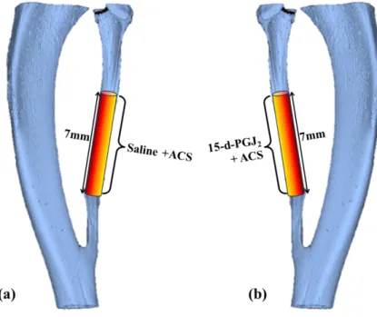
웅성 백서 16 마리의 양측 비골에 7mm 의 분절성 골 결손을 형성 하였다. 우측 골 결손부에 1mg/500 ㎕ 15deoxyΔ12,14 prostaglandinJ₂100 ㎕ (200μg)을 적신 absorbable collagen sponge 10x5x5mm 을 이식하였고, 대조군으로 좌측 골 결손부에 100 ㎕ saline 을 적신 absorbable collagen sponge 10x5x5mm 을 이식하였다. 근육의 층별봉합을 시행하여 이식편을 고정 하였고 실험 동물의 움직임은 제한하지 않았다.
실험 4 주 경과 후에 9 마리, 8 주후에 8 마리를 CO2 chamber 를
이용하여 희생하였으며, 비골과 장골을 적출하여 육안적, 방사선학적, 조직학적으로 관찰하였다.
7
Figure 3. Experiment design. a) Left control site. b) Right experimental site
Group(n) Implants
Control (Left)
4week (9) Saline soaked absorbable collagen sponge 8week (8) Saline soaked absorbable collagen sponge
Experimental (Right) 4week (9) 15deoxyΔ 12,14PGJ 2 soaked absorbable collagen sponge 8week (8) 15deoxyΔ 12,14PGJ 2 soaked absorbable collagen sponge
Table 1. Study design.
a )
b )
8
나. 실험 방법
실험동물은 연세대학교 의료원 실험동물부 지침에 따라 1 주일의 순화 기간을 거쳤다
Zoletil® (Virbac, Carros, France, 0.06mg/kg)와 Rumpun® (Bayer Korea, Korea, 0.04mg/kg)을 둔부에 근육 주사하여 전신마취 하였다. 비골 부위의 털을 깎은 후 10% povidoneiodine 용액으로 소독 후 고정하였으며, 1: 100,000 epinephrine 이 함유된 lidocaine 주사 후 약 3cm 의 피부 절개를 다리의 외측에 시행 하였다. 비골 상방의 근육을 절개 후 둔박리 하여 비골을 노출 시키고, 버니어 캘리퍼를 이용하여 비골의 골간에서 7.0mm 계측 하여 분절성 골 결손을 형성하였다. 실험군인 우측 비골에 골 결손 형성 직후 주변 근육이 박리된 상태에서 1mg/500 ㎕ 농도의 15deoxyΔ12,14prostaglandin J₂(Cayman Chemicals,
Ann Arbor, MI, USA)을 100 ㎕을 적신(총 200 ㎍) 10x5x5mm absorbable collagen sponge (CollaHealTM, Bioland, Korea)을 결손 부위에 위치시키고
근육을 층별봉합 하였다.
대조군인 좌측 비골에 동일한 골 결손을 형성하고 100 ㎕ Saline 을 적신 10x5x5mm absorbable collagen sponge 을 위치 시킨 후 동일한 층별 봉합을 시행하였다. 봉합은 absorbable 4-0 VicrylTM (Johnson & Johnson
Korea, Belgium)을 이용 하여 시행하였고 피부봉합 마무리 후 10% povidoneiodine 용액으로 소독 하였다.
실험동물은 연세대학교 의료원 임상의학연구센터 소형동물실의 지침에 따라 물과 pellet 형태의 사료를 공급받았다..
9
실험 4 주, 8 주 후 CO2 chamber 를 이용하여 각 각 9 마리, 8 마리를
희생하였고, 희생한 웅성 백서의 양측 비골과 경골을 부착 근육과 함께 절제하여 4% paraformaldehyde/0.01M phosphate-buffered saline (PBS, Ph 7.4)에 고정하였다.
Figure 4. Procedure of 15-deoxy-Δ12,14-prostaglandin J₂ applied absorbable collagen sponge grafting on SD-rat fibula 7mm defect site. a) Exposure of the fibula for the formation of the defect site. b) Measurement of 7mm SD-rat fibula with a vernier calliper. c) Grafting of the absorbable collagen sponge on the defect site. d) Appliance of Saline volume: 100 ㎕ on the Left site and 15d-PGJ2 concentration: 1mg/500 ㎕, volume:100 ㎕ on the Right site.
10
i.
방사선 학적 평가
표준 치과방사선 필름을 이용한 Plain X-ray 촬영 (Voltage(kv) = 60, Current(Ma)=70, Exposure(s)=0.08) 과 미세 컴퓨터 단층 촬영 (Micro- computed Tomography ; Micro-CT, SkyScan 1076, Belgium)을 시행하였다(Resolution=18 ㎛, Voltage(kv)= 100, Current(㎂)= 100, Filter=0.5mm Aluminium, Exposure(ms)= 1180, Rotation Step(deg) =0.500/360 Rotation).
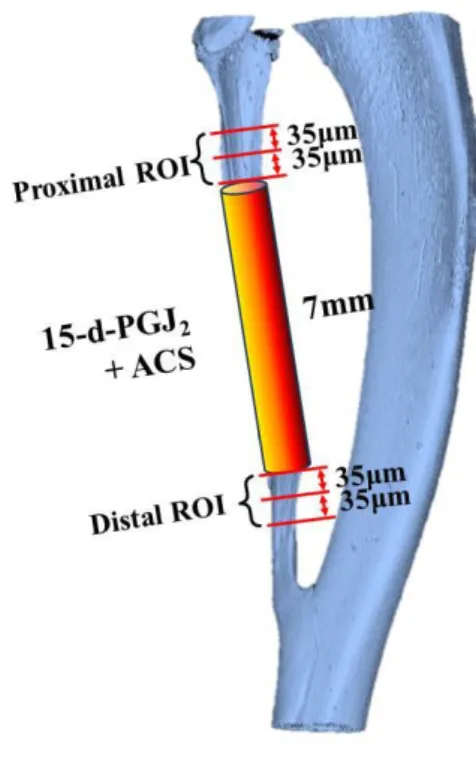
Absorbable collagen sponge 를 적용한 양 끝을 근위부, 원위부로 설정하고 각 각 35μm 간격으로 3 Layer 단면을 ROI(Region of Interest)로 설정하였다. CTAn® software(Skyscan)를 이용하여 ROI 3 Layer 의 평균 Bone fraction volume(BV/TV,%), Trabecular thickness (TbTh,μm), Trabecular number (TbN,mm), Trabecular separation (TbSp,μm), Structure model index (SMI) 을 측정 하였다.
데이터뷰어 (Dataviewer, Skyscan. Version 1.4.4 32-bit)를 통해 CT 의 단면 영상들을 관찰하였고, ONDEMAND program 으로 3 차원 영상을 재구성 하였다.
11
.
Figure 5. Region of interest (ROI). Interval of the each ROI layer is 35μm. Average value of the each layer is used for the statistical analysis.
ii.
조직학적 평가 (H-E Stain)
희생 후 비골과 경골 시편을 Paraformaldehyde 에 고정하였다. 연세대학교 의료원 임상의학연구센터 실험동물부 조직병리교실의 protocol 에 따라 탈회 및 파라핀 포매 후 H–E(Hematoxylin and Eosin) stain 한 슬라이드를 광학 현미경 관찰 하였다.
iii.
통계 분석
측정된 지표 값들은 SPSS 19.0 프로그램을 이용하여 two sample t-test 통계분석 시행하였다
12
III. 결 과
1. 육안 소견
1) 4 주 희생군
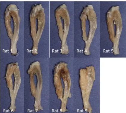
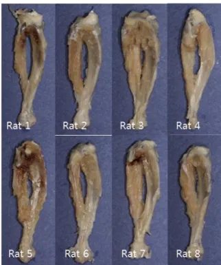
A. 대조군 결손 부 주변 조직의 변화를 관찰하기 위해, 시편 채취 시 촉진으로 골 결손 부위 조직을 유지하며 주변 근육의 박리를 시행하였다. 대부분의 시편에서 비유합이 관찰 되었으며 carrier 인 absorbable collagen sponge 는 모두 흡수되었다. 실험 과정에서 형성한 결손부 주변 근육의 형태가 잘 유지 된 시편이 (Rat3,5) 보여졌고 결손부위가 더 넓어진 시편 (Rat1,8)도 관찰 되었다. 연조직의 염증이나 감염은 관찰되지 않았다. B. 실험군 전반적으로 대조군에 비해 결손부위의 박리 시 주변 근육의 부착이 단단하였으며 외형이 더 건재한 소견을 보였다. 결손부 주변조직의 부피가 더 커진 시편이 관찰되었고 (Rat5,8,9), 이 시편들에서 골성유합이나 이소성 골 형성의 소견들을 기대 할 수 있었다. 비골의 전반적인 형태가 상실된 비유합 소견도 관찰되었다 (Rat7). 연조직의 염증이나 감염은 관찰되지 않았다.13
Figure 6. Control group at 4week
14
2) 8 주 희생군
A. 대조군
4 주군에 비해 전반적인 경골의 길이성장이 관찰되었고, 비골의 결손부 확대가 더 뚜렷하게 관찰되었다. absorbable collagen sponge 는 완전히 흡수되었으며 연조직의 염증소견은 보이지 않았다. 시편 채취 시 결손부 주변 조직을 완전 박리 하지 않고 조직소견에서 추가적 관찰을 시행하였다.
B. 실험군
대조군에 비해 비골의 외형이 더 뚜렷하게 관찰되었으며 주변 부착 근육도 더 안정적인 형태였다. 결손부위 촉진 시 단단한 감촉이 확인되었고 absorbable collage sponge 는 완전히 흡수되었다 (Rat 2,4). 4 주 실험군과 비교 시 결손부 골 유합 상태는 큰 차이가 없었다.
15
Figure 8. Control group at 8week.
16
2. 방사선학적 소견
가. Standard X-Ray
1) 4 주 희생군
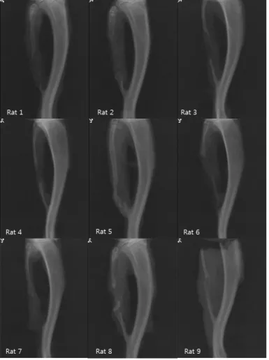
A. 대조군 대부분의 시편에서 골 결손부의 재생이 일어나지 않고 절단연의 추가적 흡수 및 변이가 보여졌다. 흡수가 대부분 이였으나 일부에서는 결손부에 얇은 불투과성 물질이 관찰 되거나 (Rat3), 근심 절단연의 두께와 부피가 증가하는 소견 (Rat5), 원심 절단연에 이소성 골 형성 (Rat 9)의 흔적이 관찰 되기도 하였다. B. 실험군 결손부의 골 재생이 대조군에 비해 현저하지는 않지만 일부 시편에서 분절성 골 결손부의 재생이 (Rat5,8,9) 관찰되었다. 근심 절단면에서 골성 유합 및 원심 절단면에서는 이소성 골 성장 양상을 보이는 시편이 (Rat5) 관찰되었고, 원심 절단면에서 골성 유합 및 결손부위 골 재생이 정상 골조직 보다는 약하지만 흡사한 광화도를 보이는 시편도 (Rat9) 관찰되었다. 하지만 형성한 결손부 크기보다 추가적 흡수가 나타난 시편들도 (Rat6,7) 관찰되어 x-ray 소견상에서 현저한 유의성이 있다고 보기는 어려웠다.17
Figure 10. Radiographic findings at 4 week control group on Left site. All defect sites remain vacant, however, each cutting site shows different bony change. Rat 3 shows very thin and sharpened calcified fragment at defect site , which infers bone formation. Widening and bulk change of the fibula at mesial cutting edge is observed at Rat5 and indistinct bone formation evidence is shown at Rat9. Natural healing process after defect formation, even though we only maintained the space with absorbable collagen sponge, is observed in a few. The majority of this group shows 6-8mm or enlarged defect site.
18
Figure 11. Radiographic findings at 4 week experimental group on Right site. Bony change at the cutting margin is observed. New bone formation and overgrowth of the defect site is observed at Rat5. Irregularity and sharpening of the cutting edge is observed at Rat 1,2,3,4, referentially Rat 2 distal cutting edge shape is like palm toward the defect site with an open medullary cavity. Extension of the defect site is observed at Rat 6,7 and shows sharpening of the remained fibula. Ectopic bony growth is observed at Rat 8, which give a guess the muscle cuff leakage. Similar x-ray opacity to the normal fibula is observed at Rat 9 defect site.
19
2) 8 주 희생군
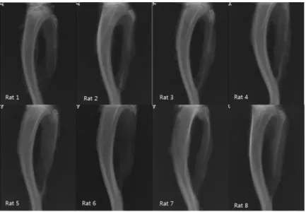
A. 대조군 결손부위의 비유합이 모든 시편에서 관찰되었다. 4 주군에 비해 비골의 절단연 주변이 얇게 흡수되고 날카로워진 양상을 보였으며 10mm 이상으로 보이는 추가적 골 흡수가 일어난 시편도 관찰 할 수 있었다 (Rat6). B. 실험군 일부 시편에서는 결손부위에 정상 골조직과 유사한 두께와 광화도를 보이는 골 조직이 형성 (Rat2) 되었다. 또한 신생골 조직으로 보이는 분절성 시편이 결손부위에 섬유성 결합 형태로 연결 된 소견도 (Rat4) 관찰되었다. 하지만 4 주군에 비해 현저하게 뚜렷한 골 유합 이나 재생 소견을 보이지는 않았다.Figure 12. Radiographic findings at 8 week control group on Left site. All of them shows non-calcified connection and cutting margin is sharpened and irregular.
20
Bone ends appeared to be pointed suggesting that the bone was undergoing significant bone resorption. Rat6 show over 10mm defect site extension.
Figure 13. Radiographic findings at 8 week experimental group on Right site. Bone regeneration is observed at Rat2 which shows similar bone density to normal fibula and continuity at mesial cutting margin. Calcified fragment is connected with a fibrous connective tissue at defect site Rat4, not continuous but position is maintained. 7mm defect site is remained vacant, cutting edge is irregular at Rat 1,3,6,7 and resorption area is extended at Rat 5,8.
21
나. Micro CT
1) 4 주 희생군
A. 실험군 일부 시편에서 신생골 형성이 관찰 되었다. Rat 5 는 근위부에서 연속적으로 신생골이 형성되었으며 원위부에서는 이소성 골 형성을 보였다. Rat 9 은 열린 골수강의 형태를 보이며 원위부에서 연속적으로 신생골이 형성 되었다. B. 대조군 대부분의 시편에서 골 결손이 그대로 남아 있었으며 절단면의 흡수도 관찰되었다. Rat 9 에서 일부 석회화 물질이 관찰되었으나 신생골 형성으로 보기에는 불충분 하였다.Figure 14. Micro CT : longitudinal & cross sectional view at 4 week Rat5 experimental site. Calcification material at the defect site is observed with irregular pattern and lower density than normal fibula. Continuity between mesial cutting margin and newly formed bone is shown at micro CT image. New bone formation is observed on the micro CT.
22
Figure 15.Micro CT : longitudinal & cross sectional view at 4 week Rat9 experimental site. At defect site, open marrow cavity bone formation is observed with a calcified margin. Continuity between the distal cutting margin and newly formed bone is shown, although the continuity is not complete.
Figure 16.. Micro CT : longitudinal & cross sectional view at 4 week Rat9 control site. Ectopic bony change is observed at the distal margin but the continuity of the fibula is irregular. In this specimen we put on supposition that only keep the muscle cuff at the original site and maintain the defect site can help the bone generation. But Rat9 was the most calcified specimen among the 4 week control group and newly formed calcification pattern is too much irregular and not enough to suggest new bone formation.
23
2) 8 주 희생군
A. 실험군 일부 시편에서 결손부위 신생골 형성을 관찰 할 수 있었다. Rat 2 에서는 일정한 두께의 피질골과 내부 해면골 영상을 보이는 신생골이 관찰되었다. Rat 4 에서는 골수강이 보이지 않는 골 조직이 결손부위에 신생되어 절단면 과 비연속적이지만 섬유성 결합으로 연결되어 있었다. B. 대조군 대부분의 시편에서 신생골 형성을 관찰하기 어려웠고, 골 흡수로 결손 부위가 확대된 시편들이 관찰되었다.Figure 17. Micro CT : longitudinal & cross sectional view at 8 week Rat2 experimental site. Micro CT images shows open medullary cavity at fibula defect site, which indicates endochondral ossification. Thickness of the newly formed bone is not enough, however , enough to predict new bone formation.
24
Figure 18. Micro CT : longitudinal & cross sectional view at 8 week Rat4 experimental site. Ossified bone formation is observed at defect site with none-medullary cavity. Length and thickness of the new bone is similar to the normal fibula but continuity is not definite. Fibrous connection between the fibula and the new bone at the cutting margin can be inferred.
Figure 19. . Micro CT : longitudinal & cross sectional view at 8 week Rat4 control site. Bone resorption of the defect area is extended over 7mm .
25
다. 3D Reconstruction
1) 4 주 희생군
A. 실험군
Figure 20. 3D Reconstruction of the 4 week experiment site. a)Rat5 experimental site. New bone formation is observed at the defect site. Continuity of the fibula is recovered between the new bone and the cutting margin. b)Rat8 experimental site. Bulky growth change and calcified material formation is observed at the distal cutting margin. c)Rat9 experimental site. New bone formation which is continuous from the distal cutting margin is observed.
B. 대조군
Figure 21. 3D Reconstruction of the 4week control site. Rat5. Change of the fibula cutting site is observed. Margin of the cutting site is changed to the bulky and sharpened edge. Only collagen sponge and saline material might be inferred to have bone growth tendency. However, Rat 5 was the only specimen showing bulky change at the cutting margin among the 4week control group.
26
2) 8 주 희생군
A. 실험군
Figure 22. 3D Reconstruction of the 8 week experiment site. a) Rat 2 experimen tal site. Full rehabilitation and new bone formation is bserved at the defect site. b) Rat4 experimental site. Not continuous but newly formed bone is observed at the defect site.
27
3. 통계분석
독립된 두 집단의 모평균 분석을 위한 독립 표본 t-검정 통계분석을 시행하였다. 신뢰수준 95%, 유의수준 5%로 설정하였고, 실험군 과 대조군의 Bone fraction volume(BV/TV%), Trabecular thickness (TbThμm), Trabecular number (TbN/mm), Trabecular separation (TbSp μm ), Structure model index (SMI) 평균값을 각각 4 주와 8 주에서 분석하였다(table 2,3).
Table 2. Statistical analysis of the 4 week experiment and control group
BV/TV(%) Mean(SD) * TbTh(μm) Mean(SD) TbN(1/mm) Mean(SD) * TbSp(μm) Mean(SD) SMI Mean(SD) Control Group 13.1(7.32) 117.58(12.89) 1.13(0.65) 137.51(3.13) 2.34 (0.32) Experimental Group 24.5(14.76) 122.71(14.78) 1.99(1.17) 134.44(8.13) 2.20 (0.32) P 0.007 0.275 0.011 0.144 0.206 *: p<0.05
Table 3. Statistical analysis of the 8 week experiment and control group
BV/TV(%) Mean(SD) * TbTh(μm) Mean(SD) TbN(1/mm) Mean(SD) * TbSp(μm) Mean(SD) * SMI Mean(SD) * Control Group 9.71(7.87) 124.68(17.2) 0.66(0.44) 140.13(0.81) 2.48 (0.28) Experimental Group 19.39(10.16) 131.69(7.24) 1.4737(0.77) 138.73(1.87) 2.04 (0.37) P 0.005 0.143 0.001 0.012 0.001 *: p<0.05 4 주의 BV/TV(%), TbN(1/mm)와 8 주의 BV/TV(%), TbN(1/mm), TbSp(μm), SMI 값이 p-value < 0.05 로 통계적으로 유의하여 15dPGJ2 가 골의 재생과 항상성 향상에 영향 준다는 결과를 얻었다
28
Figure 23. Graph of the statistical analysis. Bone fraction volume(BV/TV%), Trabecular thickness (TbTh), Trabecular number (TbN), Trabecular separation (TbSp) value is shown in the graph. (* ; The value which shows a significant difference.)
29
4. 조직형태학적 소견
1) 4 주 희생군
A. 실험군 절단연 사이에서 신생골 형성, 석회화된 기질침착, 신생혈관형성 및 다양하고 활발한 세포반응을 보이는 시편과 연조직의 개입으로 유합이 실패된 시편 들이 관찰되었다. B. 대조군 대부분의 시편에서 결손부위의 추가적 흡수가 관찰 되었으나 일부에서는 절단연의 부피가 증가하거나, 석회화 된 작은 조직들이 관찰되었다.2) 8 주 희생군
A. 실험군 신생골이 형성된 시편에서는 4 주군 보다 성숙한 석회화 기질의 피질골이 관찰되었으며 절단면에서 결손부위로 연속되는 골 형성이 보여졌다. 신생골이 형성되지 못한 시편들 역시 관찰 할 수 있었다. B. 대조군 대부분의 시편에서 현저해진 골 흡수와 연조직의 개입이 관찰되었다.30
Figure 24. Histologic findings of the 4week experiment site (Rat 5). Osteocytes surrounded by the osseous matrix and a lot of blood vessels are observed at the defect site. Although union is not complete, endochondral ossification by the chondroblast is observed at the cutting margin of the fibula, which means new bone formation .Ax50. Bx50 .Cx100 .Dx100. Ex200.
31
Figure 25. Histologic findings of the 4week experiment site (Rat 8). Mineralized matrix, fibroblasts, arteries are mixed at defect site. Although mature bone formation is not shown, active cell reaction is expected at A.C. Change of the cutting margin with enlarged new bone site is observed at B.D.E. Ax50.Bx50. Cx100.Dx100. Ex100.
32
Figure 26. Histologic findings of the 4week experiment site (Rat 9). New bone formation is observed with a lot of osteocytes and blood vessels. Medullary cavity is shown at new bone site in slide A.C.E. Change of the cutting margin, which failed to complete union, shows a lot of fibroblasts, arteries and bone matrix mixture at B.D. Ax50.Bx50.Cx100.Dx200.Ex200.
33
Figure 27. Histologic findings of the 4week control site (Rat 9). Cutting margins shows no bone formation, which is surrounded by the fibroblast and adipocyte in slide A.C.E. However, small new bone fragment is observed at the defect site with a lot of osteocyte and blood vessels in slide B.D. Ax50.Bx50.Cx100.Dx100. Ex200.
34
Figure 28. Histologic findings of the 8week experiment site (Rat 2). New bone formation at the defect site is observed in all slides. Connection between the fibula and the new bone is observed in slide B.D. Newly formed bone matrix and osteoclast lining around the new bone is observed in slide C. Ax50.Bx100.Cx200. Dx200.
35
Figure 29. Histologic findings of the 8week experiment site (Rat 4). Bone conncetion bewteen the new bone and the fibula is observed in slide A.C.D. Trabecular bone formation by the osteoblast and osteoclast with a lot of blood vessels is shown and newly formed mineralized matrix replace the defect site. Ax50.Bx50.Cx100.Dx200.Ex200.
36
Figure 30 Histologic findings of the 8week control group (Rat 4). Bone resorption of the cutting margin and ROI area is observed. Ax50.Bx100.Cx200.Dx200.
37
IV. 고 찰
이 실험에서 15d-PGJ2 가 특정 기전을 통해 골 흡수를 억제 할 뿐만
아니라 신생골 형성 능력을 지내고 있음을 확인 할 수 있었다. 골의 대사 작용은 RANKL-RANK 반응과 OPG 에 의해 조절된다. Receptor activator of nuclear factor kappa-B ligand (RANKL)은 tumor necrosis factor ligand superfamily member 11(TNFSF11), osteoprotegrin ligand(OPGL), osteoclast differentiation factor(ODF)로 불리며, osteoclast 의 Receptor activator of Nuclear Factor κB (RANK)에 작용 및 활성화 시켜 골 흡수를 야기한다(Wong BR et al, 1997. Anderson DM et al, 1997). RANKL mRNA 는 뼈와 골수강 및 림프조직에서 높은 농도로 발현되고, 뼈에서 파골세포의 분화, 활성화 및 세포예정사 억제에 영향을 미친다. 파골세포 형성 개시인자인 macrophage-colony stimulating factor(M-CSF)와 RANKL 에 의해 파골 전구세포는 성숙한 파골세포로 분화하고 골 형성과정의 개시역할을 한다 (Malyankar UM et al, 2000.Lacey DL et al, 1998. Yasuda H et al, 1998. Wong BR et al, 1997. Anderson DM et al , 1997. Fuller K et al 1998).
반면에 RANKL 이 osteoclastogenesis inhibitory factor 라고 불리는 osteoprotegrin(OPG)과 결합하면 RANKL-RANK 반응이 억제되고, 파골 전구세포가 성숙한 파골세포로 분화하지 못하여 골 흡수가 감소한다. Estrogen 이나 골다공증 치료제 일부는 OPG 를 증가시켜 파골세포의 분화를 억제하고 bone mineral density 와 volume 을 증가시키는 효과를 보인다 (Khosla S, 2001). OPG 가 RANKL 과 결합하여 NF-κB 의 주요 활성체인 RANK 의 활성이 억제되는 반응과 (Baud'huin M et al, 2007), 15d-PGJ2에
38
유사한 파골세포 활성 억제 효과를 보일 것으로 이 연구에서는 생각 해 보았다.
이 연구에서 사용한 15d-PGJ2 는 강력한 PPARγ 친화성 ligand 이다.
PPARγ는 nuclear hormone receptor(NHR) superfamily 에 속하며, peroxisome proliferator responsive element(PPRE)와 retinoid X receptor(RXR) subfamily 를 결합시키며 전사조절 인자로 기능 한다(Kliewer, S.A et al, 1992). 활성화 된 PPARγ는 지방세포 생성, 지질 대사, 인슐린 반응 및 염증반응에 관여하며 다양한 생리적 반응을 조절 하며, 조혈모세포에서 파골 전구세포로 분화하는 것을 촉진하여 골 흡수 촉진 한다는 보고도 있다(Wan, Y, 2010). 하지만 Kim 등은 15d-PGJ2 가
RANKL 에 의해 유도 된 파골세포의 생성을 억제함을 tartrate-resistant acid phosphatase(TRAP) staining 으로 확인하였다. Kim 등의 실험에서 누드마우스에 유선암종세포를 주입하여 뼈에 전이 및 파괴가 일어나게 한 후 15d-PGJ2 를 1 주일에 3 번씩 총 6 주의 기간 동안 피하주사 하였다.
15d-PGJ2 는 RANKL 에 의해 증가하는 TRAP(tartrate-resistant acid
phosphatase)-positive cells 을 감소시켜 파골세포 생성을 억제하였고, cathepsin K 와 MMP 의 단백분해 반응을 억제하여 골 흡수를 억제 하였다. 이 실험의 양성 대조군인 Bistphosphonate 계열이 Zoledronic acid 투여시 보다는 낮은 bone volume 이었지만 정상과 유사한 골 상태를 보였다. Kim 등의 두 번째 실험에서 난소를 절제한 쥐에 15d-PGJ2 를 피하주사 하여 골다공증에 의한 골 파괴 억제 효과를 확인하였다. 마이크로 CT 분석에서 15d-PGJ2를 투여한 쥐의 대퇴골소주 구조는 난소절제에 의한 골 흡수 상태 보다 회복된 결과를 보였고 에스트로겐 결핍에 의한 골 재형성 불균형을 억제 할 수 있을 것이라고 보고 하였다(Kim Ki-Rim,. 2012). 최근 Bisphosphonate 는 파골세포의 활성 억제 뿐 아니라 조골세포에 대한 anabolic 효과가 있다는 실험들이 보고되고 있다. Im 등과 Von Knoch
39
등은 bisphosphonate 가 조골세포를 증식시키고 중간엽세포를 조골세포로 분화 유도하는 골 유도 특성이 있음을 in vitro 실험에서 보고하였다(Im, 2004 ; Von Knoch et al, 2005 ; Wang et al, 2010). 이를 검증하기 위해 2011 년 연세대학교 구강악안면외과학 교실 권진일은 웅성 백서 비골 임계 크기 분절성 골 결손 모델에서 bisphosphonate 를 적용하는 실험을 시행하였고 조골세포의 분화 촉진 및 골 생성 유도 효과를 확인하였다(권진일, 2010). 이 논문에서는 15d-PGJ2 가 NF-κB 에 작용하여 RANK 를 억제하고, 성숙한 파골세포 분화를 감소시켜 골 파괴를 억제 할 뿐 아니라 조골세포에도 작용하여 bisphosphonate 처럼 골 분화 및 형성 능력이 있다는 가설을 세우고, 골 재생 및 결손부 회복 능력을 확인하기 위해 웅성 백서 비골 임계 크기 분절성 골 결손 모델 실험을 진행하였다. 일반적으로 골절 발생시 micro vessel 이 들어오면서 혈관 내벽 pericyte 들이 cartilage progenitor cell 또는 bone progenitor cell 로 분화하여 연골아세포와 조골세포로 분화하며 woven bone 과 hyaline cartilage 를 형성 하고, 가골 형성 및 lamellar bone 의 형성을 거치며 치유된다(Brighton, C. T. et al,) 이 논문에서는 주변 골막에서 분화된 연골 아세포에 의한 연골 내골화 기전이 아닌 시약 자체의 골 형성 능력을 확인하기 위해 자연재생이 어려운 7mm 임계 결손부를 형성하고 결손부의 골막을 제거 하였다. 본 연구의 육안적 소견과 X-ray 소견에서 실험군과 대조군의 극명한 차이는 보이지 않았지만, μCT 상 4 가지 계측요소인 Bone fraction volume(BV/TV), Trabecular thickness (TbTh), Trabecular number (TbN) , Trabecular separation (TbSp), Structure model index (SMI) 중 4 주군의 BV/TV, TbN 과 8 주군의 BV/TV, TbN, TbSp, SMI 값이 실험군,
40 대조군에서 통계학적으로 유의한 차이가 있음을 확인하였다. 조직학적 소견에서는 4 주 실험군 일부 시편에서 활발한 석회화 기질의 침착과 woven bone 형성을 보였다. 절단부위의 완전 한 골의 형성을 이루지 못한 시편 에서도 15d-PGJ2 를 적용한 ACS 부위에서 섬유아세포 같은 기질세포와 신생혈관형성, 부분적 석회화 기질 침착을 보였다. 또한 결손부위에서 골을 형성하지 못하고 결손부위가 그대로 남아있는 시편들도 관찰할 수 있었다. 4 주 대조군 대부분은 결손부가 그대로 남아 있었고 일부에서 부분적 석회화 물질이 형성 되었으나 절단연과 연속성을 회복하면서 골 형성을 이룬 시편은 관찰되지 않았다. 8 주 실험군 에서는 신생골이 형성된 시편에서는 석회화 기질의 농도가 짙어지고 Trabecular bone 의 양이 증가하였다. 신생골이 형성되지 못한 시편에서는 결손부위로의 근육이나 다른 연조직의 투입이 명확해 졌고 대조군 에서는 설정한 크기보다 결손 크기가 확대된 소견이 보여졌다. 임계크기 결손 모델 실험에서 신생골이 형성되지 않은 경우에 결손부위 크기가 10mm 이상으로 확대되고 절단연은 뾰족해지며 흡수가 일어난다고 알려져 있다 (ZaWaunyka W et al, 2011). 이때 절단연은 흡수를 보이며 개방된 골수강을 볼 수 없다고 알려져 있으나 우리의 실험에서는 절단연 부위들이 개방된 골수강 상태인 조직 소견들을 일부 관찰 할 수 있었다. 개방성 골수강 상태를 유지하게 한다면 이것은 골 조직의 회복과 remodeling 을 기대할 수 있다. 많은 연구들이 PG 가 강력한 골 형성 인자임을 보고해 왔고(Yoshiyuki et al, 1985), 15d-PGJ2 의 분해 전 물질인 PGD2의 조골세포 석회화 자극 효과 역시 보고되어 왔다(Koshihara Y et al, 1989). 핵 수용체에 바로 작용 하는 15d-PGJ2 의 골 형성능에 대한 정확한 기전이 확인 된다면 생산 및 적용 면에서 효율적일 것으로 생각된다. 핵 수용체에 바로 작용하며 다양한 효과를 보이는 cytokine 인 만큼 적절한 농도의 적용이 중요하여, 2 회의 예비실험을 통해 적정농도를
41 확인하였다. 첫 번째 실험에서는 15d-PGJ2 의 농도 0.5mg/500μl, 2 mg/500μl, 5 mg/500μl 인 3 그룹으로 각각 2 마리 좌,우측 총 6 마리 웅성 백서의 좌.우측 비골 12 site 에서 본 실험과 동일한 방법으로 실험을 진행하고 4 주 후에 희생하였다. Saline 대조군 없이 농도간의 비교만을 시행 하였고 육안소견과 x-ray 소견으로 결과를 분석하였다. 두 번째 실험에서는 0.5mg/500μl, 2 mg/500μl 인 2 그룹으로 각각 4 마리 웅성 백서의 좌,우측 비골 총 8 마리 16site 에서 saline 대조군 없이 동일한 방법으로 실험을 진행하였다. 4 주 후 희생하여 육안소견 및 x-ray 소견을 확인하였고 본 실험에서는 최종적으로 1mg/ 500μl 를 100 μl 씩 absorbable collagen sponge 에 적용하였다. 15d-PGJ2 가 다양한 기전을 통해 골의 결손이나 골 질환 시 항상성을 유지하는 방향으로 골 형성을 촉진한다는 결론을 실험과 논문들을 통해 확인하였다. PPARγ 수용체에 작용하여 파골세포 분화를 자극 및 골 흡수를 증가시킨다는 보고도 있지만, 골의 형성 과정이 조골세포와 파골세포의 순환기전에 의해 진행 됨으로 특정세포의 분화 촉진은 전체적 골 형성을 향상 시키게 작용 할 수 있다고 생각한다. 15d-PGJ2 가 광범위 골 결손부위에서 실질적인 이식제로 사용되기 위해서는 좀 더 반복적이고 일관성 있게 골이 형성 되는 실험결과들을 확보해야 하며 이를 위해서 결손부위 디자인의 변화, 흡수성 콜라겐 이외의 carrier 의 다양한 적용, 실험 술자의 숙련도 향상, 농도에 대한 반복 구체적 실험들이 필요하다. 또한 RT-PCR(reverse transcription polymerase chain reaction) 분석으로 골모세포 표지자를 확인하고 조직면역화학 염색을 통한 골 유도능의 구체적 확인 역시 진행되어야 할 것으로 생각된다.
42
V. 결 론
15-deoxy-Δ12,14-prostaglandin J₂ 가 웅성 백서 비골의 분절성 골 결손부위에 이식 시 통계적으로 유의한 골 형성 능력을 보임을 이 연구에서 확인 하였다. 골을 생성하는 능력이 우수하다고 보기에는 부족한 부분들이 있고 기전의 확인을 위한 추가적인 연구들이 필요하지만 핵 수용체에 바로 작용하여 조골세포와 파골세포에 영향을 줄 수 있게 때문에, 지속적으로 일정한 농도를 방출 할 수 있는 적절한 carrier 를 만난다면 더 나은 골 이식재로 사용 될 수 있을 것으로 생각된다.43
VI. REFERENCE
Sbordone, L., Toti, P., Menchini-Fabris, G. B., Sbordone, C., Piombino, P., Guidetti, F. "Volume changes of autogenous bone grafts after alveolar ridge augmentation of atrophic maxillae and mandibles". International Journal of Oral and Maxillofacial Surgery 38(10): 1059-1065, 2009. Adam S. DeConde, Matthew K. Lee, Douglas Sidell, Tara Aghaloo, Min Lee,
Sotirios Tetradis, Kyle Low,David Elashoff, Tristan Grogan, Ali . Sepahd ari, Maie St John. “Defining the Critical-Sized Defect in a Rat Segmental Mandibulectomy Model” JAMA Otolaryngol Head Neck Surg. Published online. November 14, 2013.
Rubens Moreno de Freitas, Rubens Spin-Neto, Elcio Marcantonio Junior, Luís Antônio Violin Dias Pereira, Ulf ME Wikesjö, Cristiano Susin.“Alveolar Ridge and Maxillary Sinus Augmentation Using rhBMP-2: A Systematic Review”. Clin Implant Dent Relat Res. Sep 17, 2013.
Triplett RG, Nevins M, Marx RE, Spagnoli DB, Oates TW, Moy PK, et al.“Pivotal, randomized, parallel evaluation of recombinant human bone morphogenetic protein-2/absorbable collagen sponge and autogenous bone graft for maxillary sinus floor augmentation”. J Oral Maxillofac Surg. 67(9): 1947-1960, 2009.
E.J. Woo. “Adverse events reported after the use of recombinant human bone morphogenetic protein 2”. J Oral Maxillofac Surg. Apr 70(4): 765-7, 2012.
Kim KiRim. “15deoxyΔ12,14prostaglandin J₂ inhibits bone loss of breast cancer metastasis and ovariectomyinduced osteoporosis”. 연세대학교 대학원. 서울. 2012
44
Im, G. "Osteoblast proliferation and maturation by bisphosphonates". Biomaterials 25(18): 4105-4115, 2004.
Beatriz Díez-Dacal and Dolores Pérez-Sala. “Anti-Inflammatory Prostanoids: Focus on the Interactions between Electrophile Signaling and Resolution of Inflammation”. The Scientific World JOURNAL 10, 655–675, 2010.
Shibata, T., Kondo, M., Osawa, T., Shibata, N., Kobayashi, M., and Uchida, K. “15-Deoxy-delta12,14-prostaglandin J2. A prostaglandin D2 metabolite generated during inflammatory processes”. J. Biol. Chem. 277,10459–10466, 2002.
Fukushima, M., Kato, T., Narumiya, S., Mizushima, Y., Sasaki, H., Terashima, Y., Nishiyama, Y., and Santoro, M.G. “Prostaglandin A and J: antitumor and antiviral prostaglandins”. Adv. Prostaglandin Thromboxane Leukot. Res. 19, 415–418, 1989.
Kato, T., Fukushima, M., Kurozumi, S., and Noyori, R. “Antitumor activity of delta 7-prostaglandin A1 anddelta 12-prostaglandin J2 in vitro and in vivo”. Cancer Res. 46, 3538–3542, 1986.
Kim, I.K., Lee, J.H., Sohn, H.W., Kim, H.S., and Kim, S.H. “Prostaglandin A2 and delta 12-prostaglandin J2induce apoptosis in L1210 cells”. FEBS Lett. 321, 209–214, 1993.
Santoro, M.G. “Antiviral activity of cyclopentenone prostanoids”. Trends Microbiol. 5, 276–281, 1997.
Straus, D.S., Pascual, G., Li, M., Welch, J.S., Ricote, M., Hsiang, C.H., Sengchanthalangsy, L.L., Ghosh, G., Glass, C.K. “15-deoxy-delta
45
12,14-prostaglandin J2 inhibits multiple steps in the NF-kappa B signaling pathway”. Proc Natl Acad Sci U S A, 97(9): p.4844-9, 2000.
Gilmore TD. "Introduction to NF-κB: players, pathways, perspectives". Oncogene 25 , (51): 6680–4, 2006.
Wong BR, Rho J, Arron J, Robinson E, Orlinick J, Chao M, Kalachikov S, Cayani E, Bartlett FS, Frankel WN, Lee SY, Choi Y. "TRANCE is a novel ligand of the tumor necrosis factor receptor family that activates c-Jun N-terminal kinase in T cells". J. Biol. Chem. 272 (40): 25190–4, October. 1997.
Anderson DM, Maraskovsky E, Billingsley WL, Dougall WC, Tometsko ME, Roux ER, Teepe MC, DuBose RF, Cosman D, Galibert L. "A homologue of the TNF receptor and its ligand enhance T-cell growth and dendritic-cell function". Nature. 390 (6656) : 175–9, November, 1997.
Malyankar UM, Scatena M, Suchland KL, Yun TJ, Clark EA, Giachelli CM “ Osteoprotegerin is an α v β3-induced, NF-κB-dependent survival factor for endothelial cells”. J Biol Chem 275:20959–20962, 2000.
Lacey DL, Timms E, Tan H-L, Kelly MJ, Dunstan CR, Burgess T, Elliott R, Colombero A, Elliott G, Scully S, Hsu H, Sullivan J, Hawkins N, Davy F, Capparelli C, Eli A, Qian YX, Kaufman S, Sarosi I, Shalboub V, Senaldi G, Guo J, Delaney J, Boyle WJ “Osteoprotegerin (OPG) ligand is a cytokine that regulates osteoclast differentiation and activation”. Cell 93:165– 176 , 1998.
Yasuda H, Shima N, Nakagawa N, Yamaguchi K, Kinosaki M, Mochizuki S, Tomoyasu A, Yano K, Goto M, Murakami A, Tsuda E, Morinaga T, Higashio K, Udagawa N, Takahashi N, Suda T. “Osteoclast differen
46
tiation factor is a ligand for osteoprotegerin/osteoclastogenesis-inhibitory factor and is identical to TRANCE/RANKL”. ProcNatlAcadSci USA 95:3597–3602, 1998.
Fuller K, Wong B, Fox S, Choi Y, Chambers TJ. “TRANCE is necessary and sufficient for osteoblast-mediated activation of bone resorption in osteoclasts”. J Exp Med 188:997–1001, 1998.
Khosla S , "Minireview: the OPG/RANKL/RANK system". Endocrinology 142 (12): 5050–5. Dec, 2001.
Baud'huin M, Lamoureux F, Duplomb L, Rédini F, Heymann D . "RANKL, RANK, osteoprotegerin: key partners of osteoimmunology and vascular diseases". Cell Mol Life Sci64 (18): 2334–2350.Sep, 2007.
Kliewer, S.A., et al. “Convergence of 9-cis retinoic acid and peroxisome proliferator signaling pathways through heterodimer formation of their receptor”. Nature, 358(6389): 771-4, 1992.
Wan, Y. “ PPARgamma in bone homeostasis”. Trends Endocrinol Metab, Dec; 21(12) :722-8, 2010.
Von Knoch, F., Jaquiery, C., Kowalsky, M., Schaeren, S., Alabre, C., Martin, I., Rubash, H. E., Shanbhag, A. S. "Effects of bisphosphonates on proliferation and osteoblast differentiation of human bone marrow stromal cells". Biomaterials 26(34): 6941-6949, 2005.
Wang, C.-Z., Chen, S.-M., Chen, C.-H., Wang, C.-K., Wang, G.-J., Chang, J.-K., Ho, M.-L. "The effect of the local delivery of alendronate on human
47
adipose-derived stem cell-based bone regeneration". Biomaterials 31(33): 8674-8683. 2010.
권진일: 웅성 백서 비골의 분절성 골 결손에서 bisphosphonate 의 국소 적용이 골 재 생과 미세 골 구조에 미치는 영향 =. 연세대학교 대학원, 서울 :, 2011.
Brighton CT, Hunt RM. “Early histologic and ultrastructural changes in microvessels of periosteal callus”. J Orthop Trauma. May;11(4):244-53, 1997.
ZaWaunyka W. Lazard, Michael H. Heggeness, John A. Hipp, Corinne Sonnet, Angie S. Fuentes, Rita P. Nistal, Alan R. Davis, Ronke M. Olabisi, Jennifer L. West, Elizabeth A. Olmsted-Davis. “Cell-based gene therapy for repair of critical size defects in the rat fibula”. J Cell Biochem. , 112(6):1563-71. Jun 2011.
Yoshiyuki H, Yoshinori N, Masahiki H, Eiko I, Masayoshi K. “Inductive effects of prostaglandins on alkaline phosphatase in osteoblastic cells, clone MC3T3-E1” . J Biochem , 97(1);97-104, 1985.
Koshihara Y, Kawamura M. “Prostaglandin D2 stimulates calcification of human osteoblastic cells”. Biochemical and Biophysical Research
48
Abstract
The effect of locally administered 15deoxyΔ
12,14
prostaglandin J₂on bone regeneration in segmental defect
Joo
Young Hong, D.D.S,
Department of Dental Science
The Graduate School, Yonsei University
(Directed by Professor Hyung Jun Kim, D.D.S., M.S.D., Ph.D.)
Development of the bone graft material for the reconstruction and regeneration in the oromaxillofacial defect has been continuously achieved with molecular biology. It was reported that 15deoxyΔ12,14
prostaglandin J₂ (15dPGJ₂) reduced bone destruction in the mice with metastatic breast cancer and recovered the serum levels of bone turnover markers in the ovariectomized mice in Yonsei university, Department of applied science, 2012, and we are based on it. The purpose of this study is to evaluate bone regeneration effect of 15dPGJ₂. We made 7mm defect in the fibula of 16 spraguedawley rats and grafted 15deoxyΔ12,14 prostaglandin J₂(concentration;1mg/500
49
㎕ , volume; 100 ㎕) soaked absorbable collagen in the right fibula defect site. At the control group, 100 ㎕ saline soaked absorbable collagen sponge was grafted in the left fibula defect site. Sacrifice of 9 rats and 8 rats was made after 4 weeks and 8 weeks, respectively.
Clinical observation, radiological analysis with an X-ray and micro CT, 3D reconstruction with a data viewer and histologic analysis were made. Bone fraction volume(BV/TV%), Trabecular thickness (TbThμm), Trabecular number(TbN/mm), Trabecular separation (TbSpμm), Structure model index(SMI) value were statistically evaluated. In the 4 weeks group, BV/TV, TbN were significantly different from the control group and in the 8 week group BV/TV, TbN, TbSp, SMI were significantly different.
Although significant bone formation were not displayed in clinical observation and X-ray analysis, micro CT analysis and 3D reconstruction showed continuous new bone formation at defect site in some specimens. Some specimens showed new bone formation, mineralized matrix and angiogenesis at defect site in histologic analysis.
This study evaluated bone regeneration effect of the 15dPGJ₂ in the segmental defect. Although the bone formation ability is not enough and mechanism needs more studies, it is certain that 15dPGJ₂ stimulates osteoblast and osteoclast into the bone metabolism cycle in the nucleus receptor unit. Further study for the concentration and the carrier about 15dPGJ₂is needed in order to use 15dPGJ₂ as an effective bone graft material.
Key words : 15dPGJ₂, bone regeneration, bone formation, bone graft material