Research Article
J Innate Immun 2021;13:94–106In vivo Anti-Cancer Effects of Resveratrol
Mediated by NK Cell Activation
Yoojin Lee
a, bHeewook Shin
a, bJongsun Kim
aaDepartment of Microbiology and Institute for Immunology and Immunological Diseases, Yonsei University College
of Medicine, Seoul, Republic of Korea; bBrain Korea 21 PLUS Project for Medical Science, Yonsei University College of
Medicine, Seoul, Republic of Korea
Received: December 6, 2019 Accepted: July 19, 2020
Published online: September 16, 2020
Jongsun Kim
Department of Microbiology and Institute for Immunology and Immunological Diseases, Yonsei University College of Medicine
© 2020 The Author(s) Published by S. Karger AG, Basel karger@karger.com
www.karger.com/jin
DOI: 10.1159/000510315
Keywords
Natural killer cell · Resveratrol · Interferon-gamma · Natural killer cytotoxicity · Immunotherapy
Abstract
Natural killer (NK) cells are innate immune lymphocytes that play an important role in anti-viral and anti-tumour immune responses. Several cancer immunotherapy approaches tar-geting NK cells are currently in clinical or preclinical develop-ment. Here, we aimed to find food nutrients that activate NK cells and determine their usefulness as candidates for anti-cancer and anti-metastatic drugs. Resveratrol appeared to activate NK cells most effectively among the substances test-ed and synergistically increastest-ed IFN-γ secretion and NK cell cytotoxicity with interleukin-2 (IL-2). CD107a, NKp30, and NKG2D expression levels were upregulated on the surface of NK cells upon treatment with resveratrol in combination with IL-2 compared with treatment with IL-2 alone. More-over, NK cell activity in human and mouse whole blood was enhanced upon treatment with resveratrol. Most important-ly, administration of resveratrol effectively inhibited tumour growth and metastasis in mice. In conclusion, we suggest that resveratrol may represent a candidate anti-cancer drug that acts by activating NK cells in vivo.
© 2020 The Author(s) Published by S. Karger AG, Basel
Introduction
Natural killer (NK) cells are innate immune cytes accounting for about 15% of all circulating lympho-cytes that spontaneously kill cells deemed to be danger-ous to the host in the absence of prior sensitization; they thus are presumed to be key effectors in cancer immuno-surveillance, transplantation rejection, and early viral im-munity [1–3]. Recently, it was reported that physical ac-tivity and metabolic status may alter the immune system and that physical inactivity and metabolic abnormalities are associated with reduced NK cell activity [4]. Further, NK cells are potent killers of malignant cells and are par-ticularly important in limiting tumour metastasis [5, 6]. NK cells can inhibit tumour growth by direct cytotoxicity through the release of cytolytic granules, by secretion of cytokines such as interferon-gamma (IFN-γ), or indirect-ly by the orchestration of anti-tumour immune responses [7]. NK cells are activated by Th1-type cytokines, such as interleukin (IL)-2, -12, -15, or -18, natural cytotoxicity receptors, and NKG2D [8–10]. When NK cells are acti-vated, they secrete large amounts of IFN-γ, which is es-sential in viral and tumour clearance and activates the signal transducers and activators of transcription (STAT) signalling pathway in target cells, leading to the improved control of infection [11, 12]. IFN-γ production by NK cells restricts tumour angiogenesis, stimulates adaptive
immunity, and helps shape T-cell responses in lymph nodes [13].
Third-generation immunotherapy represents a new paradigm for cancer treatment [14]. As the focus of treat-ment has shifted from the tumour itself to the host’s im-mune system, cancer immunotherapies activating the body’s immune system to fight cancer cells have been de-veloped. Most immunotherapeutic strategies focus on enhancing T-cell responses; however, there has been re-cent interest in using the relatively underexplored NK cell compartment for therapeutic interventions [15–17]. Moreover, it is becoming clear that some of the clinical approaches originally developed to increase T-cell cyto-toxicity may also activate NK cells [16].
In some clinical studies, the use of a low dose of IL-2, alone or in combination with other agents (such as low-dose IL-2 and IFN-α), results in similar clinical response rates and survival probabilities as the use of high doses of IL-2 alone [18]. Unfortunately, cytokines and other small molecules secreted by IL-2-activated effector cells induce toxicity, which offsets the efficacy of IL-2. To overcome this limitation, studies of therapies involving new cyto-kines are underway, including the use of IL-12 alone or in combination with IL-2 [19, 20]. In the last decade, the field of nutritional immunology has grown steadily. We hypothesized that, in addition to cytokines, there may be substances among natural compounds with the ability to activate NK cells [21].
Here, we screened several compounds including anti-oxidants, vitamins, and food ingredients to identify com-pounds that can activate NK cells. Furthermore, we dem-onstrated the potential of one of these compounds, resve-ratrol, as an anti-cancer and anti-metastatic drug candidate, which may act via NK cell activation. Overall, our finding enlightens the novel function of resveratrol as an NK booster, which can be the potential adjuvant for immunotherapeutics in cancers.
Materials and Methods Cell Lines and Cell Culture
The human NK cell line NK92 (ATCC® CRL-2407TM) was
maintained in the minimum essential medium-α (MEM-α) (Gibco, New York, NY, USA) supplemented with 12.5% heat-inactivated foetal bovine serum (FBS; Gibco), 12.5% heat-inactivated horse se-rum (Gibco), 0.2-mM myo-inositol (Sigma-Aldrich, St. Louis, MO, USA), 0.1-mM 2-mercaptoethanol (Sigma-Aldrich), 0.02-mM folic acid (Sigma-Aldrich), 1% penicillin/streptomycin (WelGENE, Gyeongsansi, Korea), and 5 ng/mL IL-2 (ATGen, Sungnamsi, Ko-rea). Human erythroleukemia cell line K562 (ATCC® CCL-243TM) was maintained in the RPMI 1640 medium (Gibco) supplemented
with 10% FBS and 1% penicillin/streptomycin. Mouse melanoma cell line B16F10 (ATCC® CRL-6475TM) was maintained in Dulbec-co’s modified Eagle’s medium (DMEM) (Gibco) supplemented with 10% FBS and 1% penicillin/streptomycin.
Antibodies and Reagents
Resveratrol, 3-amino-1-propanesulfonic acid (3-APS), gluta-thione, taurine, glycine, alanine, β-alanine, β-carotene, retinoic acid, retinol, retinal, tazarotene, vitamin B1, vitamin B12, vitamin D3, 2,3-butandiol, quercetin, triacetyl resveratrol, and piceatannol were purchased from Sigma-Aldrich. Lycopene and catechin were purchased from Santa Cruz Biotechnology (Santa Cruz, CA, USA). Resveratrol-3-O-glucuronide was purchased from Cayman Chem-ical (Ann Arbor, MI, USA). Resveratrol-4′-glucuronide was pur-chased from Spibio (Montigny Le Bretonneux, France). Resvera-trol-trisulphate was purchased from Carbosynth (Berkshire, UK). For flow cytometry, PE-conjugated anti-human CD107a antibody (555801) and APC-conjugated anti-human CD56 antibody (555518) were purchased from BD Pharmingen (San Diego, CA, USA). BV421-conjugated anti-human CD3 antibody (300434), APC-Cy7-conjugated anti-human CD16 antibody (302017), PE-conjugated anti-human NKG2D antibody (320806), PE-conjugat-ed anti-human NKp30 antibody (325208), PE-conjugatPE-conjugat-ed anti-hu-man NKp44 antibody (325108), PE-conjugated anti-huanti-hu-man NKp46 antibody (331908), BV650-conjugated anti-mouse TCRβ antibody (109251), PE-conjugated anti-mouse NK1.1 antibody (108708), PerCP-Cy5.5-conjugated anti-mouse IFN-γ antibody (505822), and PE-Cy7-conjugated anti-mouse CD107a antibody (121619) were purchased from BioLegend (San Diego, CA, USA).
Cell Proliferation Assay
Cell proliferation was measured using the CCK-8 assay kit (Dojindo Laboratories, Kumamoto, Japan). NK92 cells (1 × 10⁴ cells/well) (100 μL) were dispensed in triplicate into a 96-well flat-bottom plate and incubated for 4 h. After incubation, 10 μL of the CCK-8 solution was added to each well. After 4 h, absorbance was measured at 450 nm using a microplate reader.
ELISA
An IFN-γ ELISA set was purchased from BD Biosciences (San Diego, CA, USA). NK92 cells (5 × 10⁴ cells/well) were dispensed into a 24-well plate and incubated for 36 h. Then, the supernatants were harvested and dispensed in triplicate into a 96-well microplate pre-coated with capture antibody. After incubation for 2 h, each well was washed with the washing buffer (0.05% Tween 20 in PBS, pH 7.4), followed by the addition of HRP-conjugated detection an-tibody and incubation for 1 h. The substrate solution 3,3′,5,5′-tet-ramethylbenzidine (TMB) was added under protection from light. Absorbance was measured at 450 nm using a microplate reader.
CD107a Degranulation Assay
NK92 cells were treated with or without resveratrol and incu-bated for 36 h. After incubation, NK92 cells (2 × 10⁴ cells/well) were dispensed into a 96-well round-bottom plate and incubated with an equal number of K562 target cells. Then, anti-CD107a an-tibody was added, and the plate was incubated for 4 h. Cells were harvested and stained with anti-CD56 antibody for 30 min on ice. After 2–3 washes with PBS containing 1% FBS, fluorescence was measured using a BD LSR II flow cytometer (BD Biosciences). PBMCs were treated with or without resveratrol for 24 h in the
presence of IL-2. After incubation, PBMCs were incubated with K562 cells (5:1 ratio) for 4 h. Cells were harvested and stained with anti-CD3, -CD16, and -CD56 antibody in the presence of fixable live/dead stain (Invitrogen) for 30 min on ice. After 2–3 washes with PBS containing 1% FBS, fluorescence was measured using a BD LSR II flow cytometer (BD Biosciences).
Flow Cytometry
NK92 cells (5 × 10⁴ cells/500 μL) were incubated with or with-out resveratrol. After incubation, cells were washed twice with PBS containing 1% FBS and stained with fluorochrome-conjugated an-tibodies in PBS containing 1% FBS for 30 min on ice in the dark. The cells were washed twice, and then fluorescence was measured using BD LSR II.
Real-Time PCR Analysis
Total RNA was extracted from NK92 cells using TRIzol (Life Technologies, Carlsbad, CA, USA), and cDNA was synthesized using the SensiFAST cDNA synthesis kit (Bioline, Taunton, MA, USA) according to the manufacturer’s instructions. Synthesized cDNAs were used as templates for subsequent PCR amplification of the IFNG, NKp30 isoforms, and GAPDH genes. The following primers were used:
1. IFNG, 5′-GTCCAACGCAAAGCAATACA-3′ and 5′-CTCT-TCGACCTCGAAACAGC-3′
2. NKp30a, 5′-GGTGGTGGAGAAAGAACATC-3′ and 5′-CTT-TCCAGGTCAGACATTTGC-3′
3. NKp30b, 5′-GGTGGTGGAGAAAGAACATC-3′ and 5′-GAG-AGTAGATTTGGCATATTTGC-3′
4. NKp30c, 5′-GGTGGTGGAGAAAGAACATC-3′ and 5′-CAT-GTGACAGTGGCATTTGC-3′
5. GAPDH, 5′-CAGCCTCAAGATCATCAGCA-3′ and 5′-GTC-TTCTGGGTGGCAGTGAT-3′
Quantitative real-time RT-PCR was carried out using a KAPA SYBR FAST qPCR kit (KAPA Biosystems, Wilmington, MA, USA), and amplification was performed on an ABI Prism StepOnePlusTM detection system (Applied Biosystems, Foster City, CA, USA) ac-cording to the conditions recommended by the manufacturer. The experiments were performed in triplicate, and results were normal-ized to the expression of GAPDH. The relative expression levels of the target genes were calculated by the 2−ΔΔCt method.
Cytotoxicity Assay
The calcein-AM (Invitrogen, Carlsbad, CA, USA) assay was used to measure NK cell cytotoxicity against target cells. Calcein-AM was added to target cells to a final concentration of 2 μM and incubated for 30 min at 37°C and 5% CO2. Calcein-AM-labelled target cells were washed with PBS twice, and then 1 × 10⁴ cells were dispensed in quadruplicate into a 96-well round-bottom plate. NK cells were added at different effector:target (E:T) ratios and co-cultured for 4 h. The calcein-AM released from lysed target cells was measured with a spectrophotometer (ex: 485 nm/em: 535 nm). Total specific lysis was calculated by the following formula:
( )=(( - ))´
-experimental release spontaneous release
Specific cytotoxicity % 100
maximum release spontaneous release
Ex vivo NK Cell Activity Assay
Blood was obtained from healthy donors after acquiring insti-tutional review board approval and informed consent (No.
4-2019-0007). Animal experiments were approved by the Yonsei Univer-sity Health System Institutional Animal Care and Use Committee (YUHS-IACUC, No. 2017-0203).
For the human NK cell activity assay, 1 mL of whole blood was incubated with the activator (provided as part of the kit) for 24 h at 37°C under 5% CO2 (n = 13). For the analysis of mouse NK cell activity, 100 μL of whole blood was incubated with 30 μL activator for 24 h at 37°C (tumour-naïve or tumour-bearing mouse model, n = 8). After incubation, the supernatant was collected, and the released IFN-γ was measured using the murine NK Vue kit (AT-Gen). Standards and samples were then incubated in an anti-hu-man (or murine) IFN-γ antibody-coated plate for 2 h (1 h for mu-rine) at 20–25°C. After incubation, each well was washed 4 times before the addition of HRP-conjugated detection antibody and in-cubated for 1 h. Each well was washed 7 times (4 times for murine), and then the TMB substrate solution was added in the dark. After incubation for 30 min, the stop solution was added, and absor-bance was measured at 450 nm.
Animal Studies
Animal experiments were approved by the Yonsei University Health System Institutional Animal Care and Use Committee (YUHS-IACUC, No. 2017-0203). Specific pathogen-free female C57BL/6 mice, 7 weeks of age, were purchased from Orient Bio (Seoul, Korea). Mice were maintained in a temperature-controlled, air-conditioned animal house under a 12/12-h light/dark cycle and received food and water ad libitum. Animals were cared for and treat-ed in accordance with the proctreat-edures outlintreat-ed in the Guide for the Care and Use of Laboratory Animals (National Research Council, WA, DC, USA). B16F10 cells were used between passages 4 and 5.
For the anti-tumour assay, B16F10 cells (5 × 10⁵ cells) in 0.2 mL of PBS were injected subcutaneously (s.c.) into the right flanks of mice on day 0. On day 18, the mice were sacrificed, and the tumours were dissected. The activity of NK cells isolated from the tumour and draining lymph nodes was evaluated by flow cytometry. Tumour vol-ume was calculated for each mouse by measuring the length (L), width (W), and height/diameter (H) of approximately ellipsoid tu-mours using a caliper, according to the following formula:
Tumor volume (mm3) = L × W × H × 0.52
For the pulmonary tumour metastasis assay, B16F10 cells at a concentration of 1 × 10⁵ cells in 0.1 mL of PBS were injected intra-venously (i.v.) into the tail veins of mice on day 0. On day 18, the mice were sacrificed, and the lungs were extracted. The melanoma foci on the lung surface were counted macroscopically. The activity of NK cells isolated from the lungs was evaluated by flow cytometry.
To evaluate the activity of resveratrol, 0.5 mg/kg of resveratrol in 0.1 mL of PBS was i.v. injected a total of 6 times, on days −2, 0, 2, 4, 6, and 8 (n = 10 or 7).
NK Cell Depletion Model
To deplete NK cells, 25 μg anti-NK1.1 monoclonal antibody (PK136) or control mouse IgG2a (both from Bio-X-Cell) was inject-ed intraperitoneally (i.p.) on days −3, 2, and 8. B16F10 cells (5 × 10⁵ cells) in 0.2 mL of PBS were injected subcutaneously into the right flanks of mice on day 0. On day 18, the mice were sacrificed, and the tumours were dissected, and tumour volume was calculated.
Statistical Analysis
Statistical analysis was performed with GraphPad Prism soft-ware (GraphPad Softsoft-ware, San Diego, CA, USA). Data are
ex-pressed as means ± standard error of the mean (SEM). One- or two-way ANOVA was used to compare distributions between groups. p < 0.05 was considered statistically significant.
Results
Screening of NK Cell-Activating Molecules
To identify a non-cytokine natural substance capable of activating NK cells, several compounds, including known antioxidants (Fig. 1a), vitamin-based substances (Fig. 1b), and food ingredients (Fig. 1c), were tested for their ability to activate NK92 cells. In this study, the vehicle is the sol-vent control which contains the same amount of ethanol used to dissolve each compound. Since NK cells require IL-2 to proliferate, the experiment was conducted in the presence of IL-2. Most of the compounds appeared to have no effect on NK cell viability when added at a concentra-tion of up to 20 μM, whereas retinoic acid and retinal slight-ly inhibited cell proliferation (data not shown). IFN-γ se-cretion was next measured to determine whether NK cells were activated by these substances. Interestingly, while most substances appeared to have no effect on IFN-γ secre-tion, only resveratrol significantly increased IL-2-induced IFN-γ secretion about 2-fold compared to that of IL-2 treatment alone, suggesting potential roles of resveratrol in activating NK cells (Fig. 1).
Resveratrol Increased IL-2-Induced IFN-γ Secretion from NK Cells
As mentioned above, among the antioxidants, vita-mins, and food nutrients tested, only resveratrol appeared to increase the IFN-γ secretion from NK cells (Fig. 1). Moreover, as shown in Figure 2a, b, resveratrol did not significantly affect NK cell proliferation but increased IFN-γ secretion in a dose-dependent manner. Particular-ly, at a resveratrol concentration of 20 μM, IFN-γ secre-tion was increased by >50% compared to that of IL-2-alone-treated cells (Fig. 2b). Next, we measured NK cell proliferation and IFN-γ secretion after treatment with resveratrol with or without IL-2 to investigate whether resveratrol is effective as a replacement for IL-2 or has synergistic effects in combination with IL-2. As shown in Figure 2c, d, resveratrol had no effect in the absence of IL-2. Moreover, in the presence of IL-2, NK cell prolif-eration was not affected by resveratrol (Fig. 2c). While IFN-γ secretion was not altered for up to 12 h after treat-ment, it thereafter increased by about 40% or more com-pared to that observed in the group treated with IL-2 alone (Fig. 2d). We also confirmed by real-time PCR that the mRNA level of IFNG was upregulated by resveratrol treatment in the presence, but not the absence, of IL-2 (Fig. 2e). Altogether, these results indicate that resvera-trol has synergistic effects in combination with IL-2 in activating NK cells. 0 100 200 300 400 IFN-γ, pg/mL Anti-oxidant 0 100 200 300 400 IFN-γ, pg/mL Vitamins 0 100 200 300 400 *** IFN-γ, pg/mL
Vehicle Vehicle 3APS
Glutathione
Taurine Glycine Alanine
β-Alanine a +IL-2 Vehicle Vehicle β-Carot ene Retinoic acid Retinol Retinal Tazarot ene
Vitamin B1 Vitamin B12 Vitamin D3
b +IL-2
Vehicle Vehicle
Resvera
trol
2,3-Butandiol
Lycopene Catechin Quercetin
c +IL-2
Food nutrients
Fig. 1. Screening of NK cell-activating molecules. a–c Comparisons of IFN-γ secretion in NK92 cells treated with resveratrol and other compounds (with 5 ng/mL of IL-2) for 36 h. IFN-γ secretion was measured by ELISA. Data are shown as mean ± SEM of 4 independent experiments. Asterisks indicate statistical significance by one-way ANOVA: *p < 0.05; **p < 0.01; ***p < 0.001; ns, not significant (p > 0.05). IL-2, interleukin-2; IFN-γ, interferon-gamma.
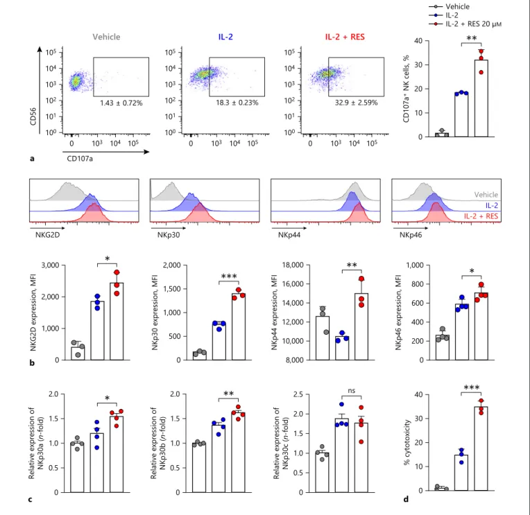
Effects of Resveratrol on NK Cell Activity and Activating Receptor Expression
We next examined the effects of resveratrol on NK cell degranulation activity by measuring CD107a expression as a functional marker of NK cell activation [22]. NK cells were co-incubated with K562 target cells, which do not express major histocompatibility complex (MHC) class I molecules and are susceptible to NK cell-mediated killing [23]. This was followed by analysis of surface levels of CD107a by flow cytometry. As shown in Figure 3a, after NK cells were pretreated with IL-2, NK cells expressing CD107a accounted for 18.3 ± 0.23% of all CD56+ NK
cells. CD107a expression on the surface of NK cells pre-treated with resveratrol and IL-2 increased 1.8-fold com-pared to that on the surface of IL-2-alone-pretreated cells, resulting in 32.9 ± 2.59% of CD56+ NK cells expressing
CD107a. Next, we measured the effects of resveratrol on the expression of NK cell-activating receptors which have central roles for NK cell cytotoxicity. As shown in Figure
3b, the expression of NK cell-activating receptors was sig-nificantly increased by resveratrol more than IL-2 alone. Since a previous study has shown immunomodulatory functions of NKp30 depending on its isoforms [24], we analyzed the change in the expression level of NKp30 iso-forms upon IL-2 with or without resveratrol treatment. While resveratrol did not affect the expression of NK-p30c, known as an inhibitory Nkp30 isoform [24], the activating isoforms including NKp30a as well as NKp30b were significantly increased by resveratrol (Fig. 3c). Alto-gether, these results suggest that resveratrol might en-hance overall cytotoxicity of NK cells. Hence, we exam-ined the effect of resveratrol on the cytotoxicity of NK cells. Consistent with previous studies, IL-2 treatment significantly enhanced the killing activity of NK cells (Fig. 3d). Intriguingly, co-treatment of resveratrol togeth-er with IL-2 furthtogeth-er heightened NK cytotoxicity by 21.3% compared with IL-2-treated cells at 1:1 ratio, indicating the increment of NK cytotoxicity by resveratrol (Fig. 3d).
0 100 200 300 400 500 *** *** *** IFN-γ, pg/mL 0 50 100 150 Cell proliferation , % Vehicle IL-2 IL-2 + RES Vehicle 0 5 10 20 +IL-2, µM b Vehicle 0 5 10 20 a +IL-2, µM 6 4 2 0 e Relative expre ssion of IFN-γ ( n-fold) *** Vehicle IL-2 + RES 20 µM IL-2 RES 20 µM 0 6 12 24 36 48 0 0.2 0.4 0.6 0.8 1.0 O.D values c ns 0 6 12 24 36 48 0 100 200 300 400 IFN-γ, pg/mL d * h h
Fig. 2. Resveratrol increased IL-2-induced IFN-γ secretion from
NK cells. a, b NK92 cells were treated with 0–20 μM resveratrol in the presence of 5 ng/mL IL-2 for 36 h. c, d NK92 cells were treated with 20 μM resveratrol for 0–48 h with or without 5 ng/ mL IL-2. e The mRNA levels of IFNG in NK92 cells after treat-ment of resveratrol for 24 h were measured by real-time PCR and
calculated by the ΔΔCT method. Gapdh was used as an internal control. Data are shown as mean ± SEM of 3 independent experi-ments. Asterisks indicate statistical significance by one- or two-way ANOVA: *p < 0.05; **p < 0.01; ***p < 0.001; ns, not significant (p > 0.05). RES, resveratrol; IL-2, interleukin-2; IFN-γ, interferon-gamma.
NKG2D NKp30 NKp44 NKp46
IL-2 + RESIL-2 Vehicle a 1.43 ± 0.72% CD56 100 101 102 103 104 105 105 CD107a 103 104 0 Vehicle 32.9 ± 2.59% 100 101 102 103 104 105 105 103 104 0 IL-2 + RES 18.3 ± 0.23% 100 101 102 103 104 105 105 103 104 0 IL-2 0 10 20 30 40 CD107a + NK cells, % ** Vehicle IL-2 IL-2 + RES 20 µM b 0 1,000 2,000 3,000 NKG2D expression, MFI * c 0 0.5 1.0 1.5 2.0
Relative expression of NKp30a (
n-fold) * 0 500 1,000 1,500 2,000 NKp30 expression, MFI *** 0 0.5 1.0 1.5 2.0 Relative expression of NKp30b ( n-fold) ** 8,000 10,000 12,000 14,000 16,000 18,000 NKp44 expression, MFI ** 0 0.5 1.0 1.5 2.0 2.5 Relative expression of NKp30c ( n-fold) ns 0 200 400 600 800 1,000 NKp46 expression, MFI * d 0 10 20 30 40 % cytotoxicity ***
Fig. 3. Effects of resveratrol on NK cell activity and activating re-ceptor expression. a CD107a expression on the surface of NK92 cells after resveratrol treatment was measured by flow cytometry. NK92 cells were treated with 20 μM resveratrol for 36 h, followed by co-culture with K562 target cells at a 1:1 ratio for 4 h. Quantita-tive analysis showing the percentages of CD56+ CD107a+ cells. Data are shown as mean ± SEM of 3 independent experiments. b Expression of natural cytotoxicity receptors and NKG2D was measured by flow cytometry. NK92 cells were treated with 20 μM resveratrol for 36 h. Data are shown as mean ± SEM of 3 indepen-dent experiments. c The mRNA levels of NKp30 isoforms in NK92
cells after treatment of resveratrol for 6 h were measured by real-time PCR and calculated by the ΔΔCT method. Gapdh was used as an internal control. Data are shown as mean ± SEM of 4 indepen-dent experiments. d NK cell cytotoxicity was determined using calcein-AM. NK92 effector (E) cells were treated with 20 μM res-veratrol for 36 h, followed by incubation with K562 target (T) cells at a 1:1 ratio for 4 h. Data are shown as mean ± SEM of 3 indepen-dent experiments. Asterisks indicate statistical significance by one-way ANOVA: *p < 0.05; **p < 0.01; ***p < 0.001; ns, not significant (p > 0.05). RES, resveratrol; IL-2, interleukin-2; NK cells, natural killer cells.
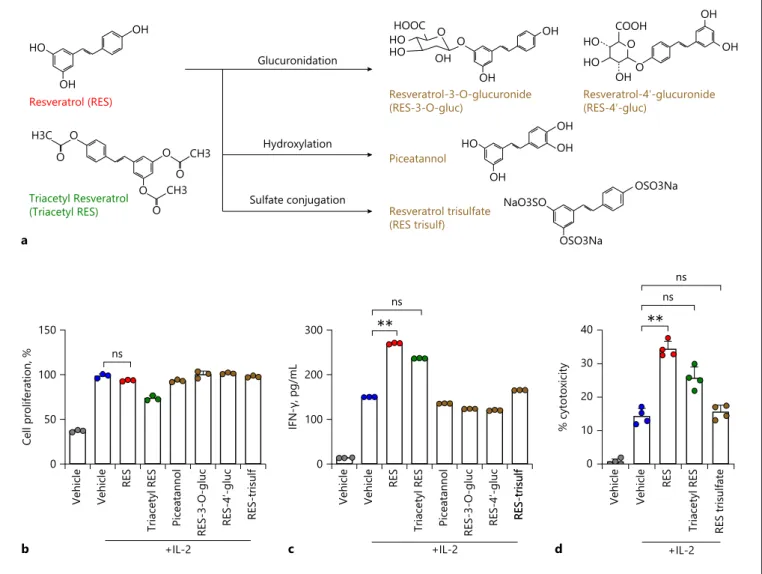
Effects of Derivatives and Metabolites of Resveratrol
Resveratrol is currently marketed as a dietary supple-ment. Triacetyl resveratrol, a derivative of resveratrol, is easily converted to resveratrol by esterase activity and ex-hibits similar bioactivity as resveratrol in cell cultures. Once resveratrol was absorbed, it is rapidly metabolized by conjugation to the hydroxyl group, glucuronic acid, and/or sulphate. The sites of glucuronidation in resvera-trol in the 3- and 4′-positions have been firmly
estab-lished after synthesis of the metabolites. Similarly, the major site of sulphate conjugation, in the 4′-position, was also established by synthesis (Fig. 4a) [25]. So, we inves-tigated whether the derivative or any of the metabolites of resveratrol could activate NK cells better than resveratrol itself. First, we investigated the effects of these com-pounds on NK cell proliferation. Triacetyl resveratrol, a derivative of resveratrol, slightly inhibited cell prolifera-tion, but the other compounds did not (Fig. 4b). Next, we
H3C O O O O CH3 O O CH3 OH OH HO OH NaO3SO OSO3Na OSO3Na OH OH HO Triacetyl Resveratrol (Triacetyl RES) Resveratrol-4ʹ-glucuronide (RES-4ʹ-gluc) Resveratrol-3-O-glucuronide (RES-3-O-gluc) Piceatannol Resveratrol trisulfate (RES trisulf) Glucuronidation Hydroxylation Sulfate conjugation a HOOC HO HO OH OH OH O O HO COOH HO OH OH OH O 0 100 200 300 IFN-γ, pg/mL c +IL-2 ** ns Vehicle Vehicle RES Tria cetyl RES Piceatannol RES-3-O-gluc RES-4 ʹ-gluc RES-trisulfRES-trisulf Vehicle Vehicle RES Tria cetyl RES Piceatannol RES-3-O-gluc RES-4 ʹ-gluc RES-trisulf 0 50 100 150 Cell prolifera tion, % b +IL-2 ns ** ns Vehicle Vehicle RES Tria cetyl RES RES trisulfate 0 10 20 30 40 % cytotoxicity d +IL-2 ns Resveratrol (RES) O
Fig. 4. Effects of derivatives and metabolites of resveratrol. a Me-tabolism of resveratrol and structures of resveratrol, triacetyl res-veratrol (derivative of resres-veratrol), and metabolites of resres-veratrol. Resveratrol is metabolized by hydroxylation or glucuronidation or sulphate conjugation to produce combined forms of sulphate and glucuronide. b, c Cell proliferation and IFN-γ secretion in NK92 cells after indicated treatments at 20 μM (with 5 ng/mL of IL-2) for 36 h were determined by CCK-8 and ELISA. Data are shown as
mean ± SEM of 3 independent experiments. d NK cell cytotoxicity was determined using calcein-AM. NK92 effector (E) cells were treated with 20 μM resveratrol or its metabolites for 36 h, followed by incubation with K562 target (T) cells at a 1:1 ratio ratio for 4 h. Data are shown as mean ± SEM of 4 independent experiments. Asterisks indicate statistical significance by one-way ANOVA: *p < 0.05; **p < 0.01; ***p < 0.001; ns, not significant (p > 0.05). IL-2, interleukin-2; IFN-γ, interferon-gamma; RES, resveratrol.
measured IFN-γ secretion in NK cells after treatment with the resveratrol derivative and metabolites. As shown in Figure 4c, the derivative and most of the metabolites did not affect IFN-γ secretion. Triacetyl resveratrol and resveratrol trisulphate increased IFN-γ secretion com-pared to that observed in IL-2-alone-treated cells, but their effect was weaker than that of resveratrol. Finally, we examined the effects of triacetyl resveratrol and resvera-trol trisulphate on NK cytotoxicity using K562 target cells and the calcein-AM method. As shown in Figure 4d, after treatment with resveratrol itself, NK cytotoxicity was in-creased by 20% at an E:T ratio of 1:1, compared to that of IL-2 treatment alone. After triacetyl resveratrol treat-ment, NK cytotoxicity was increased by 11.4% at an E:T ratio of 1:1, but not significant. Unlike resveratrol and triacetyl resveratrol, resveratrol trisulphate did not affect NK cytotoxicity. These results indicate that the intact form of resveratrol is the most effective for activating NK cells.
Anti-Cancer and Anti-Metastatic Effects of Resveratrol in Mice
We examined the in vivo efficacy of resveratrol ad-ministration in melanoma mouse models. In vivo activa-tion of NK cells by resveratrol and its anti-cancer effects were assessed by 2 different methods. First, 18 days after s.c. injection of B16F10 cells, tumour volumes were mea-sured after dissection. As shown in Figure 5a, adminis-tration of resveratrol (0.5 mg/kg) in the presence of IL-2 significantly inhibited tumour growth compared to the administration of IL-2 alone. Quantitative analysis showed that administration of IL-2 inhibited tumour growth compared to that in vehicle control mice. Fur-thermore, administration of resveratrol in the presence of IL-2 significantly inhibited tumour growth compared to the administration of IL-2 alone. Treatment of resve-ratrol together with IL-2 significantly inhibited tumour growth (Fig. 5a) accompanied with robust activation of NK cells in the tumour (Fig. 5b) as well as draining lymph
nodes (Fig. 5c) compared to the vehicle or IL-2-treated group.
Second, we investigated whether resveratrol affects tumour metastasis in the lung metastatic model as well. B16F10 cells were i.v. injected into mice; after 18 days, the lungs were extracted, and melanoma foci on lung sur-faces were counted. As shown in Figure 5d, the number of melanoma foci decreased in mice treated with resve-ratrol. Furthermore, IFN-γ production and degranula-tion levels were significantly enhanced in the lung resid-ual NK cells by the treatment of resveratrol compared with the vehicle or IL-2-treated group (Fig. 5e). These results suggest that resveratrol may prohibit tumour growth and metastasis by activating NK cells. Finally, to clarify the intrinsic roles of NK cells on anti-tumoural ef-fects of resveratrol, mice were injected with tumour and treated with vehicle, resveratrol, or resveratrol plus anti-NK1.1 to deplete NK cells in vivo. Intriguingly, as shown in Figure 5f, anti-tumoural effects of resveratrol was al-most completely abrogated by the depletion of NK cells. Consistent with previous results, resveratrol treatment with isotype antibody significantly reduced tumour growth. However, anti-tumoural effects of resveratrol were completely abrogated upon the depletion of NK cells (Fig. 5f). Altogether, these results indicate the piv-otal roles of NK cells for resveratrol-dependent anti-tu-moural effects.
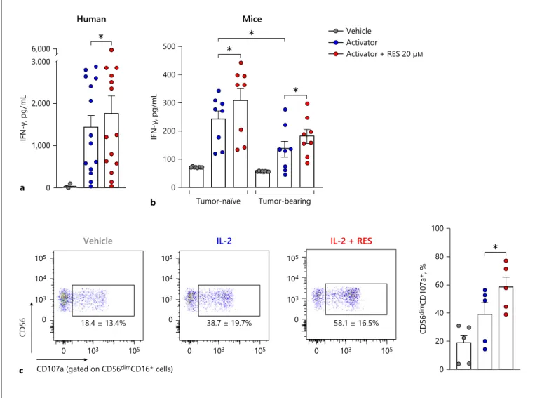
Ex vivo Effects of Resveratrol in Human and Mouse Whole Blood
Further, we investigated the effect of resveratrol on hu-man and mouse NK cell activity in whole blood using the NK Vue kit. After resveratrol treatment with the activa-tor, human NK cell activity increased by about 25% com-pared to treatment with the activator alone (Fig. 6a). Like-wise, after resveratrol treatment with the activator, tu-mour-naïve mouse NK cell activity increased by about 27.2% compared to treatment with the activator alone (Fig. 6b). Also, NK cell activity was measured in
tumour-Fig. 5. Anti-cancer and anti-metastatic effects of resveratrol in
mice. B16F10 cells (5 × 10⁵) in 0.2 mL PBS were injected subcuta-neously into C57BL/6 mice on day 0 (n = 14). a The tumour vol-ume was measured after dissection on day 18 after injection of melanoma cells. The activity of NK cells isolated from tumour (b) and draining lymph node (c) was evaluated by flow cytometry. B16F10 cells (1 × 10⁵) in 0.1 mL PBS were injected into the tail veins of C57BL/6 mice on day 0 (n = 10). d The melanoma foci on the lung surface were counted macroscopically 18 days after injec-tion of melanoma cells. e The activity of NK cells isolated from the lungs was evaluated by flow cytometry. f To deplete NK cells,
25 μg anti-NK1.1 monoclonal antibody (PK136) or control mouse IgG2a was injected intraperitoneally (i.p.) on days −3, 2, and 8. B16F10 cells (5 × 10⁵ cells) in 0.2 mL of PBS were injected subcu-taneously into the right flanks of mice on day 0. The tumour vol-ume was measured after dissection on day 18 after injection of melanoma cells. To evaluate the activity of resveratrol, mice were intravenously injected a total of 6 times, on days −2, 0, 2, 4, 6, and 8. Asterisks indicate statistical significance by one-way ANOVA: *p < 0.05; **p < 0.01; ***p < 0.001; ns, not significant (p > 0.05). RES, resveratrol; IL-2, interleukin-2; IFN-γ, interferon-gamma;
a
Velcihe IL-2 IL-2 + RES
0 500 1,000 1,500 2,000 Tumor volume, mm 3 IL-2 IL-2 + RES 20 µM Vehicle ** d
Vehicle IL-2 IL-2 + RES
0 20 40 60 80 100
Number of melanoma spot
* Tumor-infiltrating NK cells b 0 20 40 60 80 100 IFN-γ + NK cells, % * 0 20 40 60 80 CD107a + NK cells, % * e 0 20 40 60 80 100 IFN-γ + NK cells, % *** 0 10 20 30 CD107a + NK cells, % ***
Draining lymph node
c 0 20 40 60 80 100 IFN-γ + NK cells, % * 0 20 40 60 80 CD107a + NK cells, %
* Iso + Vehicle Iso + IL-2 + RES aNK1.1 + IL-2 + RES
f
Iso + Vehicle Iso + IL-2 + RES
aNK1.1 + IL-2 + RES
0 100 200 300 400 Tumor volume, mm 3 ns ** * 5
bearing mouse models. Even in the presence of an activa-tor, NK cell activity was significantly lower in the tumour-bearing mouse than in the tumour-naïve group (Fig. 6b). However, after resveratrol treatment in the presence of the activator, NK cell activity increased by about 27.3% compared to treatment with the activator alone in the tu-mour-bearing mouse (Fig. 6b). Finally, PBMCs were iso-lated from whole blood and treated with resveratrol for 24 h in the presence IL-2 and then stimulated by K562
target cells (Fig. 6c). Intriguingly, compared with IL-2-treated cells, we found that resveratrol significantly en-hanced degranulation in CD56dimCD16+ human primary
NK cells known for their cytolytic activity (Fig. 6c) [26]. Thus, these results indicate that resveratrol has potential to activate human primary NK cells and suggest that res-veratrol could be used in cancer therapy by activating NK cells. a 0 1,000 2,000 3,000 6,000 IFN-γ, pg/mL Human * b Tumor-naïve Tumor-bearing 0 100 200 300 400 500 IFN-γ, pg/mL Mice Activator + RES 20 µM Activator Vehicle * * * 0 20 40 60 80 100 CD56 dim CD107a +, % *
CD107a (gated on CD56dimCD16+ cells)
CD56 103 105 0 18.4 ± 13.4% c 0 103 104 105 38.7 ± 19.7% 103 105 0 0 103 104 105 58.1 ± 16.5%
Vehicle IL-2 IL-2 + RES
103 105 0 0 103 104 105
Fig. 6. Ex vivo effects of resveratrol in human and mouse whole
blood. a For the human NK cell activity assay, 1 mL of whole blood was incubated with or without the activator and resveratrol for 24 h at 37°C. b For the mouse NK cell activity assay, 100 μL of whole blood was incubated with or without 30 μL activator and resveratrol for 24 h at 37°C (tumour-naïve or tumour-bearing mouse model, n = 8). The supernatant was collected, and the re-leased IFN-γ was measured using the NK Vue kit. c CD107a
ex-pression of primary NK cells was measured by flow cytometry. PBMCs were treated with 20 μM resveratrol for 24 h in the presence of IL-2, followed by co-culture with K562 target cells at a 5:1 (E:T) ratio for 4 h (n= 5). Asterisks indicate statistical significance by Dunn’s multiple comparison test: *p < 0.05; **p < 0.01; ***p < 0.001; ns, not significant (p > 0.05). IL-2, interleuk2; IFN-γ, in-terferon-gamma; RES, resveratrol.
Discussion
Resveratrol is a natural compound and antioxidant found in peanuts, soy, hop, and various fruits, especially grapes and red wines [27, 28]. Resveratrol possesses vari-ous biological activities, including anti-tumour, anti-vi-ral, anti-inflammation, anti-fungal, and antibacterial ef-fects [27, 29–32]. We have demonstrated that resveratrol increases the expression of activating receptors on NK cells (Fig. 3). Interestingly, we found that resveratrol + IL-2 significantly increase NKp30 more than IL-2 alone. Especially, NKp30a and NKp30b isoforms that have im-mune-stimulatory roles were significantly increased by resveratrol more than IL-2 alone. One of the proposed approaches for tumour therapy is enhancing tumour sen-sitivity to NK cell cytolysis by upregulating the NKp30 ligand B7-H6 on tumour cells using stress inducers [33]. Moreover, preclinical approaches involve increasing tu-mour sensitivity to NK cells by upregulating the expres-sion of NKG2D ligands on cancer cells using HDAC in-hibitors [34]. Enhancement of cell surface expression of NKG2D ligands and DR4/5 on various cancer cells by res-veratrol increases TRAIL sensitivity and could facilitate NK cell-mediated killing of cancer cells [35, 36]. How-ever, there are no reports that resveratrol also increases B7-H6 expression on cancer cells. If resveratrol did in-duce the expression of B7-H6 on cancer cells, it would be a good candidate for an anti-cancer drug, acting as both an immune stimulator for NK cells and a stress inducer for cancer cells.
NK cells require IL-2 to proliferate, and resveratrol treatment without IL-2 did not affect IFN-γ secretion or NK cytotoxicity in vitro (Fig. 2). IL-2 is capable of activat-ing NK cells and is used as an anti-cancer drug in immu-notherapy [37]. According to a recent report, resveratrol has shown promising results in adjuvant therapy [27]. Resveratrol in combination with 5-fluoruracyl or cispla-tin exhibited synergistic effects in the induction of apop-tosis in cancer cells in vitro and in vivo [38, 39]. There-fore, resveratrol can be expected to exert a greater effect in combination with IL-2.
Resveratrol shows a concentration-dependent bipha-sic effect [40]. Low doses of resveratrol reportedly activate NK cells, while high doses reportedly promote cell apop-tosis via the caspase signalling pathway [40]. In one study, NK cell killing activity was enhanced at low concentra-tions of resveratrol ranging from 0.33 to 5.48 μM, with a maximum activity at 1.31 μM; however, NK cell killing activity was inhibited at higher concentrations of 21.92 and 87.68 μM resveratrol [41]. In another study by Lu and
Chen [42], 12.5 μM resveratrol induced the highest NK cell killing activity. In our study, resveratrol did not sig-nificantly affect NK cell proliferation in vitro, but it did increase IFN-γ secretion in a dose-dependent manner up to a concentration of 20 μM. We also found that resvera-trol increased IFN-γ secretion ex vivo at a concentration of 20 μM and did not affect survival when injected into mice at 0.5 mg/kg (data not shown).
Resveratrol is currently being sold as a dietary supple-ment. Resveratrol is first detectable in plasma 15 min after oral administration, reaching its peak concentra-tion after 30 min, and it is quickly metabolized in the intestine and liver into glucuronide and sulphate conju-gates [43]. According to a study on the absorption of wine-related polyphenols, the highest serum level of to-tal resveratrol (free and conjugated) was recorded after consumption of 25-mg trans-resveratrol per 70 kg body weight in 3 different matrices (wine, grape juice, and vegetable juice) [44]. Free trans-resveratrol accounted for 1.7–1.9% of the peak total serum concentration. This is considerably lower than the concentrations tested in vitro showing beneficial effects. Moreover, resveratrol is rapidly metabolized in the intestine by phase II enzymes, so it is very difficult to reach pharmacologically effective systemic plasma concentrations, which might signifi-cantly restrict the bioavailability of resveratrol [45, 46]. Our data showed that IFN-γ secretion increased upon treatment with intact resveratrol but not with its me-tabolites (Fig. 4). Accordingly, resveratrol would be more effective when administered by injection rather than by diet.
Acknowledgements
The authors would like to thank Si-Hwan Ko (Yonsei Univer-sity College of Medicine) for his technical assistance.
Statement of Ethics
Blood was obtained from healthy donors after acquiring insti-tutional review board approval and informed consent (No. 4-2019-0007). Animal experiments were approved by the Yonsei Univer-sity Health System Institutional Animal Care and Use Committee (YUHS-IACUC).
Conflict of Interest Statement
Funding Sources
This study was supported in part by the National Research Foundation of Korea (NRF) Grant (2016-31-0446) funded by the Korean government (MEST).
Author Contributions
J. Kim: conceptualization, design, supervision, project admin-istration, funding acquisition, interpretation of data, and final ap-proval of the manuscript; Y. Lee: conceptualization, design, inves-tigation, interpretation of data, validation, visualization, and man-uscript writing; H. Shin: design and interpretation of data. References
1 Cerwenka A, Lanier LL. Natural killer cells, viruses and cancer. Nat Rev Immunol. 2001 Oct;1(1):41–9.
2 Vivier E, Tomasello E, Baratin M, Walzer T, Ugolini S. Functions of natural killer cells. Nat Immunol. 2008 May;9(5):503–10.
3 Guillerey C, Huntington ND, Smyth MJ. Tar-geting natural killer cells in cancer immuno-therapy. Nat Immunol. 2016 Aug 19;17(9): 1025–36.
4 Jung YS, Park JH, Park DI, Sohn CI, Lee JM, Kim TI. Physical inactivity and unhealthy metabolic status are associated with decreased natural killer cell activity. Yonsei Med J. 2018 Jun;59(4):554–62.
5 Kim HR, Lee KH, Park SJ, Kim SY, Yang YK, Tae J, et al. Anti-cancer activity and mecha-nistic features of a NK cell activating mole-cule. Cancer Immunol Immunother. 2009 Oct;58(10):1691–700.
6 Glasner A, Ghadially H, Gur C, Stanietsky N, Tsukerman P, Enk J, et al. Recognition and prevention of tumor metastasis by the NK re-ceptor NKp46/NCR1. J Immunol. 2012 Mar 15;188(6):2509–15.
7 Morvan MG, Lanier LL. NK cells and cancer: you can teach innate cells new tricks. Nat Rev Cancer. 2016 Jan;16(1):7–19.
8 Zwirner NW, Domaica CI. Cytokine regula-tion of natural killer cell effector funcregula-tions. Biofactors. 2010 Jul–Aug;36(4):274–88. 9 Koch J, Steinle A, Watzl C, Mandelboim O.
Activating natural cytotoxicity receptors of natural killer cells in cancer and infection. Trends Immunol. 2013 Apr;34(4):182–91. 10 Abel AM, Yang C, Thakar MS, Malarkannan
S. Natural killer cells: development, matura-tion, and clinical utilization. Front Immunol. 2018;9:1869.
11 Smyth MJ, Hayakawa Y, Takeda K, Yagita H. New aspects of natural-killer-cell surveillance and therapy of cancer. Nat Rev Cancer. 2002 Nov;2(11):850–61.
12 Wang R, Jaw JJ, Stutzman NC, Zou Z, Sun PD. Natural killer cell-produced IFN-γ and TNF-α induce target cell cytolysis through up-regulation of ICAM-1. J Leukoc Biol. 2012 Feb;91(2):299–309.
13 Vivier E, Raulet DH, Moretta A, Caligiuri MA, Zitvogel L, Lanier LL, et al. Innate or adaptive immunity? The example of natural killer cells. Science. 2011 Jan 7;331(6013):44–9.
14 Dempke WCM, Fenchel K, Uciechowski P, Dale SP. Second- and third-generation drugs for immuno-oncology treatment: the more the better? Eur J Cancer. 2017 Mar;74:55–72.
15 Sharma P, Hu-Lieskovan S, Wargo JA, Ribas A. Primary, adaptive, and acquired resistance to cancer immunotherapy. Cell. 2017 Feb 9; 168(4):707–23.
16 Chiossone L, Dumas PY, Vienne M, Vivier E. Natural killer cells and other innate lymphoid cells in cancer. Nat Rev Immunol. 2018 Nov; 18(11):671–88.
17 Van Schandevyl S, Kerre T. Chimeric antigen receptor T-cell therapy: design improvements and therapeutic strategies in cancer treatment. Acta Clin Belg. 2018 Nov 13;75(1):26–32. 18 Buzio C, Andrulli S, Santi R, Pavone L,
Pas-salacqua R, Potenzoni D, et al. Long-term im-munotherapy with low-dose interleukin-2 and interferon-alpha in the treatment of pa-tients with advanced renal cell carcinoma. Cancer. 2001 Nov 1;92(9):2286–96.
19 Szczepanski MJ, Szajnik M, Welsh A, Foon KA, Whiteside TL, Boyiadzis M. Interleukin-15 en-hances natural killer cell cytotoxicity in patients with acute myeloid leukemia by upregulating the activating NK cell receptors. Cancer Immu-nol Immunother. 2010 Jan;59(1):73–9. 20 Zhu EF, Gai SA, Opel CF, Kwan BH, Surana
R, Mihm MC, et al. Synergistic innate and adaptive immune response to combination immunotherapy with anti-tumor antigen an-tibodies and extended serum half-life IL-2. Cancer Cell. 2015 Apr 13;27(4):489–501. 21 Khuda-Bukhsh AR, Das S, Saha SK.
Molecu-lar approaches toward targeted cancer pre-vention with some food plants and their prod-ucts: inflammatory and other signal path-ways. Nutr Cancer. 2014;66(2):194–205. 22 Alter G, Malenfant JM, Altfeld M. CD107a as
a functional marker for the identification of natural killer cell activity. J Immunol Meth-ods. 2004 Nov;294(1–2):15–22.
23 Lozzio BB, Lozzio CB. Properties and useful-ness of the original K-562 human myeloge-nous leukemia cell line. Leuk Res. 1979;3(6): 363–70.
24 Delahaye NF, Rusakiewicz S, Martins I, Mé-nard C, Roux S, Lyonnet L, et al. Alternatively spliced NKp30 isoforms affect the prognosis of gastrointestinal stromal tumors. Nat Med. 2011 Jun;17(6):700–7.
25 Walle T. Bioavailability of resveratrol. Ann N Y Acad Sci. 2011 Jan;1215:9–15.
26 De Maria A, Bozzano F, Cantoni C, Moretta L. Revisiting human natural killer cell subset function revealed cytolytic CD56(dim) CD16+ NK cells as rapid producers of abun-dant IFN-gamma on activation. Proc Natl Acad Sci U S A. 2011 Jan 11;108(2):728–32.
27 Varoni EM, Lo Faro AF, Sharifi-Rad J, Iriti M. Anticancer molecular mechanisms of resve-ratrol. Front Nutr. 2016;3:8.
28 Wiciński M, Socha M, Walczak M, Wódkie-wicz E, Malinowski B, Rewerski S, et al. Ben-eficial effects of resveratrol administration-focus on potential biochemical mechanisms in cardiovascular conditions. Nutrients. 2018 Nov 21;10(11):1813.
29 Kundu JK, Surh YJ. Cancer chemopreventive and therapeutic potential of resveratrol: mechanistic perspectives. Cancer Lett. 2008 Oct 8;269(2):243–61.
30 Lu CC, Lai HC, Hsieh SC, Chen JK. Resvera-trol ameliorates Serratia marcescens-induced acute pneumonia in rats. J Leukoc Biol. 2008 Apr;83(4):1028–37.
31 Wang XL, Li T, Li JH, Miao SY, Xiao XZ. The effects of resveratrol on inflammation and ox-idative stress in a rat model of chronic ob-structive pulmonary disease. Molecules. 2017 Sep 12;22(9):1529.
32 Zhao X, Tong W, Song X, Jia R, Li L, Zou Y, et al. Antiviral effect of resveratrol in piglets infected with virulent pseudorabies virus. Vi-ruses. 2018 Aug 27;10(9):457.
33 Cao G, Wang J, Zheng X, Wei H, Tian Z, Sun R. Tumor therapeutics work as stress inducers to enhance tumor sensitivity to natural killer (NK) cell cytolysis by up-regulating NKp30 ligand B7-H6. J Biol Chem. 2015 Dec 11; 290(50):29964–73.
34 Spear P, Wu MR, Sentman ML, Sentman CL. NKG2D ligands as therapeutic targets. Can-cer Immun. 2013;13:8.
35 Fulda S, Debatin KM. Resveratrol-mediated sensitisation to TRAIL-induced apoptosis de-pends on death receptor and mitochondrial sig-nalling. Eur J Cancer. 2005 Mar;41(5):786–98. 36 Hu L, Cao D, Li Y, He Y, Guo K. Resveratrol
sensitized leukemia stem cell-like KG-1a cells to cytokine-induced killer cells-mediated cytolysis through NKG2D ligands and TRAIL receptors. Cancer Biol Ther. 2012 May;13(7):516–26. 37 Burns LJ, Weisdorf DJ, DeFor TE, Vesole DH,
Repka TL, Blazar BR, et al. IL-2-based immu-notherapy after autologous transplantation for lymphoma and breast cancer induces im-mune activation and cytokine release: a phase I/II trial. Bone Marrow Transplant. 2003 Jul; 32(2):177–86.
38 Cosco D, Paolino D, Maiuolo J, Marzio LD, Carafa M, Ventura CA, et al. Ultradeformable liposomes as multidrug carrier of resveratrol and 5-fluorouracil for their topical delivery. Int J Pharm. 2015 Jul 15;489(1–2):1–10.
39 Ma L, Li W, Wang R, Nan Y, Wang Q, Liu W, et al. Resveratrol enhanced anticancer effects of cisplatin on non-small cell lung cancer cell lines by inducing mitochondrial dysfunction and cell apoptosis. Int J Oncol. 2015 Oct; 47(4):1460–8.
40 Li Q, Huyan T, Ye LJ, Li J, Shi JL, Huang QS. Concentration-dependent biphasic effects of resveratrol on human natural killer cells in vi-tro. J Agric Food Chem. 2014 Nov 12;62(45): 10928–35.
41 Falchetti R, Fuggetta MP, Lanzilli G, Tricarico M, Ravagnan G. Effects of resveratrol on hu-man immune cell function. Life Sci. 2001 Nov 21;70(1):81–96.
42 Lu CC, Chen JK. Resveratrol enhances perfo-rin expression and NK cell cytotoxicity through NKG2D-dependent pathways. J Cell Physiol. 2010 May;223(2):343–51.
43 Lee AM, Shandala T, Nguyen L, Muhlhausler BS, Chen KM, Howe PR, et al. Effects of res-veratrol supplementation on bone growth in young rats and microarchitecture and remod-eling in ageing rats. Nutrients. 2014 Dec 16; 6(12):5871–87.
44 Goldberg DM, Yan J, Soleas GJ. Absorption of three wine-related polyphenols in three dif-ferent matrices by healthy subjects. Clin Bio-chem. 2003 Feb;36(1):79–87.
45 Wenzel E, Somoza V. Metabolism and bio-availability of trans-resveratrol. Mol Nutr Food Res. 2005 May;49(5):472–81.
46 Leischner C, Burkard M, Pfeiffer MM, Lauer UM, Busch C, Venturelli S. Nutritional immu-nology: function of natural killer cells and their modulation by resveratrol for cancer preven-tion and treatment. Nutr J. 2016 May 4;15(1):47.