68
Korean J Ophthalmol Vol.29, No.1, 2015
Recurred Adenoid Cystic Carcinoma
of Lacrimal Gland with Aggressive
Local Invasion to the Maxillary Bone
Marrow without Increased Uptake in
PET-CT
Dear Editor,
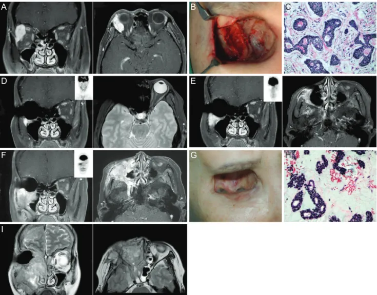
This report describes a patient with locally aggressive ad-enoid cystic carcinoma (ACC) of the lacrimal gland that re-curred after neoadjuvant intra-arterial chemotherapy (IAC), local surgery and postoperative radiotherapy. A 47-year-old female presented with painless swelling of her right eyelid that had persisted for 3 months. Physical examination re-vealed 3-mm exophthalmos of the right eye. Orbit magnetic resonance imaging (MRI) with contrast revealed an ap-proximately 3-cm-sized enhancing lesion in the right lacri-mal gland with bony invasion (Fig. 1A). Incisional biopsy verified ACC. Neoadjuvant intracarotid adriamycin and cis-platinum was started, but the patient was switched to the intravenous regimen due to facial swelling and tender-ness. After two chemoreduction cycles, the patient under-went orbital exenteration. Because the resected margin of the lateral exenterated orbit and zygoma were positive for carcinoma, additional lateral zygoma bone was removed (Fig. 1B). The pathology report verified T4bN0M0 stage ACC, cribriform type, with lymphovascular invasion (Fig. 1C). The patient then received postoperative 59.4 Gy radio-therapy. An MRI taken 1 year after exenteration revealed an approximately 7-mm, focal enhancing lesion in the right anterior maxilla bone, suggestive of possible tumor recur-rence. However, positron emission tomography-computed tomography (PET-CT) did not show increased uptake of fludeoxyglucose (FDG) (Fig. 1D). The patient refused to have a bone biopsy. A repeat MRI performed 2 years after surgery indicated that there was an increase in the extent of the enhancing lesion inferior to the right orbit and right pterygomaxillary fissure and suspicious tumor spread along the right foramen rotundum, but again no increased FDG uptake was observed on PET-CT (Fig. 1E). A bone biopsy was recommended which the patient refused. A PET-CT taken 3 years postoperatively again showed a stable disease state, however, the patient presented with an elevated
pig-mented mass along the inferolateral margin of the exenter-ated orbit after 6 months. An MRI revealed a markedly in-creased infiltrating mass along the entire maxillary sinus wall with perineural spread to the cavernous sinus via the foramen rotundum. At that time, PET-CT finally showed consistent findings of high FDG uptake (Fig. 1F). An exci-sional biopsy of the elevated pigmented lesion (Fig. 1G) confirmed the pathology as recurred ACC (Fig. 1H). The patient was referred to the head and neck surgery depart-ment and a salvage operation was planned, however the pa-tient refused to undergo the operation and she did not re-turn until she presented to the emergency room in a confused mental status with blindness in her left eye a year later. An MRI revealed an increased right maxillary sinus mass with intracranial extension, brain edema, and infiltra-tion into the left orbital apex compressing the optic nerve (Fig. 1I). Upon refusal of further treatment, the patient died within 1 month.
ACC is an aggressive malignant neoplasm with locore-gional recurrence being the most common cause of disease progression and death [1]. Surgery and radiotherapy are the conventional treatment modalities, but whether the addition of IAC improves survival and whether the risk of potential toxicities should be taken remain controversial. Tse et al. [2] reported that the cumulative 5-year carcinoma cause-specific death rate was 16.7% in the group treated with IAC and exenteration. In contrast, Fellman et al. [3] reported 2 cases of ACC recurrence and distant metastasis after IAC followed by surgery and high-dose radiation therapy. We report a case of recurring ACC that involved the contral-ateral eye and brain, despite multimodality treatment. The condition was not detectable on PET-CT scan for several years. ACC has been reported to be FDG avid, manifesting low FDG uptake compared to size-matched squamous cell carcinoma [4]. Cribriform or tubular histologic subtypes show even lower FDG uptake compared to the solid pattern, and therefore physicians should be meticulous when interpreting PET-CT results in ACC patients.
The locally aggressive nature of the cancer and the incon-sistent radiologic findings [5] which was apparent in this case highlight the need for serial MRI follow-up for moni-toring of disease progression, for prompt biopsy when re-currence is suspected, and for radical local control of the disease. Also, the potential complications of IAC, such as facial tenderness and swelling that were experienced by our patient, and other factors that lead to the optimal treatment
Korean J Ophthalmol 2015;29(1):68-70 http://dx.doi.org/10.3341/kjo.2015.29.1.68
69
response, should be taken into consideration when deciding the treatment regimen for ACC.
Moonjung Choi
Department of Ophthalmology, Severance Hospital, Institute of Vision Research, Yonsei University College of Medicine, Seoul, Korea
Ja Seung Koo
Department of Pathology, Yonsei University College of Medicine, Seoul, Korea
Jin Sook Yoon
Department of Ophthalmology, Severance Hospital, Institute of Vision Research, Yonsei University College of Medicine, Seoul, Korea
E-mail: yoonjs@yuhs.ac
Fig. 1. (A) Coronal and axial views of orbit magnetic resonance imaging (MRI) taken preoperatively, revealing an approximately 3-cm-sized enhancing lesion in the right lacrimal gland with bony invasion. (B) Photograph of the exposed lateral zygoma bone during operation which was pathologically proven to have cancer infiltration. (C) Histologic section of primary cancer of stage T4bN0M0, cribriform type with lymphovascular invasion (H&E, ×100). (D) Coronal and axial views of orbit MRI with positron emission tomog-raphy-computed tomography (PET-CT) scan contrast coronal images in the middle taken 1 year after exenteration, showing a focal en-hancing lesion in the right anterior maxilla bone without uptake in the PET-CT scan. (E) Two years postoperatively, with increased extent of the enhancing lesion, yet still no visible uptake in the PET-CT scan. (F) Three and half years after local surgery, showing a markedly increased infiltrating mass along the entire maxillary sinus wall with corresponding fludeoxyglucose uptake in the PET-CT scan. (G) Photograph of an elevated pigmented mass along the inferolateral margin of the exenterated orbit 3.5 years after exenteration. (H) Histo-logic section of recurred cancer (H&E, ×100). The pathoHisto-logic grade was not different, but the proportion of the mesenchymal component differed in that the primary cancer contained more fibrous components whereas the recurred cancer had more myxoid components. (I) Coronal and axial views of orbit MRI 4.5 years postoperatively, showing the right maxillary sinus mass with intracranial extension, brain edema, and infiltration into the left orbital apex.
A D F I B E G C H
70
Korean J Ophthalmol Vol.29, No.1, 2015
Conflict of Interest
No potential conflict of interest relevant to this article was reported.
References
1. Esmaeli B, Ahmadi MA, Youssef A, et al. Outcomes in pa-tients with adenoid cystic carcinoma of the lacrimal gland.
Ophthal Plast Reconstr Surg 2004;20:22-6.
2. Tse DT, Kossler AL, Feuer WJ, Benedetto PW. Long-term outcomes of neoadjuvant intra-arterial cytoreductive
che-motherapy for lacrimal gland adenoid cystic carcinoma.
Ophthalmology 2013;120:1313-23.
3. Fellman M, Carter K, Call CB, Esmaeli B. Disease recur-rence after intraarterial chemotherapy in 2 patients with ad-enoid cystic carcinoma of lacrimal gland. Can J Ophthalmol 2013;48:e17-8.
4. Lee JY, Choi JY, Ko YH, et al. 18F-FDG PET/CT in pa-tients with initially diagnosed adenoid cystic carcinoma of the head and neck: clinicoplathologic correlation. Nucl Med
Mol Imaging 2009;43:395-401.
5. Qin W, Chong R, Huang X, et al. Adenoid cystic carcinoma of the lacrimal gland: CT and MRI findings. Eur J
Oph-thalmol 2012;22:316-9.
Successful Treatment of Orbital
Lymphangioma with Intralesional
Bleomycin and Application of Cont i
n uous Negative Pressure
Dear Editor,
Recently, we experienced a case of pediatric orbital lymphangioma successfully treated with intralesional bleo-mycin injection. To our knowledge, this is the first such case report in Korea of this treatment used for this condition.
An 8-year-old male visited our clinic complaining of eyelid swelling and proptosis in his left eye for 5 days (Fig. 1A and 1B). He was diagnosed with an external stye and was administered antibiotics 4 days ago, but showed no improvement. The best-corrected visual acuity of the left eye was 20 / 30 and intraocular pressure was 32 mmHg. The patient complained of severe subconjunctival hemor-rhage and pain. Initial computed tomography (CT) and magnetic resonance (MR) images showed a 22.2 × 25.4 mm-sized macrocystic lymphangioma in the left infero-temporal orbit (Fig. 1C and 1D).
An inferior lower eyelid incision was made to facilitate the approach to the macrocyst. An opening was made on the cystic wall and the internal wall of the cyst was ex-posed using Sewall retractors. Blood evacuation was
per-formed with a suction catheter. Bleomycin (1 IU per milli-liter saline) was injected into the cyst for exactly 5 minutes (Fig. 1E). Barovac (100 mL; Sewoon Medical, Seoul, Ko-rea) was inserted into the cyst to provide continuous nega-tive pressure to induce cyst shrinkage and reduce the risk of delayed or recurrent bleeding (Fig. 1F). On the third day after the operation, the visual acuity had returned to 20 / 20, and intraocular pressure remained at 20 mmHg. At this point, 10 mg of prednisolone was administered for 2 weeks with a tapering schedule. At 1 month of follow-up, the pa-tient’s symptoms and signs were completely resolved (Fig. 1G and 1H). CT and MR imaging results showed no resid-ual macrocystic lymphangioma, and there was no sign of bleeding (Fig. 1I and 1J). There were also no signs of re-currence at 6 months of follow-up.
Lymphangioma is a congenital malformation of the lym-phatic system that frequently occurs in the head and neck [1]. Orbital lymphangioma manifests in proptosis and re-striction of ocular motility following minor trauma or up-per respiratory tract infection. Its mass effect may lead to compressive optic neuropathy or exposure keratopathy. Surgery is considered when there are symptoms such as de-creased visual acuity, amblyopia, and severe cosmetic dis-figurement. However, complete resection of the lesion is difficult to achieve, increasing the risk of local recurrence and complications involving injury to surrounding struc-tures. Multiple treatment modalities including radiotherapy, intralesional/oral corticosteroid and intralesional sclerosing therapy can be adopted in conjunction with surgery [1].
Korean J Ophthalmol 2015;29(1):70-72 http://dx.doi.org/10.3341/kjo.2015.29.1.70