www.e-arms.org 23 Neurofibromatosis type 1 (NF1) leads to a greater risk of
de-velopment of malignancies, such as leukemia, gliomas, and ma-lignant peripheral nerve sheath tumors (MPNSTs).1
In some cross-sectional studies, the condition in approximately 1% to 2% of patients with NF1 progresses to MPNSTs.2
The most effec-tive treatment for MPNST is ‘early diagnosis and fast surgery’ performed by plastic surgeons. However, early MPNST diag-nosis in patients with NF1 is difficult because of the frequent accompaniment with preexisting conditions and growths of additional masses.
CASE REPORT
A 54-year-old woman with a history of NF1 came to our out-patient department with newly growing masses on her left up-per arm and forearm that had been growing at the same time for several months (Fig. 1). The masses were painless with
ulcer-ative bleeding in the forearm mass, it occurred 1 week ago with no neurologic symptoms. On enhanced computed tomography
A Case of Malignant Peripheral Nerve Sheath Tumor with
Neurofibromatosis Type 1
Sang Kyu Choi, Cheol Keun Kim, Soon Heum Kim, Dong In Jo*
Department of Plastic and Reconstructive Surgery, Konkuk University Chungju Hospital, Konkuk University School of Medicine, Chungju, Korea
CC This is an open-access article distributed under the terms of the Creative Commons Attribution Non-Commercial License (http://creativecommons.org/licenses/by-nc/4.0) which permits
unrestricted noncommercial use, distribution, and reproduction in any medium, provided the original work is properly cited. Copyright © 2017 by the Korean Society for Microsurgery. All Rights Reserved.
Received April 29, 2017 Accepted April 29, 2017 *Correspondence to: Dong In Jo Department of Plastic and Reconstructive Surgery, Konkuk University Chungju Hospital, Konkuk University School of Medicine, 82 Gugwon-daero, Chungju 27376, Korea
Tel: +82-43-840-8860 Fax: +82-43-840-8962 E-mail: [email protected]
ORCID: http://orcid.org/0000-0002-3075-4482 Financial support: None.
Conflict of interest: None.
The malignant peripheral nerve sheath tumor (MPNST) originates from neurofibromatosis type 1 (NF1). Because NF1 patients have many accompaniments with growth of additional masses, they usually overlook potential malignant changes in their masses. Our patient had two growing mass near the left elbow for several months; however, she ignored these masses until 7 days prior to writing this article, at which time they began bleeding. Tra-ditionally, sarcoma including MPNST treatment consisted of amputation of the involved extremity. However, treatment now consists of surgical resection with adjuvant therapy. Therefore, we conducted resection of the mass and subsequent coverage with a local ad-vancement flap. We believe that the most effective treatment for MPNST is early diagnosis and fast surgery, coupled with notification that there is always potential for malignant change in NF1 patient’s masses.
Key Words: Malignant peripheral nerve sheath tumor, Diagnosis, Treatment, Chemotherapy
ARMS
Archieves of Reconstructive MicrosurgeryCase Report
pISSN 2383-5257 eISSN 2288-6184 Arch Reconstr Microsurg 2017;26(1):23-25 https://doi.org/10.15596/ARMS.2017.26.1.23
Fig. 1. A 54-year-old woman presented with newly growing masses on her left upper arm and forearm that had been growing at the same time for several months.
Arch Reconstr Microsurg Vol. 26. No. 1. May 2017
24
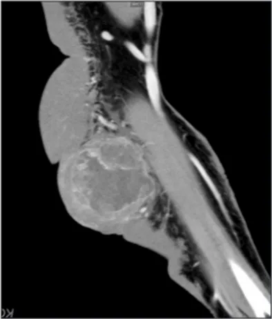
(CT), the forearm mass was a lobulated, cystic lesion with het-erogeneous enhancement, and dilatation of radial artery was seen (Fig. 2). Malignancy could not be ruled out on CT. Surgi-cal excision was subsequently performed. Under general anes-thesia, the masses were excised. The upper arm mass measured 15×15×7 cm (Fig. 3). Because it was not encapsulated, debulk-ing surgery was performed. The remaindebulk-ing defect was covered via local advancement flap. The forearm mass, including the fas-cial layer, was resected. The safety margins were made approxi-mately 2 cm from the mass contour. The mass was firm and round, and it measured 7×7×6 cm (Fig. 4). Histopathologically, the forearm mass was diagnosed as high-grade sarcoma with possible MPNST with frequent mitosis (about 53 mitosis/10 high-power fields) and necrosis (<60%). The forearm mass margins contained clear cancer cells. The upper arm mass was
diagnosed as a pigmented neurofibroma. The margins of the excised upper arm mass involved a benign nerurofibroma (Fig. 5). A whole-body positron emission tomography/computed tomography (PET/CT) was performed to detect any possible metastasis. 18F-fluorodeoxyglucose uptake was increased in operative sites. Abnormal uptake and suspicious metastatic le-sions were not observed. After postoperative day 14, the patient was discharged without complications. During the 6-month follow-up period, the patient received radiotherapy and 5 cycles of chemotherapy (doxorubicin, ifosfamide), and there was no evidence of reoccurrence 1 year after surgery.
DISCUSSION
NF1 incidence is reported in 1 in 25,000 to 30,000 live
Fig. 3. Intraoperative photo. The upper arm mass. Fig. 5. The forearm mass was diagnosed as high-grade sarcoma with pos-sible malignant peripheral nerve sheath tumor with frequent mitosis (about 53 mitosis/10 high-power fields) and necrosis (<60%) (H&E, ×400).
Fig. 2. A enhanced forearm computed tomography represented the forearm mass was a lobulated, cystic lesion with heterogeneous en-hancement, and dilatation of radial artery was seen.
Sang Kyu Choi, et al. A Case of Malignant Peripheral Nerve Sheath Tumor with Neurofibromatosis Type 1
www.e-arms.org 25
births. MPNST incidence rate is 0.0004% in the total popula-tion. However, MPNST incidence rate in patients with NF1 is 1% to 2%.2
Therefore, most MPNST originates from NF1, and additionally, NF1 predisposes patients to malignancy Because MPSNT is originates from peripheral nerves, it presents with a variety of clinical features, such as rapidly growing and pain-ful masses. There may also be local neurological symptoms, such as weakness or paresthesia. Most MPNST occurs in large peripheral nerves (sciatic nerve, brachial plexus, and sacral nerve).3 In our patient, one neurofibroma was a soft mass that was large and grew slowly in her upper arm. Another MPNST was a firm mass that was small and grew rapidly with ulcerated bleeding in her forearm. However, the mass was not painful, and there were no local neurological symptoms. Magnetic resonance imaging and CT are the standard imaging modality choice,3 but we used enhanced CT imaging. CT imaging was useful in detecting the extent of the mass and feeding vessels. As an investigation tool and due to its diagnostic and prognostic implications, PET-CT was used. Increased metabolic activity on PET-CT, which was evaluated by quantitatively assessing intracellular glucose use, was expected for metastatic lesions.1 Staging describes the most pertinent patient tumor characteris-tics and in turn can be used to perform adequate planning and appropriate treatment. According the American Joint Commit-tee on Cancer staging system for soft tissue sarcomas, staging depends on histologic grade, tumor size, depth, and metastatic lesions. Biopsies are used for histological studies.3
However, we suspected MPNST for our patient due to strong clinical features. After histologic study and treatment, we performed a surgical resection. Our case was diagnosed as stage 2, as the tu-mors were small but grade and large but superficially high-grade without evidence of metastasis. Traditionally, sarcoma treatment was amputation of the involved extremity.1
However, recent treatment has included surgical resection with adjuvant therapy. Surgical resection must achieve complete tumor exci-sion with negative margins. Postoperative radiation therapy is administered following surgical resection. Radiation therapy offers local and overall survival rates similar to those following amputations.1
Treatment of soft-tissue sarcomas with adjuvant
radiation therapy has yielded a statistically significant reduc-tion in local disease recurrence rates.4 Many researchers have shown a survival benefit with adjuvant radiation therapy.4
The role of chemotherapy in MPNST treatment is unclear, and che-motherapy is not typically administered in the case of smaller lesions, which are defined as less than 5 to 8 cm in the maximal dimension.5
The recently reported phase II trial conducted by the European Organization for Research and Treatment of Cancer showed similar outcomes for MPNST and other soft tissue sarcoma. These data suggest that doxorubicin-isosfamide combination tended to have a lower risk of recurrence and a better response rate than other regimen.5
Large tumor size (typically >5 cm), high tumor grade, trun-cal location, positive surgitrun-cal margins, lotrun-cal recurrence, and heterologous rhabdomyoblastic differentiation are poor prog-nostic factors in patients with MPNST.4 At patient presentation, almost all MPST tumors are large because of its rapid growth. Therefore, MPNST has a poor prognosis. In general, MPNST is uncommon. However, it is known to have high malignant and metastatic potential, making early detection and treatment of MPNST very important. We should not overlook new changes in patients with NF1, and the patients should visit a doctor even when they notice small changes in their bodies.
REFERENCES
1. Farid M, Demicco EG, Garcia R, Ahn L, Merola PR, Cioffi A, et al. Malignant peripheral nerve sheath tumors. Oncologist 2014;19:193-201.
2. Evans DG, Baser ME, McGaughran J, Sharif S, Howard E, Mo-ran A. Malignant peripheral nerve sheath tumors in neurofibro-matosis 1. J Med Genet 2002;39:311-4.
3. Geller DS, Gebhardt M. Malignant peripheral nerve sheath tu-mors. Ossining: Liddy Shriver Sarcoma Initiative; 2006. p. 1-11. 4. Korf BR. Malignancy in neurofibromatosis type 1. Oncologist
2005;5:477-85.
5. Kroep JR, Ouali M, Gelderblom H, Le Cesne A, Dekker TJ, Van Glabbeke M, et al. First-line chemotherapy for malignant peripheral nerve sheath tumor (MPNST) versus other histologi-cal soft tissue sarcoma subtypes and as a prognostic factor for MPNST: an EORTC soft tissue and bone sarcoma group study. Ann Oncol 2011;22:207-14.