Received on June 23, 2015. Revised on July 27, 2015. Accepted on July 30, 2015.
CC This is an open access article distributed under the terms of the Creative Commons Attribution Non-Commercial License (http://creativecommons.org/licenses/by-nc/4.0) which permits unrestricted non-commercial use, distribution, and reproduction in any me-dium, provided the original work is properly cited.
*Corresponding Authors. Hong R. Cho, Department of Surgery, Ulsan University Hospital, University of Ulsan College of Medicine, 877 Bangeojinsunwhando-ro, Dong-gu, Ulsan, Korea. Tel: 82-52-250-7100; Fax: 82-52-236-5417; E-mail: hrcho@uuh.ulsan.kr, Byungsuk Kwon, School of Biological Sciences, University of Ulsan, 93 Daehak-ro, Nam-gu, Ulsan, Korea. Tel: 82-52-259-2860; Fax: 82-52-259-2740; E-mail: bkwon@ulsan.ac.kr
#These authors contributed equally to this work.
Abbreviations: ALI, acute lung injury; BAL, bronchoalveolar lavage; EBD, Evans Blue dye; KO, knockout; KC, keratinocyte chemoattractant; PKC-δ, protein kinase C-δ; WT, wild-type
Involvement of Protein Kinase C-δ in Vascular Permeability
in Acute Lung Injury
Jong J. Ahn
1#, Jong P. Jung
2#, Soon E. Park
3, Minhyun Lee
3, Byungsuk Kwon
4,5* and Hong R. Cho
5,6*
1Department of Internal Medicine, Ulsan University Hospital, School of Medicine, University of Ulsan, Departments of 2Thoracic Surgery, 3Anesthesiology and Pain Medicine, Ulsan University Hospital, University of Ulsan College of Medicine,Ulsan 44033, 4School of Biological Sciences, University of Ulsan, Ulsan 44610, 5Biomedical Research Center, 6Department of
Surgery, Ulsan University Hospital, University of Ulsan College of Medicine, Ulsan 44033, Korea
Pulmonary edema is a major cause of mortality due to acute lung injury (ALI). The involvement of protein kinase C-δ (PKC-δ) in ALI has been a controversial topic. Here we investigated PKC-δ function in ALI using PKC-δ knockout (KO) mice and PKC inhibitors. Our results in-dicated that although the ability to produce proin-flammatory mediators in response to LPS injury in PKC-δ KO mice was similar to that of control mice, they showed enhanced recruitment of neutrophils to the lung and more severe pulmonary edema. PKC-δ inhibition promoted bar-rier dysfunction in an endothelial cell layer in vitro, and ad-ministration of a PKC-δ-specific inhibitor significantly in-creased steady state vascular permeability. A neutrophil transmigration assay indicated that the PKC-δ inhibition increased neutrophil transmigration through an endothelial monolayer. This suggests that PKC-δ inhibition induces structural changes in endothelial cells, allowing extravasa-tion of proteins and neutrophils.
[Immune Network 2015;15(4):206-211]
Keywords: Acute lung injury, Protein kinase C-δ, Vascular permeability
INTRODUCTION
Acute respiratory distress syndrome (ARDS) is caused by a variety of inflammatory insults, which result in acute in-flammation and subsequent impairment of gas exchange and lung mechanics (1). In experimental models, ARDS is commonly called acute lung injury (ALI). Alveolar mac-rophages are rapidly activated in the LPS-induced ALI model and play a central role in orchestrating inflam-mation. They recruit neutrophils and inflammatory mono-cytes to the sites of pulmonary injury, a process critical for tissue damage. In particular, acute inflammation causes damage to lung epithelia and endothelia, resulting in an impaired alveolar-capillary barrier. Inflammatory media-tors also increase vascular permeability. Disruption of this barrier and increased vessel permeability induce pulmo-nary edema.
Protein kinase C-δ (PKC-δ) belongs to the PKC sub-family and is activated by diacyglycerol (DAG) but not by calcium (2). The role of PKC-δ in ALI is controversial. An early study by Harrington’s group (3) showed that PKC-δ regulates endothelial barrier function through the Rho-GTPase pathway and that PKC-δ inhibition by
rot-tlerin results in endothelial barrier dysfunction and pulmo-nary edema formation. Similar functions of PKC-δ were observed in the coronary artery and brain-derived endothe-lial monolayers (4). However, the opposite results were re-ported for the function of PKC-δ in endothelial perme-ability (5). In this study, we revisited this issue and showed that PKC-δ regulates homeostatic endothelial perme-ability, as well as inflammation-induced pulmonary edema.
MATERIALS AND METHODS Mice
PKC-δ knockout (KO) mice were obtained from Dr. Keiichi I. Nakayama (6) and backcrossed to C57BL/6 mice for ten generations. Wild-type (WT) C57BL mice were ob-tained from Orient (Seoul, Korea). In some experiments, heterozygous littermate (PKC-δ+/−) mice were used as controls. Their response to LPS was intermediate between that of WT and PKC-δ KO mice.
Induction of ALI
Mice were anesthetized by i.p. injection of a mixture of ketamine (0.6 mg/mouse; Yuhan, Seoul, Korea) and xyla-zine (0.4 mg/mouse; Bayer Korea, Seoul, Korea), and LPS (60 μg in 60 μl of PBS; Sigma-Aldrich, St. Louis, MO) was administered intratracheally. Bronchoalveolar lavage (BAL) was performed using 1 ml of PBS.
ELISA
Levels of IL-6, TNF-α (R&D System, Minneapolis, MN, USA), and the chemokine attractants KC, MIP-2, and MCP-1 (Peprotech, Norwood, MA, USA) in BAL fluid were determined by ELISA according to the manu-facturer’s instructions.
Histology
Lung tissue was fixed in 10% (v/v) formalin, embedded in paraffin, sectioned (5 μm), stained with H&E, and analyzed.
Determination of myeloperoxidase activity
Myeloperoxidase activity was determined as previously de-scribed (7,8). In brief, lungs were harvested after BAL and stored at −70oC until use. For testing, lung tissue was ho-mogenized in 1 ml of 20 mM potassium phosphate buffer (pH 7.4) and centrifuged at 17,000 rpm for 30 min. The pellet was resuspended in 1 ml of 50 mM potassium
phos-phate buffer (pH 6) with 0.5% hexadecyltrimethylammonium bromide (Sigma-Aldrich), sonicated, incubated at 60oC for
2 h, and then centrifuged at 10,000 rpm for 10 min. Supernatants were then assayed in reaction buffer (530 nmol/L o-dianisidine and 150 nmol/L H2O2 in 50 mM
po-tassium phosphate buffer), and absorbance was read at 490 nm.
Measurement of pulmonary edema and in vivo vascular permeability
For measurement of pulmonary edema, concentrations of total proteins in BAL fluid and the lung wet/dry ratio were determined. Total proteins were quantified using Micro BCA Protein Assay Kit (Pierce, Rockford, IL, USA). Lungs were weighed immediately after harvest, and after drying at 55oC overnight, and the wet/dry ratio was
determined. In vivo vascular permeability was measured as previously described (9). In brief, Evans Blue dye (EBD; 600 μg/mouse; Sigma- Aldrich) was i.v.-injected 4 h after LPS infusion. Lungs were perfused with PBS 30 min after EBD injection, harvested, homogenized in 1 ml of for-mamide and incubated at 60oC for 48 h. Supernatants were
obtained via centrifugation at 10,000 rpm for 10 min and absorbance was read at 600 nm. In some experiments, cell-permeable PKC-δ activator peptideδv1-1 or inhibitor peptide ψδRACK (0.2 μg/mouse) was intratracheally in-jected just before LPS infusion. Cell-permeableδv1-1 and ψδRACK peptides (10) were synthesized by Peptron (Daejeon, Korea)
Endothelial monolayer permeability and neutrophil migration
HUVEC cells were isolated as described previously (11) and low (2∼3) passage cells were used for permeability and neutrophil transmigration assays. HUVEC cells (5×10⁴ cells/plate) were seeded into 12-mm transwell plates with a 0.4-μm pore polyester membrane, and were cultured to confluence at 72 h. PKC-δ inhibitors (rottlerin: 20 μM; ψδRACK: 1 μg/ml) were applied 30 min before treat-ment with LPS (1 μg/ml). For the vascular permeability assay, HRP (0.2 μg/well; Sigma-Aldrich) was added 4 h after LPS treatment. Media were harvested from the bot-tom well 30 min after incubation. The quantity of HRP was calculated by measuring its enzymatic activity using ABTS [2,2’-azino-bis(3-ethylbenzthiazoline-6-sulfonic acid); Sigma- Aldrich] as a substrate. The neutrophil transmigration assay
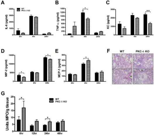
Figure 1. PKC-δ KO mice had no impairment in the production of proinflammatory mediators, but had severe neutrophil infiltra-tion and perivascular edema. (A ∼E) BAL fluid was harvested at various time points after LPS infusion and levels of proinflam-matory cytokines, TNF-α (A) and IL-6 (B), and chemokines, KC (C), MIP-2 (D) and MCP-1 (E), were measured by ELISA. n=4−10 for each group. *p< 0.05 and ***p<0.001 between the indicated groups. (F) Lungs were harvested 12 h after LPS infusion and histological analysis was performed. Neutrophil infiltra-tion and perivascular edema were more prominent in PKC-δ KO mice (right column). *perivas-cular cuff, V: blood vessel, Br: bronchus. Original magnifica-tion: ×40 (upper column) and ×200 (lower column). (G) Deter-mination of myeloperoxidase activity in lung tissue. n=7−12 for each group. *p<0.05 and ***p<0.001 between the indi-cated groups.
was performed using a QCM Chemotaxis Assay Kit (Millipore, Billerica, MA, USA). In brief, neutrophils (2×10⁵ cells/well) isolated from human PBMC using a sta-ndard Ficoll-Hypaque density centrifugation method were added to the upper well, and either KC (50 ng/ml) or MIP-2 (5 ng/ml) was added to the bottom well. Neutrophil migra-tion was allowed for 15 h, and then the extent of migrating neutrophils was determined colorimetrically.
Statistical analysis
All data were analyzed using GraphPad Prism 5 (La Jolla, CA, USA). Unpaired data were analyzed using the t test. Results are expressed as mean±SEM. Statistical sig-nificance was accepted for p values of <0.05.
RESULTS AND DISCUSSION
The involvement of PKC-δ in ALI is controversial. In this study, we compared the responses of WT and PKC-δ KO mice to LPS-induced ALI. To investigate the severity
of pulmonary inflammation after LPS infusion, we meas-ured levels of proinflammatory cytokines and chemokines in BAL fluid at various time points after LPS infusion. Levels of IL-6 and TNF-α in BAL fluid were markedly increased 6 h after LPS infusion and decreased to a basal level at 24 h after LPS infusion (Fig. 1A and B). Generally, these levels were not different between WT and PKC-δ KO mice at any of the time points investigated, even though levels of TNF-α were lower in PKC-δ KO mice than in WT mice 6 h after LPS infusion. Similarly, WT and PKC-δ KO mice did not show a distinguishable dif-ference in levels of KC, MIP-2, or MCP-1 in BAL fluid (Fig. 1C∼E). Overall, these results indicated that genetic deletion of PKC-δ did not show evident effects on LPS- induced pulmonary inflammation. However, histological observations revealed heavier neutrophil infiltration into the lungs of PKC-δ KO mice (Fig. 1F). In addition, there was severe perivascular edema in PKC-δ KO mice (Fig. 1F). Myeloperoxidase activity in lung tissue also indicated more rapid, heavier infiltration of neutrophils into PKC-δ KO
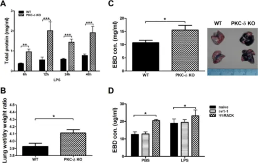
Figure 2. Inhibition of PKC-δ promotes pulmonary edema and vascular permeability. (A) Total protein levels in BAL fluid at various time points after LPS infusion. n=3−14 for each group. *p<0.05, **p<0.01, and ***p<0.001 between the two groups. (B) The lung wet/dry weight ratio at 24 h after LPS infusion. n=12−13 for each group. *p<0.05. (C and D) EBD was injected 4 h after LPS infusion and lungs were harvested 30 min later. Gross observation (left column) and concentrations of EBD in extracted lung tissue (right column). n=12−13 for each group. *p<0.05. (D)δV1-1 or ψδRACK peptide was intratracheally injected 30 min before LPS infusion. Lungs were harvested 30 min after EBD injection. n=6−8 for each group. *p<0.05 between the indicated groups.
lungs after LPS infusion (Fig. 1G). As levels of KC and MIP-2, major neutrophil chemoattractants, in PKC-δ mouse lungs were not so different from those in WT mouse lungs after the induction of ALI (Fig. 1C and D), enhanced neutrophil recruitment to the injured lungs of PKC-δ KO mice might not have been caused by dysregula-tion of neutrophil-chemotactic chemokine producdysregula-tion. More-over, PKC-δ KO neutrophils showed a similar responsive-ness to KC and MIP-2 in an in vitro migration assay com-pared to WT neutrophils (data not shown). PKC-δ KO neutrophils also did not have any survival advantage com-pared to WT neutrophils when they were challenged with LPS (data not shown). Therefore, these results, along with the observation of increased pulmonary edema in PKC-δ KO mice, indicated that dysregulation of vascular perme-ability rather than uncontrolled inflammation is likely to be the main cause of increased neutrophil recruitment to the lungs in PKC-δ KO mice.
To test the above hypothesis, we measured the amount of total proteins in BAL fluid at various time points after LPS infusion as a parameter for protein leakage from vessels. As expected, the BAL fluid of PKC-δ KO mice
contained a greater amount of total proteins at all time points investigated, relative to that of WT mice (Fig. 2A). The lung wet/dry ratio showed that the increased perme-ability in PKC-δ KO mice indeed resulted in severe pul-monary edema following LPS infusion (Fig. 2B). To di-rectly show that PKC-δ KO vessels had a higher capacity for permeability, we i.v.-injected EBD 4 h after LPS in-fusion and measured the quantity of EBD that extravasated from vessels 30 min after EBD injection. We could easily distinguish the thicker blue lungs in PKC-δ KO mice by gross observation (Fig. 2C) and quantification of EBD ex-tracted from lungs confirmed this result (Fig. 2C). We re-peated the same EBD assay using cell-permeable PKC-δ inhibitor (10). Interestingly, in vivo injection of PKC-δ in-hibitor enhanced not only steady state vascular permeability, but also LPS-initiated permeability (Fig. 2F). However, the PKC-δ activator had no effect on vascular permeability (Fig. 2F).
Finally, we explored the direct involvement of PKC-δ in permeability of an endothelial monolayer. For this, we cultured HUVEC cells in transwell plates until they reached confluence, and then performed a permeability assay. As
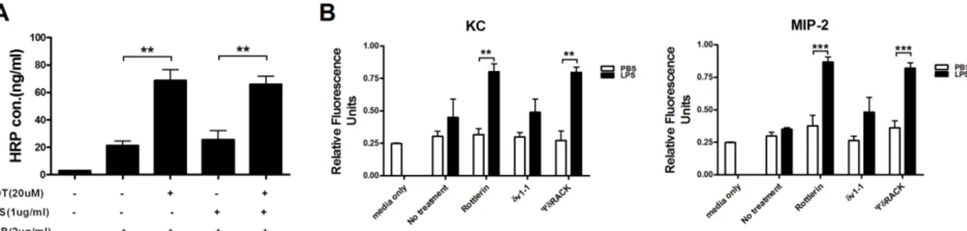
Figure 3. Inhibition of PKC-δ promotes extravasation of proteins and neutrophils through an endothelial monolayer. (A) A confluent monolayer of HUVEC cells was treated with rottlerin 30 min before the addition of LPS. Four hour later, HRP was added to HUVEC cells and extravasation was allowed to proceed for 30 min. n=3 for each group. *p<0.05 and ***p<0.001 between the indicated groups. (B) PKC-δ inhibitors and LPS were treated as described in A and neutrophil transmigration was allowed for 15 h. n=3 for each group. **p<0.01 and ***p<0.001 between the indicated groups.
seen in Fig. 3A, rottlerin, a purported PKC-δ inhibitor, increased permeability to a similar degree, regardless of whether or not HUVEC cells were pre-activated by LPS, indicating again that PKC-δ regulated steady state vas-cular permeability. Using this system, we further examined whether transendothelial migration of neutrophils could be regulated by δ. In this case, we showed that PKC-δ inhibitors (rottlerin and ψPKC-δRACK peptide) enhanced KC- and MIP-2-induced transendothelial migration of neu-trophils through the HUVEC cell monolayer only when HUVEC cells were pre-activated by LPS (Fig. 3B). These results indicated that LPS-induced upregulation of cell ad-hesion molecules might be required for interactions with neutrophils before their endothelial transmigration. As HEVEC cells remained healthy until the end of our experi-ments, it was unlikely that there was a physical lesion in the HEVEC cell monolayer.
In this study, our results suggest that PKC-δ is not in-volved in regulating the production of proinflammatory mediators, but that it plays a critical role in vascular per-meability and transmigration of neutrophils through an en-dothelial cell layer. Interestingly, inhibition of PKC-δ was shown to be essential to the regulation of steady state en-dothelial barrier function. This regulatory function of PKC-δ is maintained during ALI. As more severe pulmo-nary edema occurred in PKC-δ KO mice, PKC-δ also seems to be involved in inflammation-induced vascular permeability. Even though there are contradictory reports regarding the function of PKC-δ in ALI (3,5), our data, obtained using a PKC-δ gene deletion mouse model and PKC-δ-specific chemical and peptide inhibitors, all support
PKC-δ to be a negative regulator of vascular permeability. Further research will be needed to define the molecular mechanisms behind these observations.
ACKNOWLEDGEMENTS
This work was funded by Ulsan University Hospital (Bio-medical Research Center Promotion Fund 11-02).
CONFLICTS OF INTEREST
The authors have no financial conflict of interest.
REFERENCES
1. Han, S., and R. K. Mallampalli. 2015. The acute respiratory dis-tress syndrome: from mechanism to translation. J. Immunol. 194: 855-860.
2. Siflinger-Birnboim A., and A. Johnson. 2003. Protein kinase C modulates pulmonary endothelial permeability: a paradigm for acute lung injury. Am. J. Physiol. Lung Cell. Mol. Physiol. 284: L435-L451.
3. Klinger, J. R., J. D. Murray, B. Sasserly, D. F. Alvarez, J. A. King S. S. An, G. Choudhary, A. N. Owusu-Sarfo, R. Warburton, and E. O. Harrington. 2007. Rottlerin causes pulmonary edema in vivo: a possible role for PKCδ. J. Appl. Physiol. 103: 2084-2094.
4. Gaudreault, N. R. M. Perrin, M. Guo, C. P. clanton, M. H. Wu and S. Y. Yuan. 2008. Counter regulatory effects of PKCβII and PKCδ on coronary endothelial permeability. Arterioscler. Thromb. Vasc. Biol. 28: 1527-1533.
5. Chichger, H. K. L. Grinnell, B. Casserly, C. S. chung, J. Brazaz, J. Lomas-Neira, A. Ayala, S. Rounds, J. R. Klinger, and E. O. Harrington. 2012. Genetic disruption of protein of protein kinase Cδ reduces endotoxin-induced lung injury. Am. J. Physiol. Lung
Cell. Mol. Physiol. 303: L880-L888.
6. Miyamoto, A., K. Nakayama, H. Imaki, S. Hirose, Y. Jiang, M. Abe, T. Tsukiyama, H. Nagahama, S. Ohno, S. Hatakeyama, and K. I. Nakayama. 2002. Increased proliferation of B cells and au-to-immunity in mice lacking protein kinase C delta. Nature 416:8 65-869.
7. Lomas-Neira, J., C. S. Chung, P. S. Grutkoski, A. Dunican, H. H. Simms, W. G. Cioffi, and A. Ayala. 2005. Divergent roles of murine neutrophil chemokines in hemorrhage induced priming for acute lung injury. Cytokine 31: 169-179.
8. Neff, T. A,, R. F. Guo, S. B. Neff, J. V. Sarma, C. L. Speyer, H. Gao, K. D. Bernacki, M. Huber-Lang, S. McGuire, L. M. Hoesel, N. C. Riedemann, B. Beck-Schimmer, F. S. Zetoune, and
P. A. Ward. 2005. Relationship of acute lung inflammatory injury to Fas/FasL system. Am. J. Pathol. 166: 685-694.
9. Patterson, C. E., R. A. Rhoades, and J. G. Garcia. 1992. Evans blue dye as a marker of albumin clearance in cultured endothelial monolayer and isolated lung. J. Appl. Physiol. 72: 865-873. 10. Chen, L., H. Hahn, G. Wu, C. H. Chen, T. Liron, D. Schechtman,
G. Cavallaro, L. Banci, Y. Guo, R. Bolli, G. W. Dorn 2nd, and D. Mochly-Rosen. 2001. Opposing cardioprotective actions and parallel hypertrophic effects of delta PKC and epsilon PKC. Proc. Natl. Acad. Sci. U. S. A. 98: 11114-11119.
11. Baudin, B., A. Bruneel, N. Bosselut, and M. Vaubourdolle. 2007. A protocol for isolation and culture of human umbilical vein endo-thelial cells. Nat. Protoc. 2: 481-485.