The potential roles of T-type Ca
2
+
channels in motor
coordination
Young-Gyun Park
1, Jeongjin Kim
1and Daesoo Kim
1,2*
1Department of Biological Sciences, Korea Advanced Institute of Science and Technology, Daejeon, South Korea 2
KAIST Institute for the BioCentury, Korea Advanced Institute of Science and Technology, Daejeon, South Korea
Edited by:
G. J. Augustine, KIST, South Korea Reviewed by:
Michael Nitabach, Yale University School of Medicine, USA
Chris I. De Zeeuw, Erasmus Medical Center, Netherlands
*Correspondence: Daesoo Kim, Department of Biological Sciences, KAIST Institute for the BioCentury, Korea Advanced Institute of Science and Technology, 291 Daehak-Ro, Daejeon 305-701, South Korea
e-mail: daesoo@kaist.ac.kr
Specific behavioral patterns are expressed by complex combinations of muscle
coordination. Tremors are simple behavioral patterns and are the focus of studies
investigating motor coordination mechanisms in the brain. T-type Ca
2+channels mediate
intrinsic neuronal oscillations and rhythmic burst spiking, and facilitate the generation of
tremor rhythms in motor circuits. Despite substantial evidence that T-type Ca
2+channels
mediate pathological tremors, their roles in physiological motor coordination and behavior
remain unknown. Here, we review recent progress in understanding the roles that T-type
Ca
2+channels play under pathological conditions, and discuss the potential relevance of
these channels in mediating physiological motor coordination.
Keywords: T-type Ca2+channels, tremors, motor coordination, inferior olive, thalamocortical neurons
INTRODUCTION
Unraveling the mechanisms that underlie specific motor patterns
is a challenging issue in neuroscience. Motor coordination is a
complex process that determines the timing and sequence of both
activation and relaxation of a huge number of muscles (reviewed
by
Mauk et al., 2000; De Zeeuw et al., 2011
). More than one
gigahertz of information processing is required for the optimal
execution of even the simplest motor behavior, such as holding
a cup.
Tremors are one of the simplest forms of motor
coordina-tion and are characterized by involuntary rhythmic movements of
either the whole body or of body parts (reviewed by
Findley, 1995;
Grimaldi and Manto, 2008
). Low amplitude tremor, also known
as physiological tremor, exist in humans and animals during
nor-mal states and may function to help behavioral control (
Elble
et al., 1984
). High amplitude tremors that interfere with voluntary
movements are observed in pathological conditions (reviewed by
Deuschl et al., 2001
). These pathological tremors are classified
into different types, and are associated with specific neural
mech-anisms and circuits (Table 1) (reviewed by
Wilms et al., 1999;
Deuschl et al., 2001
).
T-type Ca
2+channels (Ca
V3.1, 3.2, and 3.3) modulate both
physiological and pathological rhythms in the brain (
Crunelli
et al., 1989
; reviewed by
Huguenard, 1996
). These channels
medi-ate the generation of low-threshold spikes (LTS) in response
to hyperpolarizing membrane potentials elicited by inhibitory
inputs. LTS regulate neural oscillations, resonance, and synchrony
(
Llinas and Yarom, 1986; Crunelli et al., 1989; Kim et al., 2001,
2011; Mangoni et al., 2006
) (Figure 1, left). Pharmacological and
genetic studies show that T-type Ca
2+channels are also involved
in the generation of pathological tremors (
Sinton et al., 1989;
Handforth et al., 2010
). However, the precise roles of T-type
Ca
2+channels in physiological tremors and in normal motor
coordination remain unknown. Here, we propose potential roles
for T-type Ca
2+channels in physiological motor functions based
on their pathological roles.
ROLES OF T-TYPE CA
2+CHANNELS IN GENERATING
PATHOLOGICAL TREMORS
T-TYPE CA2+CHANNELS IN ESSENTIAL TREMOR
Essential tremor is the most common form of movement
disor-der (
Kurtzke, 1982
; reviewed by
Louis, 2005
) and is characterized
by postural and kinetic tremors at 4–12 Hz (
Bain et al., 2000;
Brennan et al., 2002
). Harmaline is a plant-derived metabolite
(
Lutomski et al., 1974
) that induces ET-like tremors and
tremor-related neural oscillations in both humans and animals (
Battista
et al., 1969; de Montigny and Lamarre, 1973; Llinas and Volkind,
1973; Ahmed and Taylor, 2012
). Since harmaline binds to
vari-ous ion channels, including glutamate receptors, GABA receptors,
and voltage-gated Ca
2+channels (
Du et al., 1997; Glennon et al.,
2000; Splettstoesser et al., 2005
), specific molecular mechanism
that underlies harmaline tremor have been unclear.
Inferior olive (IO) neurons are also implicated in the
gener-ation of harmaline tremor. IO lesions reduce harmaline tremors
in rats (
Simantov et al., 1976
). Harmaline also alters the
intrin-sic properties of IO neurons: it increases LTS and amplifies
sub-threshold oscillations (STOs). Both of these properties are
dependent upon the conductance of T-type Ca
2+channels (
Llinas
and Yarom, 1986; Crunelli et al., 1989; Park et al., 2010
). A
non-selective T-type Ca
2+channel inhibitor, 1-octanol, reduces
harmaline-induced tremors in rats (
Sinton et al., 1989
),
sup-porting the role of T-type Ca
2+channels in harmaline-induced
tremor.
There are three distinct isoforms of T-type Ca
2+channels,
Ca
V3.1, 3.2, and 3.3 (
Cribbs et al., 1998; Perez-Reyes et al., 1998;
Lee et al., 1999
). Ca
V3.1 is the major isoform expressed in the
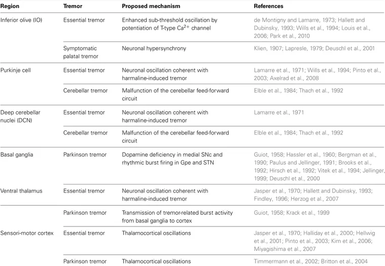
Table 1 | Mechanisms of pathological tremor.
Region Tremor Proposed mechanism References
Inferior olive (IO) Essential tremor Enhanced sub-threshold oscillation by potentiation of T-type Ca2+channel
de Montigny and Lamarre, 1973; Hallett and Dubinsky, 1993; Wills et al., 1994; Louis et al., 2006; Park et al., 2010
Symptomatic palatal tremor
Neuronal hypersynchrony Klien, 1907; Lapresle, 1979; Deuschl et al., 2001
Purkinje cell Essential tremor Neuronal oscillation coherent with harmaline-induced tremor
Lamarre et al., 1971; Wills et al., 1994; Pinto et al., 2003; Axelrad et al., 2008
Cerebellar tremor Malfunction of the cerebellar feed-forward circuit
Elble et al., 1984; Thach et al., 1992
Deep cerebellar nuclei (DCN)
Essential tremor Neuronal oscillation coherent with harmaline-induced tremor
Lamarre et al., 1971
Cerebellar tremor Malfunction of the cerebellar feed-forward circuit
Elble et al., 1984; Thach et al., 1992
Basal ganglia Parkinson tremor Dopamine deficiency in medial SNc and rhythmic burst firing in Gpe and STN
Guiot, 1958; Hassler et al., 1960; Bergman et al., 1990; Paulus and Jellinger, 1991; Brooks et al., 1992; Hirsch et al., 1992; Vitek et al., 1994; Jellinger, 1999; Deuschl et al., 2000
Ventral thalamus Essential tremor Neuronal oscillation coherent with harmaline-induced tremor
Jasper et al., 1970; Hallett and Dubinsky, 1993; Findley, 1996; Herzog et al., 2007
Parkinson tremor Transmission of tremor-related burst activity from basal ganglia to cortex
Guiot, 1958; Krack et al., 1999
Sensori-motor cortex Essential tremor Thalamocortical oscillations Jasper et al., 1970; Halliday et al., 2000; Hellwig et al., 2001; Pinto et al., 2003; Kim et al., 2006; Miyagishima et al., 2007
Parkinson tremor Thalamocortical oscillations Timmermann et al., 2002; Britton et al., 2004
IO, Purkinje cells, and deep cerebellar nuclei (DCN) (Figure 1,
right) (
Talley et al., 1999; Lein et al., 2006
). Ca
V3.1
−/−mice
treated with harmaline (dose: 9 mg/kg) do not display behavioral
tremors, 4–10 Hz tremor-related oscillation in the olivocerebellar
pathway, or STOs in IO neurons (
Park et al., 2010
). Patch clamp
recording revealed that harmaline inhibits the activation of
Ca
V3.1 channels while also promoting their de-inactivation.
These effects result in a net potentiation of Ca
V3.1 channels
under physiological conditions (
Park et al., 2010
).
Other ionic mechanisms and their interactions with Ca
V3.1
channel could also contribute to the generation of harmaline
tremor. A Ca
2+activated K
+channel isoform (KCa1.1) is known
to form a complex with Ca
V3 channels and be activated in
response to Ca
V3—mediated calcium influx (
Rehak et al., 2013
).
Interaction between Ca
V3 and Ca
2+activated K
+channels could
possibly be involved in the generation of STO and its exaggeration
during harmaline tremor. Hyperpolarization-activated cation
channel is another candidate. Activation of this channel could
contribute to slow rebound potential in STOs. This channel is
also involved in rhythmic thalamic oscillation and the blockade
of the channel ameliorates oscillation induced by hyperpolarizing
current injection into IO neurons (
Bal and McCormick, 1997
).
Investigation of harmaline tremor and tremor rhythm in Ca
2+activated K
+channel or hyperpolarization-activated cation
channel knockout mice is required to test these possibilities.
GABA-A1 receptor knockout mice (a1
−/−) are a genetic
model of essential tremor. These mutant mice display
∼25 Hz
ET-like tremors (
Kralic et al., 2005
). A subset of non-selective
T-type Ca
2+channel antagonists (ethosuximide, zonisamide,
ECN, KYS05064, and NNC 55-0396) ameliorate both a1
−/−mouse tremors and harmaline tremor (
Handforth et al., 2010
).
While Ca
V3.1 knockout mice display reduced harmaline tremor
(
Park et al., 2010
), double knockout mice (Ca
V3.1
−/−and
a1
−/−) exhibit exacerbated tremor behavior (
Chang et al., 2011
).
Thus, these two distinct animal models of essential tremor
(a1
−/−and harmaline-induced tremors) likely result from
dif-ferent mechanisms, such as the involvement of distinct T-type
Ca
2+channel isoforms (e.g., Harmaline tremor by Ca
V3.1 and
a1
−/−tremor by Ca
V3.2 or 3.3). The heterogeneity of
essen-tial tremor is well-described by clinical studies (
Kovach et al.,
2001; Louis et al., 2007
). Future studies are necessary to
deter-mine how the other T-type isoforms contribute to essential
tremor.
T-TYPE CA2+CHANNELS IN PARKINSON TREMORS
Resting tremor is one of the most detrimental symptoms
expe-rienced by Parkinson’s disease (PD) patients. PD is caused by a
dopamine deficiency in the brain. Rhythmic stimulation of the
motor cortex via subcortical pathway is thought to underlie
rest-ing tremor of PD patients (
Plenz and Kital, 1999; Magnin et al.,
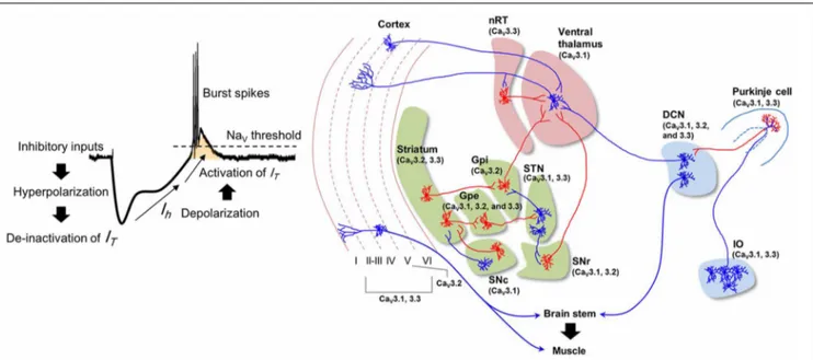
FIGURE 1 | T-type Ca2+channels and pathological tremor pathways. Potentiation of T-type Ca2+conductance and the generation of low-threshold
spikes (LTS) (left). Hyperpolarization and subsequent depolarization by HCN channel-mediated currents (Ih) open T-type Ca2+channels. IT: T-type calcium
current; NaV: voltage-gated sodium channel. Neural pathways involved in
pathological tremors (right). Blue indicates excitatory neurons, and red indicates inhibitory neurons. The blue regions represent the olivocerebellar
pathway, the green regions represent basal ganglia circuits, and the red regions indicate thalamocortical pathways. The T-type Ca2+channel isoforms
(CaV3.1, 3.2, and 3.3) expressed in these regions are indicated (Lein et al.,
2006). IO: Inferior olive; DCN: deep cerebellar nuclei; SNc: substantia nigra compacta; Gpe: globus pallidus externa; Gpi: globus pallidus interna; STN: subthalamic nucleus; SNr: substantia nigra reticulata; nRT: nucleus reticularis of the thalamus.
2000; Chan et al., 2011
). There are several hypotheses about the
origin of the tremor.
The basal ganglia circuit hypothesis states that rhythmic burst
firings of neurons in subthalamic nuclei (STN) underlie the
rest-ing tremor in PD patients (
Magnin et al., 2000; Chan et al., 2011
).
Suppressing STN activity using deep brain stimulation
amelio-rates resting tremor in PD patient (
Kumar et al., 1998; Sturman
et al., 2004; Amtage et al., 2008
). STN burst activity is associated
with the activation of T-type Ca
2+channels (
Beurrier et al., 1999;
Tai et al., 2011
). Moreover, pharmacological inhibition of T-type
Ca
2+channels in the STN rescues locomotor deficits in rat PD
models, while effect on the resting tremor was not accessed (
Tai
et al., 2011
). Isoforms of T-type Ca
2+channels that express in
STN (Ca
V3.1 and 3.3) (Figure 1) could possibly play a role in the
generation of resting tremor in PD patients.
The ventrolateral (VL) thalamus is another candidate for PD
resting tremor. Dopamine deficiency in PD would result in the
activation of globus pallidus interna (Gpi) and substantia nigra
reticulata (SNr) neurons that provides GABAergic input to the
VL thalamus (
Vitek, 2002
). Subsequent hyperpolarization of the
neurons may induce rhythmic LTS in VL thalamocortical (TC)
neurons and thus generates the resting tremor. Consistently,
tremor-related rhythmic LTS is observed in VL thalamic neurons
(
Zirh et al., 1998; Magnin et al., 2000; Pifl et al., 2012
). These
results suggest that Ca
V3.1 expressing in VL thalamus (Figure 1)
could be involved in the tremor generation. However, some other
studies in PD patients also reports that LTS in VL neurons do not
coincide with ongoing resting tremor (
Zirh et al., 1998; Magnin
et al., 2000
). Thus, the role of thalamic burst firing in the resting
tremor is still controversial.
A third hypothesis is the “dimmer-switch model” which
states that core tremor activities are expressed by the
cerebello-thalamo-cortical circuit (Figure 1, right) (
Helmich et al., 2012
).
Hyperactivity in the cerebellum of PD patients is reported
(
Ghaemi et al., 2002; Timmermann et al., 2002
), which is
con-comitant with the activation of sensory and motor cortices
responsible for hand exhibiting resting tremor (
Timmermann
et al., 2004; Pollok et al., 2009
). Deep brain stimulation of the
STN or Gpi, or the administration of Levodopa normalizes
cere-bellar activity and improves tremor in PD patients (
Payoux et al.,
2008; Wu et al., 2009
). This may be due to the ventral intermediate
thalamus (which receives excitatory inputs from the cerebellum)
acting as a critical relay station in the cerebello-thalamo-cortical
circuit (
Lenz et al., 1995; Tarsy et al., 2008
). Consistently, PD
resting tremor is suppressed by stimulation of the ventral
inter-mediate thalamus, with decreased blood flow in cerebellar cortex
(
Deiber et al., 1993
).
In spite of these evidences that T-type Ca
2+channels are
involved in neuronal burst activity and oscillations in PD tremor
circuits, there is no direct evidence that links T-type Ca
2+chan-nels to resting tremor. One of the main obstacles on defining the
role of T-type Ca
2+channels in PD tremor is a lack of robust
rodent models that display resting tremor (
Potashkin et al., 2010
).
Developing a robust resting tremor model and modulating T-type
Ca
2+channels in the model might unravel the mechanism of PD
resting tremor.
T-TYPE CA2+CHANNELS IN PALATAL TREMOR
Palatal tremor, also called palatomyoclonus, is characterized by
rhythmic movement of soft palate and sometimes of other
mus-cles (
Deuschl et al., 2001
). Hypertrophy of IO neurons has been
proposed as a pathologic substrate of the tremor (
Deuschl et al.,
2000; Pearce, 2008
). This condition develops after lesions in the
brainstem or cerebellum, manifesting as tremor in body parts
contralateral to the region of damage in both human and
ani-mals (
De Zeeuw et al., 1998; Deuschl et al., 2000
). Studies of
hemicerebellectomized animals reveal that hypertrophic IO
neu-rons show failure in after-depolarization of action potential and
have decreased numbers of GABAergic boutons in their dendrites
(
Ruigrok et al., 1990; De Zeeuw et al., 1998
).
Because GABAergic input to IO neuron modulates
electro-tonic coupling of IO neurons (
Sotelo et al., 1986; Leznik et al.,
2002
), it can be inferred that synchrony between IO neurons
would be enhanced in the hypertrophic IO (
Deuschl et al., 2000
).
This might entrain larger range of IO neurons with synchronized
STOs, resulting rhythmic activations required for palatal tremor
(
Deuschl et al., 2000
).
Ca
V3.1 could also be involved in palatal tremor generation,
in consideration of its role in STOs (
Llinas and Yarom, 1986;
Choi et al., 2010; Park et al., 2010
). However, the
contribu-tion of Ca
V3.1 would be different from the case of harmaline
tremor. Potentiation of the Ca
V3.1 channel is required for
harma-line tremor (
Park et al., 2010
). Hyperpolarization of IO neurons
contributes Ca
V3.1 potentiation which amplifies STOs and
sub-sequently generates tremor rhythm. (
Park et al., 2010
). In palatal
tremor, however, hypertrophic IO neurons seem to be
depolar-ized, meaning that Ca
V3.1 might not be potentiated (
Crunelli
et al., 1989
). Instead, increased firing rate with enhanced
syn-chrony in IO neurons could, when combined with basal STOs
generate a rhythmic activity for palatal tremor. Investigation of
palatal tremor in Ca
V3.1
−/−mice and changes in the Ca
V3.1
activity may shed light on the differential mechanisms of
IO-dependent tremor generation by T-type Ca
2+channel.
THE ROLE OF T-TYPE CA
2+CHANNELS IN GENERATING
PHYSIOLOGICAL MOTOR FUNCTIONS
While their association with pathological tremors (Table 1) is
well-understood, whether T-type Ca
2+channels contribute to
physiological motor functions is unclear. Both Ca
V3.1
−/−(
Park
et al., 2010
) and Ca
V3.2
−/−mice (
Choi et al., 2006
) do not
have significant motor defects. In addition, overexpression of the
Ca
V3.1 gene in mouse brain does not result in motor dysfunction
(
Ernst et al., 2009
). These results might be due to compensatory
expressions between Ca
V3 channels, or the possibility that motor
defects in Ca
V3 mutants are not be able to be examined by
con-ventional motor tests. Below, we summarize the potential roles
of T-type Ca
2+channels in physiological motor behavior and
describe how to study these potential roles.
GENERATION OF PHYSIOLOGICAL TREMORS BY T-TYPE CA2+ CHANNELS
Physiological tremors are induced by extrinsic factors such as
gravity force (
Marsden et al., 1969; Young and Hagbarth, 1980
),
or by central mechanisms such as a tremor rhythm pacemaker
in the brain (
Hagbarth et al., 1983; Vallbo and Wessberg, 1993;
Llinas and Pare, 1994
). 8–12 Hz component of physiological
tremor is associated with central mechanisms because this
com-ponent is unaffected by extrinsic factors (
Elble and Randall, 1976;
Vallbo and Wessberg, 1993
).
The intrinsic rhythmicity of IO neurons may be one of these
central mechanisms (
de Montigny and Lamarre, 1973; Llinas
and Pare, 1994; Findley, 1995
), since the frequencies of STOs
are around 10 Hz (
Llinas and Yarom, 1986; Chorev et al., 2007;
Khosrovani et al., 2007
). These frequencies are similar to those of
physiological tremors of humans and animals (
Elble and Randall,
1976; Elble et al., 1984; Vallbo and Wessberg, 1993
). Moreover,
vibrissal movement generated at around 10 Hz (
Fukuda et al.,
1989
) is abolished by electrolytic lesions of the IO in rats (
Semba
and Komisaruk, 1984
), supporting that STOs in IO neurons could
be the origin of the physiological tremor.
While the frequencies of STOs are
∼10 Hz, the average firing
rate of IO neuron is about 1 Hz, suggesting that individual IO
neurons are insufficient to generate signal for 10 Hz
physiologi-cal tremor (
Keating and Thach, 1997; Lang et al., 1999; Chorev
et al., 2007
). However, presence of gap junction in IO and
prop-erty of their connectivity with descending motor pathway support
that STOs in IO neurons could be responsible for physiological
tremor. Gap junctions synchronize IO neurons with a 10 Hz STO
rhythm (
Long et al., 2002; Van Der Giessen et al., 2008
), ensuring
that some IO neurons fire with the 10 Hz cycle at the population
level (
Chorev et al., 2007; Park et al., 2010
). Since multiple IO
neu-rons innervate a DCN neuron via Purkinje cells (
Van der Want
et al., 2004
), an individual DCN neuron may receive 10Hz
rhyth-mic input, as well as pharmacologically induced synchronization
of IO rhythmic activity evoke rhythmic modulation of DCN
fir-ing with same frequency (
Lamarre et al., 1971; Park et al., 2010
).
The 10 Hz oscillation in DCN may recruit brainstem nuclei (e.g.,
the red nucleus and the lateral reticular formation) and motor
neurons, resulting in 10 Hz physiological tremor (Figure 2, left).
One simple way to link STOs and physiological tremor would
be to examine physiological tremors in Ca
V3.1
−/−mice that lack
STOs of IO neurons (
Park et al., 2010
). Unfortunately,
physiolog-ical tremors are not well documented in mice. One study reported
that 20–35 Hz forelimb vibration may reflect physiological tremor
in mice (
Kralic et al., 2005
). However, the 20–35 Hz vibration
could be an artifact of resonant frequencies in the recording
sys-tem of the study. Application of more sensitive techniques, such as
electromagnetic or optoelectronic detection methods (
Grimaldi
and Manto, 2008
), might be necessary for future studies on the
mechanism of physiological tremors in mice.
AROUSAL-INDUCED ENHANCEMENT OF PHYSIOLOGICAL TREMORS
Physiological tremors in both humans and animals are
ampli-fied in response to various alerting stimuli such as anger, novelty,
or stress (
Günther et al., 1983; Duan et al., 1996; Klein, 2002;
Siniscalchi et al., 2013
). The enhanced physiological tremor is
probably important for providing optimal muscular coordination
during arousal. Because the hypothalamus controls arousal (
Lin
et al., 1988; Adamantidis et al., 2007; Tsunematsu et al., 2011
)
and its projections to IO neurons are revealed by anterograde
tracing with AAV virus (
Lein et al., 2006
), we here propose that
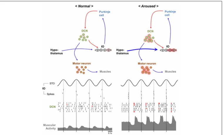
FIGURE 2 | Hypothetical mechanisms underlying normal and
arousal-enhanced physiological tremors. Physiological tremor generation (left). Adjacent IO neurons share∼10 Hz STOs via gap junctions, which results in∼10 Hz rhythms in the population of cells. Action potentials propagate from IO neurons to Purkinje cells and then to the DCN, thereby causing inhibitory responses and then rebound excitation in DCN neurons.
The rhythmicity of the IO neuron population is converted into the rhythmicity of individual DCN neurons, which results in 10 Hz rhythmic muscle contractions. Increased excitatory inputs from hypothalamic neurons depolarize the membrane potentials of IO neurons upon arousal (right), which increases the∼10 Hz rhythmic LTS (red bars) in DCN neurons and muscle contractions.
arousal-induced physiological tremors could be dependent upon
the excitation of IO neurons (Figure 2, right) by hypothalamic
input.
Increased excitatory input to the IO may enhance synchrony
between IO neurons, since infusion of a glutamate receptor
antag-onist into the IO decreases complex spike synchrony in the
mediolateral direction (
Lang, 2002
). Otherwise, the increased
firing rate of IO neurons might also raises the probability of
syn-chronous firing among IO neurons. Hypersynsyn-chronous IO firing
would be translated into rhythmic LTS in individual DCN
neu-rons (
Hoebeek et al., 2010; De Zeeuw et al., 2011
) that might
cause high amplitude physiological tremor during arousal (
Witter
et al., 2013
) (Figure 2, right).
Among the three isoforms of T-type Ca
2+channels expressed
in DCN neurons (Figure 1), Ca
V3.1 is thought to serve a role
in the tremor, as Ca
V3.1 is responsible for generating LTS with
multiple sodium spikes in DCN neurons (
Molineux et al., 2006
).
Analysis of muscular activities of Ca
V3.1
−/−mice in response to
novel contexts is required to address this possibility.
MODULATION OF MOVEMENT INITIATION TIMINGIn humans movement initiation is connected to the timing of
physiological tremor in some respects (
Travis, 1929; Goodman
and Kelso, 1983
). For example, the 10 Hz periodicity of motor
initiation timing is observed in various parts of human body
(
Harter and White, 1968
). While this restriction may reduce
the temporal precision of movement initiation (Figure 3, upper)
(
Lakie and Combes, 2000; Lakie, 2010
), physiological tremor also
helps overcome muscular friction prior to action initiation and
permits more powerful and faster movements (
Adamovich et al.,
1994; de Rugy and Sternad, 2003
). Modulation of Cav3.1 in IO
neurons hypothesized to be involved in physiological tremor and
is expected to affect the kinetics of movement inhibition.
The basal ganglia-thalamocortical circuit is important for
movement initiation in both rodents and primates (
Yin and
Knowlton, 2006; Bédard and Sanes, 2011
). Studies show that
the GABAergic outputs of medium spiny neurons expressing
dopamine receptor 1 or 2 in the dorsal striatum play inhibitory
or facilitatory roles, respectively, in movement initiation (
Kravitz
and Kreitzer, 2012
). However, in vivo imaging study shows that
the both types of medium spiny neurons are activated during
movement initiation in mice (
Cui et al., 2013
). Therefore, the
role of medium spiny neurons in movement initiation is still
controversial (
Surmeier, 2013
).
During resting states, there is increased inhibition of VL
neurons by the basal ganglia, which in turn raises the threshold for
the onset of movement initiation signals in the thalamocortical
pathway. VL neurons also receive excitatory signals from DCN
through monosynaptic connections between them (
Shinoda
et al., 1985, 1993; Lein et al., 2006
). Consistent with their
sup-posed role in generating 10 Hz DCN rhythms (Figure 2, left),
T-type Ca
2+channels may also play a role in movement
initi-ation. 10 Hz rhythmic signals in DCN neurons may reduce the
action potential threshold in VL neurons. It will be necessary to
selectively inhibit DCN-VL circuits in future studies.
PHYSIOLOGICAL TREMORS PROVIDE PREPARATION FOR EMERGENT MOTOR RESPONSES
Emergent motor responses are critical for the survival of animals
in nature. The unexpectancy hypothesis of IO function (
Devor,
2002
) states that IO neurons reliably respond to unexpected
motor disturbances. One example is the increased IO neuronal
excitability when cat misses a step on a ladder by unexpected
rung down (
Andersson and Armstrong, 1987
). Consistent with
this idea, IO neuronal excitability decreases after rodents learn
the timing of air puffs (
Kim et al., 1998
). As with
hypothala-mic control of the IO, unexpected external stimuli could activate
hypothalamic arousal pathways and amplify 10 Hz
physiologi-cal tremors through the action of Ca
V3.1 in the DCN (Figure 2,
right). This enhanced tremor may help overcome inertial
resis-tances and synchronize muscles when emergent motor responses
are required (
Greene, 1972
).
Meanwhile, T-type Ca
2+channel also could contribute to the
generation of emergent movement through a DCN-dependent
mechanism. LTS in DCN neurons is proposed to provide
syn-chronous and strong output to descending motor pathways (
De
Zeeuw et al., 2011
). Recently, a study with optogenetic
modula-tion of Purkinje cells reveals that induced LTS in DCN neurons
can evoke emergent movement (
Witter et al., 2013
). Cessation of
induced Purkinje cell ensemble activity induces rebound
activ-ity in DCN and timed movement whose amplitude is dependent
on the degree of Purkinje cell activation. As Ca
V3.1 is responsible
for generating LTS with multiple sodium spikes in DCN neurons
(
Molineux et al., 2006
), analysis of emergent motor responses
generated by Ca
V3.1
−/−mice would help to access this idea.
SENSORY SENSITIZATION HYPOTHESIS
Studies of human sensory perception suggest that
physiologi-cal tremors can facilitate sensory functions. For example, eyeball
tremors sensitize visual function (
Hennig et al., 2002
). Intentional
suppression of physiological limb tremors reduces visual
cue-tracking abilities (
Daneault et al., 2011
). Moreover, artificial
vibrations of foot muscles, which could mimic sensory feedback
by physiological tremors, increase somatosensory sensitivity (
Liu
et al., 2002
).
Common sense suggests that proprioceptive feedback signals
resulted from physiological tremors act as “noise” that might
interfere with sensation. However, the stochastic resonance theory
(
McNamara et al., 1988; Wiesenfeld and Moss, 1995
) states that
moderate levels of “noise” actually facilitate signal detection in
nervous system (Figure 3, lower) (
Douglass et al., 1993; Levin and
Miller, 1996
). Therefore, by providing moderate noise,
physio-logical tremors could enhance sensory detection (Figure 3, lower)
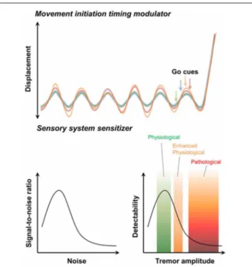
FIGURE 3 | Possible contribution of physiological tremors to motor function. The movement initiation timing modulator hypothesis (upper ). Initiation of voluntary movement in response to “Go” cues depends on the phase of the ongoing physiological tremor. The sensory system sensitivity hypothesis (lower ). The stochastic resonance signature (left). “Noise” caused by physiological tremors increases the ability to detect signals. By contrast, pathological tremors decrease the ability of the sensory system to detect signals (right).
and subsequently raise motor performances. The role of Ca
V3.1
in the IO in generating physiological tremor would be critical in
this process.
T-type Ca
2+channels in the thalamus might also be associated
with sensory sensitization. VL thalamocortical relay neurons may
receive tremor signals generated by the cerebellum through
DCN-VL connections. Rhythmic activation of the DCN-VL neurons activates
GABAergic nRT neurons through reciprocal connections between
TC and nRT neurons (
Huguenard and Prince, 1994
). Activation
of nRT neurons can in turn induces rhythmic inhibition and thus
LTS in VL thalamic neurons, while non-specifically inhibit TC
neurons with other modalities. This may increase sensory
cod-ing in TC neurons: intensifycod-ing VL thalamic input with LTS while
filtering out weak intensity sensory stimuli from other thalamic
nuclei (Pinault and Deschênes, 2001). The role of LTS in
sen-sory processing is controversial (
Beurrier et al., 1999; Perez-Reyes,
2003
), and studies evaluating sensory processing in the knockout
mice of Ca
V3.1 knockout mice would verify this hypothesis.
MUSCULAR ACTIVITY CHANGES ACCORDING TO EMOTION
Emotions play an essential role in modulating motor functions.
Anger or panic increases physiological tremor (
Duan et al., 1996;
Klein, 2002; Siniscalchi et al., 2013
) and can severely impair
motor coordination in human (
Parker et al., 1993; Allgulander
et al., 2003
). In the patient with dystonia which is characterized
by sustained synchronous muscle contractions and twisting body
parts (
Herz, 1944
), dystonic symptoms become exaggerated in
response to fear or stress (
Burgyone et al., 2004; Jabusch and
Altenmuller, 2004; Calderon et al., 2011
)
The hypothalamus is activated by either fear or stress (
Porter,
1952; Yokoo et al., 1990; Tsunematsu et al., 2011
) and
connec-tions between the hypothalamus and the IO (
Lein et al., 2006
)
lead some to speculate that the activation of IO neurons may
mediate increased dystonic responses as a result of fear or stress.
Subsequent activation of LTS in DCN neurons by synchronous
inputs from IO neurons may increase muscular synchrony and
symptoms of dystonia (Figure 2, right). Because Ca
V3.1 channel
majorly generate LTS in DCN neurons, knockout of Ca
V3.1, or
the application of T-type Ca
2+channel blockers in the DCN may
ameliorate emotion-dependent motor symptoms.
CONCLUSION
T-type Ca
2+channels are expressed in neurons that comprise
motor circuits, but the roles of these channels in physiological
motor functions remain unknown. Because T-type Ca
2+chan-nels are involved in generating pathological tremors, we propose
that these channels may also play important roles in various
phys-iological motor functions by enhancing physphys-iological tremors
or muscle tone. Optogenetic techniques (
Boyden et al., 2005;
Deisseroth, 2010
) may be useful for identifying the neural
cir-cuits and cell types that underlie each of these physiological motor
functions. Once the neural circuits are defined, then
isoform-specific knockdowns of T-type Ca
2+channels (
Park et al., 2010
)
can be applied to identified circuits. Future studies into
physiolog-ical tremors and T-type Ca
2+channels using advanced
technolo-gies will improve our understanding of the neural mechanisms
underlying higher motor coordination.
ACKNOWLEDGMENTS
This work was supported by the National Research Foundation
(NRF) and the KAIST Future Systems Healthcare Project from the
Ministry of Science, ICT and Future Planning (No. 20120008795,
2012K001117)
REFERENCES
Adamantidis, A. R., Zhang, F., Aravanis, A. M., Deisseroth, K., and de Lecea, L. (2007). Neural substrates of awakening probed with opto-genetic control of hypocretin neu-rons. Nature 450, 420–424. doi: 10.1038/nature06310
Adamovich, S., Levin, M., and Feldman, A. (1994). Merging different motor patterns: coordi-nation between rhythmical and discrete single-joint movements.
Exp. Brain Res. 99, 325–337. doi:
10.1007/BF00239599
Ahmed, A., and Taylor, N. (2012). The analysis of drug-induced tremor in mice. Br. J. Pharmacol. Chemother. 14, 350–354. doi: 10.1111/j.1476-5381.1959.tb00255.x
Allgulander, C., Bandelow, B., Hollander, E., Montgomery, S., Nutt, D., Okasha, A., et al. (2003). WCA recommendations for the long-term treatment of generalized anxiety disorder. CNS Spectr. 8, 53. Amtage, F., Henschel, K., Schelter, B.,
Vesper, J., Timmer, J., Lucking, C. H., et al. (2008). Tremor-correlated neuronal activity in the subthalamic nucleus of Parkinsonian patients.
Neurosci. Lett. 442, 195–199. doi:
10.1016/j.neulet.2008.06.087 Andersson, G., and Armstrong, D. M.
(1987). Complex spikes in Purkinje cells in the lateral vermis (b zone) of the cat cerebellum during locomo-tion. J. Physiol. 385, 107–134. Axelrad, J. E., Louis, E. D., Honig, L.
S., Flores, I., Ross, G. W., Pahwa, R. et al. (2008). Reduced Purkinje cell number in essential tremor: a post-mortem study. Arch. Neurol. 65, 101. doi: 10.1001/archneurol.2007.8
Bain, P., Brin, M., Deuschl, G., Elble, R., Jankovic, J., Findley, L., et al. (2000). Criteria for the diagnosis of essential tremor. Neurology 54, S7. Bal, T., and McCormick, D. A. (1997).
Synchronized oscillations in the inferior olive are controlled by the hyperpolarization-activated cation current I h. J. Neurophysiol. 77, 3145–3156.
Battista, A., Goldstein, M., Nakatani, S., and Anagnoste, B. (1969). Drug induced changes of abnormal move-ments in monkeys with central nervous system lesions. Stereotact.
Funct. Neurosurg. 31, 135–144. doi:
10.1159/000103474
Bédard, P., and Sanes, J. N. (2011). Basal ganglia-dependent processes in recalling learned visual-motor adaptations. Exp. Brain Res. 209, 385–393. doi: 10.1007/s00221-011-2561-y
Bergman, H., Wichmann, T., and Delong, M. R. (1990). Reversal of experimental parkinsonism by lesions of the subthalamic nucleus. Science 249, 1436–1438. doi: 10.1126/science.2402638 Beurrier, C., Congar, P., Bioulac,
B., and Hammond, C. (1999). Subthalamic nucleus neurons switch from single-spike activity to burst-firing mode. J. Neurosci. 19, 599–609.
Boyden, E. S., Zhang, F., Bamberg, E., Nagel, G., and Deisseroth, K. (2005). Millisecond-timescale, genetically targeted optical control of neural activity. Nat. Neurosci. 8, 1263–1268. doi: 10.1038/ nn1525
Brennan, K. C., Jurewicz, E. C., Ford, B., Pullman, S. L., and Louis,
E. D. (2002). Is essential tremor predominantly a kinetic or a postural tremor? A clinical and electrophysiological study. Mov. Disord. 17, 313–316. doi: 10.1002/
mds.10003
Britton, T., Thompson, P., Day, B., Rothwell, J., Findley, L., and Marsden, C. (2004). Modulation of postural wrist tremors by magnetic stimulation of the motor cortex in patients with Parkinson’s disease or essential tremor and in nor-mal subjects mimicking tremor.
Ann. Neurol. 33, 473–479. doi:
10.1002/ana.410330510
Brooks, D., Playford, E., Ibanez, V., Sawle, G., Thompson, P., Findley, L. et al. (1992). Isolated tremor and disruption of the nigrostriatal dopaminergic sys-tem an 18F−dopa PET study.
Neurology 42, 1554–1554. doi: 10.1212/WNL.42.8.1554
Burgyone, K., Aduri, K., Ananth, J., and Parameswaran, S. (2004). The use of antiparkinsonian agents in the management of drug-induced extrapyramidal symptoms. Curr.
Pharm. Des. 10, 2239–2248. doi:
10.2174/1381612043384123 Calderon, D. P., Fremont, R., Kraenzlin,
F., and Khodakhah, K. (2011). The neural substrates of rapid-onset Dystonia-Parkinsonism.
Nat. Neurosci. 14, 357–365. doi:
10.1038/nn.2753
Chan, C. S., Glajch, K. E., Gertler, T. S., Guzman, J. N., Mercer, J. N., Lewis, A. S., et al. (2011). HCN channelopathy in external globus pallidus neurons in models of Parkinson’s disease. Nat. Neurosci. 14, 85–92. doi: 10.1038/nn.2692
Chang, K. Y., Park, Y. G., Park, H. Y., Homanics, G. E., Kim, J., and Kim, D. (2011). Lack of CaV3.1 channels causes severe motor coordination defects and an age-dependent cere-bellar atrophy in a genetic model of essential tremor. Biochem. Biophys.
Res. Commun. 410, 19–23. doi:
10.1016/j.bbrc.2011.05.082 Choi, S., Na, H., Kim, J., Lee, J.,
Lee, S., Kim, D., et al. (2006). Attenuated pain responses in mice lacking CaV3. 2 T−type channels.
Genes Brain Behav. 6, 425–431. doi:
10.1111/j.1601-183X.2006.00268.x Choi, S., Yu, E., Kim, D., Urbano,
F. J., Makarenko, V., Shin, H. S., et al. (2010). Subthreshold mem-brane potential oscillations in infe-rior olive neurons are dynamically regulated by P/Q-and T-type cal-cium channels: a study in mutant mice. J. Physiol. 588, 3031–3043. doi: 10.1113/jphysiol.2009.184705 Chorev, E., Yarom, Y., and Lampl,
I. (2007). Rhythmic episodes of subthreshold membrane potential oscillations in the rat inferior olive nuclei in vivo. J. Neurosci. 27, 5043–5052. doi:
10.1523/JNEUROSCI.5187-06.2007 Cribbs, L. L., Lee, J. H., Yang, J., Satin, J., Zhang, Y., Daud, A., et al. (1998). Cloning and characteriza-tion ofα1H from human heart, a member of the T-type Ca2+ channel gene family. Circ. Res. 83, 103–109. doi: 10.1161/01.RES.83.1.103 Crunelli, V., Lightowler, S., and Pollard,
C. E. (1989). A T-type Ca2+ cur-rent underlies low-threshold Ca2+ potentials in cells of the cat and rat lateral geniculate nucleus. J. Physiol. 413, 543–561.
Cui, G., Jun, S. B., Jin, X., Pham, M. D., Vogel, S. S., Lovinger, D. M., et al. (2013). Concurrent activa-tion of striatal direct and indirect pathways during action initiation.
Nature 494, 238–241. doi: 10.1038/
nature11846
Daneault, J. F., Carignan, B., and Duval, C. (2011). Finger tremor can be vol-untarily reduced during a tracking task. Brain Res. 1370, 164–174. doi: 10.1016/j.brainres.2010.11.047 Deisseroth, K. (2010). Optogenetics.
Nat. Methods 8, 26–29. doi: 10.1038/nmeth.f.324
de Montigny, C., and Lamarre, Y. (1973). Rhythmic activity induced by harmaline in the olivo-cerebello-bulbar system of the cat. Brain
Res. 53, 81–95. doi:
10.1016/0006-8993(73)90768-3
de Rugy, A., and Sternad, D. (2003). Interaction between discrete and rhythmic movements: reaction time and phase of discrete movement initiation during oscillatory move-ments. Brain Res. 994, 160–174. doi: 10.1016/j.brainres.2003.09.031 Deiber, M. P., Pollak, P., Passingham,
R., Landais, P., Gervason, C., Cinotti, L., et al. (1993). Thalamic stimulation and suppression of parkinsonian tremor. Evidence of a cerebellar deactivation using positron emission tomography.
Brain 116, 267–279. doi: 10.1093/
brain/116.1.267
Deuschl, G., Raethjen, J., Baron, R., Lindemann, M., Wilms, H., and Krack, P. (2000). The pathophys-iology of parkinsonian tremor: a review. J. Neurol. 247, 33–48. doi: 10.1007/PL00007781
Deuschl, G., Raethjen, J., Lindemann, M., and Krack, P. (2001). The patho-physiology of tremor. Muscle Nerve 24, 716–735. doi: 10.1002/mus.1063 Devor, A. (2002). The great gate: control of sensory informa-tion flow to the cerebellum.
Cerebellum 1, 27–34. doi:
10.1080/147342202753203069 De Zeeuw, C. I., Hoebeek, F. E.,
Bosman, L. W., Schonewille, M., Witter, L., and Koekkoek, S. K. (2011). Spatiotemporal firing patterns in the cerebellum. Nat.
Rev. Neurosci. 12, 327–344. doi:
10.1038/nrn3011
De Zeeuw, C. I., Hoogenraad, C. C., Koekkoek, S., Ruigrok, T. J., Galjart, N., and Simpson, J. I. (1998). Microcircuitry and function of the inferior olive. Trends Neurosci. 21, 391–400. doi: 10.1016/S0166-2236(98)01310-1
Douglass, J. K., Wilkens, L., Pantazelou, E., and Moss, F. (1993). Noise enhancement of information
transfer in crayfish mechanore-ceptors by stochastic resonance.
Nature 365, 337–340. doi: 10.1038/
365337a0
Du, W., Aloyo, V. J., and Harvey, J. A. (1997). Harmaline competi-tively inhibits [3H]MK-801 binding to the NMDA receptor in rabbit brain. Brain Res. 770, 26–29. doi: 10.1016/S0006-8993(97)00606-9 Duan, Y. F., Winters, R., McCabe,
P. M., Green, E. J., Huang, Y., and Schneiderman, N. (1996). Behavioral characteristics of defense and vigilance reactions elicited by electrical stimulation of the hypothalamus in rabbits.
Behav. Brain Res. 81, 33–41. doi:
10.1016/S0166-4328(96)00042-3 Elble, R. J., and Randall, J. E. (1976).
Motor-unit activity responsible for 8-to 12-Hz component of human physiological finger tremor.
J. Neurophysiol. 39, 370–383.
Elble, R. J., Schieber, M. H., and Thach, W. T. Jr. (1984). Activity of muscle spindles, motor cortex and cerebellar nuclei during action tremor. Brain Res. 323, 330–334. doi: 10.1016/0006-8993(84)90308-1 Ernst, W. L., Zhang, Y., Yoo, J. W., Ernst, S. J., and Noebels, J. L. (2009). Genetic enhancement of thalamocortical network activity by elevating α1G-mediated low-voltage-activated calcium current induces pure absence epilepsy.
J. Neurosci. 29, 1615–1625. doi:
10.1523/JNEUROSCI.2081-08.2009 Findley, L. J. (1995). Handbook of
Tremor Disorders. New York, NY:
Informa Healthcare.
Findley, L. J. (1996). Classification of tremors. J. Clin. Neurophysiol. 13, 122–132. doi: 10.1097/00004691-199603000-00003
Fukuda, M., Yamamoto, T., and Llinas, R. (1989). Simultaneous recordings from Purkinje cells in rat cerebel-lum, and their relation to move-ment. Neurosci. Res. Suppl. 9, 99. Ghaemi, M., Raethjen, J., Hilker, R.,
Rudolf, J., Sobesky, J., Deuschl, G., et al. (2002). Monosymptomatic resting tremor and Parkinson’s disease: a multitracer positron emission tomographic study.
Mov. Disord. 17, 782–788. doi:
10.1002/mds.10125
Glennon, R. A., Dukat, M., Grella, B., Hong, S., Costantino, L., Teitler, M., et al. (2000). Binding of beta-carbolines and related agents at serotonin (5-HT(2) and 5-HT(1A), dopamine (D(2) and benzodiazepine recep-tors. Drug Alcohol Depend. 60, 121–132. doi: 10.1016/S0376-8716 (99)00148-9
Goodman, D., and Kelso, J. A. S. (1983). Exploring the functional significance of physiological tremor: a biospectroscopic approach. Exp.
Brain Res. 49, 419–431. doi: 10.1007/BF00238783
Greene, P. H. (1972). Problems of orga-nization of motor systems. Prog.
Theor. Biol. 2, 123–145.
Grimaldi, G., and Manto, M. (2008). Tremor: from pathogenesis to treatment. Synth. Lect. Biomed.
Eng. 3, 1–212. doi: 10.2200/S00129
ED1V01Y200807BME020 Guiot, G. (1958). Le traitement
des syndromes parkinsoniens par la destruction du pal-lidum interne. Neurochirurgia 1, 94–98.
Günther, H., Brunner, R., and Klussmann, F. (1983). Spectral analysis of tremorine and cold tremor electromyograms in ani-mal species of different size.
Pflügers Arch. 399, 180–185. doi:
10.1007/BF00656712
Hagbarth, K. E., Jessop, J., Eklund, G., and Wallin, E. (1983). The Piper rhythm–a phenomenon related to muscle resonance char-acteristics? Acta Physiol. Scand. 117, 263–271. doi: 10.1111/j.1748-1716.1983.tb07205.x
Hallett, M., and Dubinsky, R. M. (1993). Glucose metabolism in the brain of patients with essen-tial tremor. J. Neurol. Sci. 114, 45–48. doi: 10.1016/0022-510X(93) 90047-3
Halliday, D., Conway, B., Farmer, S., Shahani, U., Russell, A., and Rosenberg, J. (2000). Coherence between low-frequency activa-tion of the motor cortex and tremor in patients with essential tremor. Lancet 355, 1149–1153. doi: 10.1016/S0140-6736(00)02064-X Handforth, A., Homanics, G. E.,
Covey, D. F., Krishnan, K., Lee, J. Y., Sakimura, K., et al. (2010). T-type calcium channel antag-onists suppress tremor in two mouse models of essential tremor.
Neuropharmacology 59, 380–387.
doi: 10.1016/j.neuropharm.2010. 05.012
Harter, M. R., and White, C. (1968). Periodicity within reaction time distributions and electromyo-grams. Q. J. Exp. Psychol. 20, 157–166. doi: 10.1080/1464074680 8400144
Hassler, R., Riechert, T., Mundinger, F., Umbach, W., and Ganglberger, J. (1960). Physiological observa-tions in stereotaxic operations in extrapyramidal motor distur-bances. Brain 83, 337–350. doi: 10.1093/brain/83.2.337
Hellwig, B., Häußler, S., Schelter, B., Lauk, M., Guschlbauer, B., Timmer, J. et al. (2001). Tremor-correlated cortical activity in essential tremor. Lancet 357, 519–523. doi: 10.1016/S0140-6736(00)04044-7 Helmich, R. C., Hallett, M., Deuschl,
G., Toni, I., and Bloem, B. R. (2012). Cerebral causes and con-sequences of parkinsonian resting tremor: a tale of two circuits? Brain 135, 3206–3226. doi: 10.1093/brain/ aws023
Hennig, M. H., Kerscher, N. J., Funke, K., and Wörgötter, F. (2002). Stochastic resonance in visual cor-tical neurons: does the eye-tremor actually improve visual acuity?
Neurocomputing 44, 115–120. doi:
10.1016/S0925-2312(02)00371-5 Herz, E. (1944). Dystoniai. Histological
review; analysis of dystonic symp-toms and physiologic mechanisms involved. Arch. Neurol. Psychiatry 51, 305–318. doi: 10.1001/arch-neurpsyc.1944.02290280003001 Herzog, J., Hamel, W., Wenzelburger,
R., Pötter, M., Pinsker, M. O., Bartussek, J. et al. (2007). Kinematic analysis of thalamic versus subthalamic neurostimu-lation in postural and intention tremor. Brain 130, 1608–1625. doi: 10.1093/brain/awm077
Hirsch, E. C., Mouatt, A., Faucheux, B., Bonnet, A. M., Javoy-Agid, F., Graybiel, A. M. et al. (1992). Dopamine, tremor, and Parkinson’s disease. Lancet 340, 125. doi: 10.1016/0140-6736(92)90457-E Hoebeek, F. E., Witter, L., Ruigrok, T.
J. H., and De Zeeuw, C. I. (2010). Differential olivo-cerebellar cortical control of rebound activity in the cerebellar nuclei. Proc. Natl. Acad.
Sci. U.S.A. 107, 8410–8415. doi:
10.1073/pnas.0907118107 Huguenard, J. (1996). Low-threshold
calcium currents in central nervous system neurons. Annu. Rev. Physiol. 58, 329–348. doi: 10.1146/annurev. ph.58.030196.001553
Huguenard, J., and Prince, D. (1994). Intrathalamic rhythmicity stud-ied in vitro: nominal T-current modulation causes robust antioscil-latory effects. J. Neurosci. 14, 5485–5502.
Jabusch, H. C., and Altenmuller, E. (2004). Anxiety as an aggravating factor during onset of focal dystonia in musicians. Med. Probl. Perform.
Art. 19, 75–81.
Jasper, H. H., Shacter, D. G., and Montplaisir, J. (1970). The effect of local cooling upon spontaneous and evoked electrical activity of cerebral cortex. Can. J. Physiol. Pharmacol. 48, 640–652. doi: 10.1139/y70-094
Jellinger, K. (1999). Post mortem stud-ies in Parkinson’s disease–is it pos-sible to detect brain areas for spe-cific symptoms? J. Neural Transm.
Suppl. 56, 1. doi:
10.1007/978-3-7091-6360-3_1
Keating, J., and Thach, W. (1997). No clock signal in the discharge of neu-rons in the deep cerebellar nuclei.
J. Neurophysiol. 77, 2232–2234.
Khosrovani, S., Van Der Giessen, R., De Zeeuw, C., and De Jeu, M. (2007). In vivo mouse inferior olive neurons exhibit heteroge-neous subthreshold oscillations and spiking patterns. Proc. Natl. Acad.
Sci. U.S.A. 104, 15911–15916. doi:
10.1073/pnas.0702727104 Kim, D., Song, I., Keum, S., Lee,
T., Jeong, M. J., Kim, S. S., et al. (2001). Lack of the burst firing of thalamocortical relay neu-rons and resistance to absence seizures in mice lacking α1G T-type Ca2+ channels. Neuron 31, 35–45. doi: 10.1016/S0896-6273 (01)00343-9
Kim, J., Woo, J., Park, Y. G., Chae, S., Jo, S., Choi, J. W., et al. (2011). Thalamic T-type Ca2+ channels mediate frontal lobe dysfunc-tions caused by a hypoxia-like damage in the prefrontal cortex.
J. Neurosci. 31, 4063–4073. doi:
10.1523/JNEUROSCI.4493-10.2011 Kim, J. J., Krupa, D. J., and Thompson, R. F. (1998). Inhibitory cerebello-olivary projections and blocking effect in classical condition-ing. Science 279, 570–573. doi: 10.1126/science.279.5350.570 Kim, J. S., Park, J. W., Kim, W. J., Kim,
H. T., Kim, Y. I., and Lee, K. S. (2006). Disappearance of essential tremor after frontal cortical infarct.
Mov. Disord. 21, 1284–1285. doi:
10.1002/mds.20894
Klein, D. F. (2002). Historical aspects of anxiety. Dialogues Clin. Neurosci. 4, 295.
Klien, H. (1907). Zur pathologie der kontinuierlichen rhythmischen Krämpfe der Schlingmuskulatur (2 Fälle von Erweichungsherden im Kleinhirn). Neurologisches Centralblatt 26, 245–254.
Kovach, M. J., Ruiz, J., Kimonis, K., Mueed, S., Sinha, S., Higgins, C., et al. (2001). Genetic heterogene-ity in autosomal dominant essen-tial tremor. Genet. Med. 3, 197–199. doi: 10.1097/00125817-200105000-00009
Krack, P., Hamel, W., Mehdorn, H. M., and Deuschl, G. (1999). Surgical treatment of Parkinson’s disease.
Curr. Opin. Neurol. 12, 417–425.
doi: 10.1097/00019052-199908000-00008
Kralic, J. E., Criswell, H. E., Osterman, J. L., O’Buckley, T. K., Wilkie, M. E., Matthews, D. B., et al. (2005). Genetic essential tremor in gamma-aminobutyric acidA recep-tor alpha1 subunit knockout mice.
J. Clin. Invest. 115, 774–779. doi:
10.1172/JCI23625
Kravitz, A. V., and Kreitzer, A. C. (2012). Striatal mechanisms underlying movement, rein-forcement, and punishment.
Physiology 27, 167–177. doi: 10.1152/physiol.00004.2012 Kumar, R., Lozano, A. M., Kim, Y.
J., Hutchison, W. D., Sime, E., Halket, E., et al. (1998). Double-blind evaluation of subthalamic nucleus deep brain stimulation in advanced Parkinson’s dis-ease. Neurology 51, 850–855. doi: 10.1212/WNL.51.3.850
Kurtzke, J. F. (1982). The current neurologic burden of illness and injury in the United States.
Neurology 32, 1207–1207. doi: 10.1212/WNL.32.11.1207 Lakie, M. (2010). The
influ-ence of muscle tremor on shooting performance. Exp. Physiol. 95, 441–450. doi: 10.1113/expphysiol.2009.047555 Lakie, M., and Combes, N. (2000).
There is no simple temporal rela-tionship between the initiation of rapid reactive hand movements and the phase of an enhanced physio-logical tremor in man. J. Physiol. 523, 515–522. doi: 10.1111/j.1469-7793.2000.t01-2-00515.x
Lamarre, Y., de Montigny, C., Dumont, M., and Weiss, M. (1971). Harmaline-induced rhythmic activity of cerebellar and lower brain stem neurons. Brain Res. 32, 246. doi: 10.1016/0006-8993(71) 90174-0
Lang, E. J. (2002). GABAergic and glutamatergic modulation of spontaneous and motor-cortex-evoked complex spike activity.
J. Neurophysiol. 87, 1993–2008. doi:
10.1152/jn.00477.2001
Lang, E. J., Sugihara, I., Welsh, J. P., and Llinás, R. (1999). Patterns of spon-taneous Purkinje cell complex spike activity in the awake rat. J. Neurosci. 19, 2728–2739.
Lapresle, J. (1979). Rhythmic palatal myoclonus and the dentato-olivary pathway. J. Neurol. 220, 223–230. doi: 10.1007/BF00314146 Lee, J. H., Daud, A. N., Cribbs, L.
L., Lacerda, A. E., Pereverzev, A., Klöckner, U., et al. (1999). Cloning and expression of a novel member of the low voltage-activated T-type calcium channel family. J. Neurosci. 19, 1912–1921.
Lenz, F., Normand, S., Kwan, H., Andrews, D., Rowland, L., Jones, M., et al. (1995). Statistical predic-tion of the optimal site for thalamo-tomy in parkinsonian tremor. Mov.
Disord. 10, 318–328. doi: 10.1002/
mds.870100315
Lein, E. S., Hawrylycz, M. J., Ao, N., Ayres, M., Bensinger, A., Bernard, A., et al. (2006). Genome-wide atlas of gene expression in the adult mouse brain. Nature 445, 168–176. doi: 10.1038/nature05453 Levin, J. E., and Miller, J. P. (1996).
Broadband neural encoding in the cricket cereal sensory system enhanced by stochastic reso-nance. Nature 380, 165–168. doi: 10.1038/380165a0
Leznik, E., Makarenko, V., and Llinás, R. (2002). Electrotonically medi-ated oscillatory patterns in neuronal ensembles: an in vitro voltage-dependent dye-imaging study in the inferior olive. J. Neurosci. 22, 2804–2815.
Lin, J. S., Sakai, K., and Jouvet, M. (1988). Evidence for histaminergic arousal mechanisms in the hypotha-lamus of cat. Neuropharmacology 27, 111–122. doi: 10.1016/0028-3908(88)90159-1
Liu, W., Lipsitz, L. A., Montero-Odasso, M., Bean, J., Kerrigan, D. C., and Collins, J. J. (2002). Noise-enhanced vibrotactile sen-sitivity in older adults, patients with stroke, and patients with diabetic neuropathy. Arch. Phys.
Med. Rehabil. 83, 171–176. doi:
10.1053/apmr.2002.28025 Llinas, R., and Pare, D. (1994). Role of
intrinsic neuronal oscillations and network ensembles in the genesis of normal and pathological tremors.
Neurol. Dis. Ther. 30, 7–36.
Llinas, R., and Volkind, R. (1973). The olivo-cerebellar system: functional properties as revealed by harmaline-induced tremor. Exp. Brain Res. 18, 69–87. doi: 10.1007/BF00236557 Llinas, R., and Yarom, Y. (1986).
Oscillatory properties of guinea-pig inferior olivary neurones and their pharmacological modulation: an in vitro study. J. Physiol. 376, 163–182.
Long, M. A., Deans, M. R., Paul, D. L., and Connors, B. W. (2002). Rhythmicity without synchrony in the electrically uncoupled inferior olive. J. Neurosci. 22, 10898–10905.
Louis, E., Vonsattel, J., Honig, L., Ross, G., Lyons, K., and Pahwa, R. (2006). Neuropathologic find-ings in essential tremor. Neurology 66, 1756–1759. doi: 10.1212/01.wnl. 0000218162.80315.b9
Louis, E. D. (2005). Essential tremor.
Lancet Neurol. 4, 100–110. doi:
10.1016/S1474-4422(05)00991-9 Louis, E. D., Faust, P. L., Vonsattel,
J. P. G., Honig, L. S., Rajput, A., Robinson, C. A., et al. (2007). Neuropathological changes in essential tremor: 33 cases compared with 21 controls.
Brain 130, 3297–3307. doi: 10.1093/brain/awm266
Lutomski, J., Malek, B., and
Stachowiak, Z. (1974).
Pharmacochemical investiga-tion of the raw materials from passiflora genus. 1. New method of chromatographic separation and fluorometric-planimetric determi-nation of alkaloids and flavonoids in harman raw materials (author’s transl)]. Planta Med. 26, 311. doi: 10.1055/s-0028-1099393
Magnin, M., Morel, A., and Jeanmonod, D. (2000). Single-unit analysis of the pallidum, thalamus and subthalamic nucleus in parkinsonian patients.
Neuroscience 96, 549–564. doi: 10.1016/S0306-4522(99)00583-7 Mangoni, M. E., Traboulsie, A.,
Leoni, A. L., Couette, B., Marger, L., Le Quang, K., et al. (2006). Bradycardia and slowing of the atrioventricular conduction in mice lacking CaV3. 1/α1G T-type calcium channels. Circ. Res. 98, 1422–1430. doi: 10.1161/01.RES. 0000225862.14314.49
Marsden, C., Meadows, J., Lange, G., and Watson, R. (1969). The role of the ballistocardiac impulse in the genesis of physiological tremor. Brain 92, 647–662. doi: 10.1093/brain/92.3.647
Mauk, M., Medina, J., Nores, W., and Ohyama, T. (2000). Cerebellar func-tion: coordination, learning or tim-ing? Curr. Biol. 10, R522–R525. doi: 10.1016/S0960-9822(00)00584-4 McNamara, B., Wiesenfeld, K., and
Roy, R. (1988). Observation of stochastic resonance in a ring laser.
Phys. Rev. Lett. 60, 2626–2629. doi:
10.1103/PhysRevLett.60.2626 Miyagishima, T., Takahashi, A.,
Kikuchi, S., Watanabe, K., Hirato, M., Saito, N. et al. (2007). Effect of ventralis intermedius thalamotomy on the area in the sensorimotor cortex activated by passive hand movements: fMR imaging study.
Stereotact. Funct. Neurosurg.
85, 225–234. doi: 10.1159/ 000103261
Molineux, M. L., McRory, J. E., McKay, B. E., Hamid, J., Mehaffey, W. H., Rehak, R., et al. (2006). Specific T-type calcium channel isoforms are associated with distinct burst
phenotypes in deep cerebellar nuclear neurons. Proc. Natl. Acad.
Sci. U.S.A. 103, 5555–5560. doi:
10.1073/pnas.0601261103 Park, Y. G., Park, H. Y., Lee, C. J.,
Choi, S., Jo, S., Choi, H., et al. (2010). CaV3. 1 is a tremor rhythm pacemaker in the infe-rior olive. Proc. Natl. Acad. Sci.
U.S.A. 107, 10731–10736. doi: 10.1073/pnas.1002995107 Parker, G., Hadzi-Pavlovic, D., Brodaty,
H., Boyce, P., Mitchell, P., Wilhelm, K., et al. (1993). Psychomotor dis-turbance in depression: defining the constructs. J. Affect. Disord. 27, 255–265. doi: 10.1016/0165-0327(93)90049-P
Paulus, W., and Jellinger, K. (1991). The neuropathologic basis of differ-ent clinical subgroups of Parkinson’s disease. J. Neuropathol. Exp. Neurol. 50, 743. doi: 10.1097/00005072-199111000-00006
Payoux, P., Remy, P., Miloudi, M., Houeto, J.-L., Stadler, C., Bejjani, B.-P., et al. (2008). Contrasting changes in cortical activation induced by acute high-frequency stimulation within the globus pal-lidus in Parkinson’s disease. J. Cereb.
Blood Flow Metab. 29, 235–243. doi:
10.1038/jcbfm.2008.107
Pearce, J. (2008). Palatal myoclonus (syn. palatal tremor). Eur.
Neurol. 60, 312–315. doi:
10.1159/000159929
Perez-Reyes, E. (2003). Molecular phys-iology of low-voltage-activated T-type calcium channels. Physiol. Rev. 83, 117–161. doi: 10.1152/phys-rev.00018.2002
Perez-Reyes, E., Cribbs, L. L., Daud, A., Lacerda, A. E., Barclay, J., Williamson, M. P., et al. (1998). Molecular characterization of a neuronal low-voltage-activated T-type calcium channel. Nature 391, 896–900. doi: 10.1038/36110 Pifl, C., Kish, S. J., and Hornykiewicz,
O. (2012). Thalamic noradrenaline in Parkinson’s disease: deficits suggest role in motor and non-motor symptoms. Mov. Disord. 27, 1618–1624. doi: 10.1002/mds.25109 Pinault, D., and Deschenes, M. (2001). Anatomical evidence for a mechanism of lateral inhibi-tion in the rat thalamus. Eur.
J. Neurosci. 10, 3462–3469. doi:
10.1046/j.1460-9568.1998.00362.x Pinto, A. D., Lang, A. E., and Chen,
R. (2003). The cerebellothala-mocortical pathway in essential tremor. Neurology 60, 1985–1987. doi: 10.1212/01.WNL.0000065890. 75790.29
Plenz, D., and Kital, S. T. (1999). A basal ganglia pacemaker formed by
the subthalamic nucleus and exter-nal globus pallidus. Nature 400, 677–682. doi: 10.1038/23281 Pollok, B., Makhloufi, H., Butz,
M., Gross, J., Timmermann, L., Wojtecki, L., et al. (2009). Levodopa affects functional brain networks in Parkinsonian resting tremor. Mov.
Disord. 24, 91–98. doi: 10.1002/
mds.22318
Porter, R. (1952). Alterations in elec-trical activity of the hypothala-mus induced by stress stimuli.
Am. J. Physiol. Legacy Content 169,
629–637.
Potashkin, J. A., Blume, S. R., and Runkle, N. K. (2010). Limitations of animal models of Parkinson’s dis-ease. Parkinsons Dis. 2011, 658083. doi: 10.4061/2011/658083 Rehak, R., Bartoletti, T. M., Engbers, J.
D., Berecki, G., Turner, R. W., and Zamponi, G. W. (2013). Low volt-age activation of KCa1. 1 current by Cav3-KCa1. 1 complexes. PLoS
ONE 8:e61844. doi:
10.1371/jour-nal.pone.0061844
Ruigrok, T., de Zeeuw, C., and Voogd, J. (1990). Hypertrophy of inferior oli-vary neurons: a degenerative, regen-erative or plasticity phenomenon.
Eur. J. Morphol. 28, 224.
Semba, K., and Komisaruk, B. (1984). Neural substrates of two different rhythmical vibrissal movements in the rat. Neuroscience 12, 761–774. doi: 10.1016/0306-4522(84)90168-4 Shinoda, Y., Futami, T., and Kakei, S. (1993). Input-output orga-nization of the ventrolateral nucleus of the thalamus. Stereotact.
Funct. Neurosurg. 60, 17–31. doi:
10.1159/000100587
Shinoda, Y., Futami, T., and Kano, M. (1985). Synaptic organization of the cerebello-thalamo-cerebral pathway in the cat. II. Input-output orga-nization of single thalamocortical neurons in the ventrolateral thala-mus. Neurosci. Res. 2, 157–180. doi: 10.1016/0168-0102(85)90010-0 Simantov, R., Snyder, S. H., and
Oster-Granite, M. L. (1976). Harmaline-induced tremor in the rat: aboli-tion by 3-acetylpyridine destrucaboli-tion of cerebellar climbing fibers. Brain
Res. 114, 144. doi:
10.1016/0006-8993(76)91016-7
Siniscalchi, M., McFarlane, J., Kauter, K., Quaranta, A., and Rogers, L. (2013). Cortisol levels in hair reflect behavioural reactivity of dogs to acoustic stimuli. Res. Vet. Sci. 94, 49–54. doi: 10.1016/j.rvsc.2012. 02.017
Sinton, C. M., Krosser, B. I., Walton, K. D., and Llinás, R. R. (1989). The effectiveness of different iso-mers of octanol as blockers of
harmaline-induced tremor. Pflügers
Arch. Eur. J. Physiol. 414, 31–36. doi:
10.1007/BF00585623
Sotelo, C., Gotow, T., and Wassef, M. (1986). Localization of glutamic−acid−decarboxylase−im-munoreactive axon terminals in the inferior olive of the rat, with special emphasis on anatomical relations between GABAergic synapses and dendrodendritic gap junctions.
J. Comp. Neurol. 252, 32–50. doi:
10.1002/cne.902520103
Splettstoesser, F., Bonnet, U., Wiemann, M., Bingmann, D., and Busselberg, D. (2005). Modulation of voltage-gated channel currents by harmaline and harmane. Br. J. Pharmacol. 144, 52–58. doi: 10.1038/ sj.bjp.0706024
Sturman, M. M., Vaillancourt, D. E., Metman, L. V., Bakay, R. A. E., and Corcos, D. M. (2004). Effects of subthalamic nucleus stimulation and medication on resting and pos-tural tremor in Parkinson’s dis-ease. Brain 127, 2131–2143. doi: 10.1093/brain/awh237
Surmeier, D. J. (2013). Neuroscience: to go or not to go. Nature 494, 178–179. doi: 10.1038/nature11856 Tai, C. H., Yang, Y. C., Pan, M. K.,
Huang, C. S., and Kuo, C. C. (2011). Modulation of subthalamic T-type Ca2+ channels remedies locomotor deficits in a rat model of Parkinson disease. J. Clin. Invest. 121, 3289. doi: 10.1172/JCI46482
Talley, E. M., Cribbs, L. L., Lee, J. H., Daud, A., Perez-Reyes, E., and Bayliss, D. A. (1999). Differential distribution of three members of a gene family encoding low voltage-activated (T-type) calcium chan-nels. J. Neurosci. 19, 1895–1911. Tarsy, D., Papavassiliou, E., Lyons,
K. E., and Pahwa, R. (2008). “Thalamic Deep Brain Stimulation for Parkinson’s Disease Tremor,” in Deep brain stimulation in
neu-rological and psychiatric disorders.
(Springer), 229–241.
Thach, W. T., Goodkin, H., and Keating, J. (1992). The cerebellum and the adaptive coordination of movement. Annu. Rev. Neurosci. 15, 403–442. doi: 10.1146/annurev.ne. 15.030192.002155
Timmermann, L., Gross, J., Butz, M., Kircheis, G., Haussinger, D., and Schnitzler, A. (2004). Pathological oscillatory coupling within the human motor system in different tremor syndromes as revealed by magnetoencephalography. Neurol. Clin. Neurophysiol. 26.
Timmermann, L., Gross, J., Dirks, M., Volkmann, J., Freund, H. J., and Schnitzler, A. (2002). The cerebral
oscillatory network of parkinsonian resting tremor. Brain 126, 199–212. doi: 10.1093/brain/awg022 Travis, L. E. (1929). The relation of
voluntary movements to tremors.
J. Exp. Psychol. 12, 515. doi:
10.1037/h0073785
Tsunematsu, T., Kilduff, T. S., Boyden, E. S., Takahashi, S., Tominaga, M., and Yamanaka, A. (2011). Acute optogenetic silencing of orexin/hypocretin neurons induces slow-wave sleep in mice.
J. Neurosci. 31, 10529–10539. doi:
10.1523/JNEUROSCI.0784-11.2011 Vallbo, A., and Wessberg, J. (1993). Organization of motor output in slow finger movements in man.
J. Physiol. 469, 673–691.
Van Der Giessen, R. S., Koekkoek, S. K., van Dorp, S., De Gruijl, J. R., Cupido, A., Khosrovani, S., et al. (2008). Role of olivary elec-trical coupling in cerebellar motor learning. Neuron 58, 599–612. doi: 10.1016/j.neuron.2008.03.016 Van der Want, J., Wiklund, L., Guegan,
M., Ruigrok, T., and Voogd, J. (2004). Anterograde tracing of the rat olivocerebellar system with
phaseolus vulgaris leucoagglutinin
(PHA−L). Demonstration of climbing fiber collateral inner-vation of the cerebellar nuclei.
J. Comp. Neurol. 288, 1–18. doi:
10.1002/cne.902880102
Vitek, J. L. (2002). Mechanisms of deep brain stimulation: excitation or inhibition. Mov. Disord. 17, S69–S72. doi: 10.1002/mds.10144 Vitek, J. L., Wichmann, T., and Delong,
M. R. (1994). Current concepts of basal ganglia neurophysiology rela-tive to tremorgenesis. Neurol. Dis.
Ther. 30, 37.
Wiesenfeld, K., and Moss, F. (1995). Stochastic resonance and the bene-fits of noise: from ice ages to crayfish and SQUIDs. Nature 373, 33–36. doi: 10.1038/373033a0
Wills, A., Jenkins, I., Thompson, P., Findley, L., and Brooks, D. (1994). Red nuclear and cere-bellar but no olivary activation associated with essential tremor: a positron emission tomoraphic study. Ann. Neurol. 36, 636–642. doi: 10.1002/ana.410360413 Wilms, H., Sievers, J., and Deuschl, G.
(1999). Animal models of tremor.
Mov. Disord. 14, 557–571.
Witter, L., Canto, C. B., Hoogland, T. M., de Gruijl, J. R., and De Zeeuw, C. I. (2013). Strength and timing of motor responses mediated by rebound firing in the cerebel-lar nuclei after Purkinje cell acti-vation. Front. Neural Circuits 7:133. doi: 10.3389/fncir.2013.00133