Chemicals that modulate stem cell differentiation
Ki-Chul Hwang*
†, Ji Young Kim
‡§, Woochul Chang*, Dae-Sung Kim
‡§, Soyeon Lim*, Sang-Moon Kang
‡,
Byeong-Wook Song*, Hye-Yeong Ha
‡, Yong Joon Huh
‡§, In-Geol Choi
¶, Dong-Youn Hwang
储, Heesang Song**,
Yangsoo Jang*, Namsik Chung*, Sung-Hou Kim
†,††, and Dong-Wook Kim
†‡§‡‡*Cardiovascular Research Institute,‡Department of Physiology, and§Brain Korea 21 Project for Medical Science, Yonsei University College of Medicine,
Seoul 120-752, Korea;¶Division of Biotechnology, College of Life Sciences and Biotechnology, Korea University, Seoul 136-701, Korea;储CHA Stem Cell
Institute, Pochon CHA University College of Medicine, Seoul 135-081, Korea; **Department of Pediatrics, Washington University School of Medicine, 606 South Euclid Avenue, St. Louis, MO 63108;††Department of Chemistry, University of California, Berkeley, CA 94720; and‡‡Stem Cell Research Center, Seoul
120-752, Korea
Contributed by Sung-Hou Kim, March 24, 2008 (sent for review November 15, 2007)
Important cellular processes such as cell fate are likely to be
controlled by an elaborate orchestration of multiple signaling
pathways, many of which are still not well understood or known.
Because protein kinases, the members of a large family of proteins
involved in modulating many known signaling pathways, are likely
to play important roles in balancing multiple signals to modulate
cell fate, we focused our initial search for chemical reagents that
regulate stem cell fate among known inhibitors of protein kinases.
We have screened 41 characterized inhibitors of six major protein
kinase subfamilies to alter the orchestration of multiple signaling
pathways involved in differentiation of stem cells. We found that
some of them cause recognizable changes in the differentiation
rates of two types of stem cells, rat mesenchymal stem cells (MSCs)
and mouse embryonic stem cells (ESCs). Among many, we describe
the two most effective derivatives of the same scaffold compound,
isoquinolinesulfonamide, on the stem cell differentiation: rat MSCs
to chondrocytes and mouse ESCs to dopaminergic neurons.
chemical modulation兩 differentiation agonists 兩 dopaminergic neurons 兩 chondrocytesF
or practical use of stem cells for regeneration therapy there
are at least three prerequisites: (i) the directed differentiation
of stem cell to specific cell types, (ii) achieving high survival of
the cells after transplantation, and (iii) prevention of
undiffer-entiated stem cells that are prone to form teratomas or cancers.
Because such important cellular processes are likely to be
controlled by a complicated orchestration of many signaling
pathways, including many as-yet-undiscovered pathways,
com-mon signal modulators, such as protein kinases, are likely to play
an important role in balancing multiple signals affecting several
aspects of the above prerequisites. Protein kinases belong to one
of the largest protein families in the human genome, and they
play critical roles in signaling pathways implicated in
develop-ment, differentiation, proliferation, and death of cells. Several
signaling pathways have been known to induce or suppress
differentiation of stem cells, such as the pathways involving
mitogen-activated kinases, glycogen synthase kinase-3,
PI3-kinase, and others (1–5), and a glycogen synthase kinase 3

(GSK-3
) inhibitor synthesized by combinatorial chemistry has
been shown to induce neurogenesis (5).
To broadly test whether kinase inhibitors can induce
differ-entiation of stem cells to a specific cell type, we assembled a small
library (41 compounds) of characterized and commercially
available inhibitors of six major subfamilies (6) of protein kinases
that are known to inhibit various cellular processes: TK (tyrosine
kinase families), TKL (tyrosine kinase-like families), CMGC
(CDK, MAPK, GSK3, CLK families), CAMK
(Ca/calmodulin-dependent protein kinase), AGC (PKA, PKG, and PKC
fami-lies), and CKI (Casein kinase family). In this study, these
compounds were screened for their ability to alter differentiation
rates of rat mesenchymal stem cells (MSCs) to chondrocytes and
mouse embryonic stem cells (ESCs) to dopaminergic neurons.
Results
Under proper stimulation, MSCs can be induced to differentiate
into chondrocytes, myocytes, adipocytes, osteoblasts, tenocytes,
and hematopoietic-supporting stroma (7). To examine the effect
of these kinase inhibitors on the differentiation of rat MSCs into
various cell types, the MSCs were seeded and treated every 3
days with the kinase inhibitors for up to 11 days.
Among the 41 inhibitors tested, 14 showed recognizable
indications of inducing various cell types at different degrees. To
assess the effectiveness of these 14 inhibitors for directed target
cell development, we constructed a profile matrix, where the
rows and columns represent target cell markers and kinase
inhibitors, respectively (Fig. 1A). Using the principal component
analysis (PCA) method on the profile matrix, we examined the
relative strength of the induction and the cross-relationship
among them. The size of the profile matrix used in PCA was 7
⫻
15 in which we obtained the coordinates of inhibitors and target
cell types by using the first three principal components from PCA
and scaled the coordinates to plot them together in the map (Fig.
1B) for visualization. From the examination of Fig. 1 A and B,
we found several good candidates that drive rat MSCs into
specific cell types. Among them, H-89, an inhibitor of protein
kinase A, was found to be potentially implicated in
chondro-genesis of the MSCs. H-89 is a derivative of
isoquinolinesulfon-amide, N-[2-((p-bromocinnamyl)
amino)ethyl]-5-isoquinoline-sulfonamide, 2HCl (Calbiochem/EMD Biosciences). The
compound is cell-permeable and known to be selective and a
potent inhibitor of protein kinase A (K
i⫽ 48 nM). Among the
kinases tested, it inhibited other kinases only at much higher
concentrations: CaM kinase II (K
i⫽ 29.7
M), casein kinase I
(K
i⫽ 38.3
M), myosin light chain kinase (K
i⫽ 28.3
M),
protein kinase C (K
i⫽ 31.7
M), and Rho-associated kinase
(ROCK)-II (IC
50⫽ 270 nM) (8–11).
To examine the capability of H-89 in inducing chondrogenesis
and mechanisms underlying the process, we carried out several
experiments with cell culture assays: (i) Quantification of
chon-drogenesis by measuring absorbance of Alcian blue extract
indicated that H-89 enhanced chondrogenesis
⬎2-fold over the
control (Fig. 1C). (ii) Rat MSCs treated with various
concen-trations of H-89 (0.1–1
M) were differentiated into
chondro-cytes in a dose-dependent manner as judged by the up-regulated
expression of aggrecan, a chondrocyte marker (Fig. 1D). We
used H-89 at 1
M concentration for the rest of our studies. (iii)
The induction of chondrogenesis by H-89 is thought to be
Author contributions: K.-C.H., S.-H.K., and D.-W.K. designed research; J.Y.K., W.C., D.-S.K., S.L., S.-M.K., B.-W.S., H.-Y.H., Y.J.H., D.-Y.H., H.S., Y.J., and N.C. performed research; K.-C.H., S.-H.K., and D.-W.K. contributed new reagents/analytic tools; K.-C.H., J.Y.K., W.C., D.-S.K., S.L., S.-M.K., B.-W.S., H.-Y.H., Y.J.H., I.-G.C., D.-Y.H., H.S., Y.J., N.C., S.-H.K., and D.-W.K. analyzed data; and K.-C.H., S.-H.K., and D.-W.K. wrote the paper.
The authors declare no conflict of interest.
†To whom correspondence may be addressed. E-mail: kchwang@yuhs.ac, shkim@cchem.
berkeley.edu, or dwkim2@yuhs.ac.
© 2008 by The National Academy of Sciences of the USA
CELL
mediated by the (ERK) MAP kinase signaling pathway. H-89
treatment increased the phosphorylation of ERK through
un-known pathways (Fig. 1E). (iv) Furthermore, the treatment with
U0126, a selective inhibitor of MEK, was shown to block the
phosphorylation of ERK even in the presence of H-89 and
nullified the chondrogenic inducibility of H-89 (Fig. 1E). (v).
Expression level of N-cadherin, which mediates cell–cell
inter-action, was highest on the first day of H-89 treatment, but
decreased as the differentiation of MSCs to chondrocytes
pro-ceeded (Fig. 1F). (vi). Because the interaction of cell with
extracellular matrix (ECM) in addition to cell–cell interaction is
involved in the regulation of chondrogensis, potential
involve-ment of H-89 in the regulation of integrin
␣51 and its ligand
fibronectin was also examined. During the chondrogenic
differ-entiation of MSCs treated with H-89, the fibronectin-receptor
(integrin
␣51) and fibronectin were down-regulated (Fig. 1F).
(vii) During chondrogenesis, H-89 treatment reduced the
pro-liferation of MSCs compared with nontreated cells, indicating
that H-89 might modulate the balance between differentiation
and proliferation (data not shown).
We have also examined the effect of the 41 kinase inhibitors
on the differentiation of mouse ESCs into dopamine neurons.
We differentiated J1, a mouse ESC line, into midbrain dopamine
neurons on a PA6 feeder layer for 14 days (12, 13). During this
procedure, the ESCs were treated with 41 different kinase
inhibitors at two different concentrations (0.1 and 1
M) during
days 5–9, which correspond to neural precursor stage. J1 ESCs
without the treatment with kinase inhibitors were used as a
control. After 14 days, expression of tyrosine hydroxylase (TH),
a catecholaminergic neuronal marker, was measured from
dif-ferentiated cells by sandwich ELISA. Some inhibitors decreased
the expression level of TH, whereas others increased the
ex-pression level of TH compared with control (data not shown).
Among the 41 compounds tested, H-1152 (Calbiochem/EMD
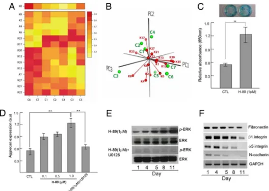
Fig. 1. Use of protein kinase inhibitors for the differentiation of rat MSCs. (A) The relative expression level of seven target cell markers (C1–C7) in the presence
of various kinase inhibitors are detected by sandwich ELISA, and normalized in the range of 0 to 1 for standardization. Kinase inhibitors used (at 1M) are from Calbiochem and are distinguished by the numerals after K. The kinases they preferentially inhibit are in parentheses: K0, no inhibitor; K1 (AKT 1,2), 1,3-dihydro-1-(1-((4-(6-phenyl-1H-imidazo[4,5-g]quinoxalin-7-yl)phenyl)methyl)- 4-piperidinyl)-2H-benzimidazol-2-one; K2 (AKT), 1L6-hydroxymethyl-chiro-inositol-2-(R)-2-O-methyl-3-O-octadecyl-sn-glycerocarbonate; K4 (CaMK II), Lavendustin (5-(N-2⬘,5⬘-dihydroxybenzyl) aminosalicylic acid); K6 (calcium channel), HA 1077 (Fasudil, 5-isoquinolinesulfonyl)homopiperazine, 2HCl); K8 (CaseinK I), D4476(4-(4-(2,3-dihydrobenzo[1,4]dioxin-6-yl)-5-pyridin-2-yl-1H-imidazol -2-yl)benzamide; K12 (CDK 1,2), NU6102 [6-cyclohexylmethoxy-2-(4⬘-sulfamoylanilino)purine]; K13 (TGF-R I kinase), [3-(pyridin-2-yl)-4-(4-quinonyl)]-1H-pyrazole; K17 (PKA), H-89 [N-[2-((p-bromocinnamyl)amino)ethyl]-5-isoquinolinesulfonamide, 2HCl]; K21 (PKC), Go¨ 6983 (2-[1-(3-dimethylaminopropyl)-5-methoxyindol-3-yl]-3-(1H-indol-3-yl)) maleimide; K22 (PKG-1␣,), guanosine 3⬘,5⬘-cyclic monophosphorothioate, -phenyl-1, N2-etheno-8-bromo-, Rp-isomer, sodium salt; K23 (EGFR PTK), compound 56 (4-[(3-bromophenyl)amino]-6,7-diethoxyquinazoline); K27 (ROCK), N- (4-pyridyl)-N⬘-(2,4,6-trichlorophenyl) urea; K30 (DNA-PK), 4,5-dimethoxy-2-nitrobenzaldehyde; K35 (p38 MAPK), SB 202190 [4-(4-fluorophenyl)-2-(4-hydroxyphenyl)-5-(4- pyridyl)1H-imidazole]. The target cells tested for each cell type were: C1, MSC positive; C2, MSC negative; C3, osteocyte; C4, chondrocyte; C5, cardiomycyte; C6, hepatocyte; C7, endothelium. The target cell markers assayed for each cell type were: C1, CD105; C2, CD34; C3, osteopontin (OPN); C4, aggrecan (AGG); C5, cardiac troponin T (CTnT); C6, CK-18, C7: CD31. (B) The effectiveness of inhibitors relative to the directed target cell development was analyzed by the principal component analysis (PCA) method using numerical values used to generate A. We obtained the coordinates of inhibitors and target cell types by using the first three principal components (for visualization) from PCA and scaled the two sets of the coordinates to plot them together in the map. The three largest principal components of PCA analysis are represented as PC1, PC2, and PC3. The inhibitors are indicated by red balls and the cell types are shown as green balls. The balls attached to the longer vectors are more efficient differentiation inducers for the cell types nearest to the balls. (C) Chondrogenesis of MSCs in vitro was significantly improved in cells treated with 1M H-89 for 11 days. The data of Alcian blue staining for sulfated proteoglycan indicate the quantitation of chondrogenesis. (D) Aggrecan, one of the extracellular matrix genes in chondrocytes, was induced in MSCs treated with various concentrations of H-89 for 11 days (0.1–1M). The effect of U0126, a selective inhibitor of MEK, was examined on the expression of aggrecan. Cotreatment with 1M H-89 and 10 M U0126 did not lead to a change of expression level. During chondrogenesis of MSC, the change of expression level in aggrecan was examined by sandwich ELISA. (E) Activation of ERKs was increased in MSCs treated with 1M H-89 but cotreatment with 1 M H-89 and 10 M U0126 did not lead to a change in ERK activation for 11 days. (F) Semiquantitative RT-PCR showed a significantly changed level of cell adhesion molecule during chondrogenesis, using primers to the RNAs indicated. MSCs were cultured for the indicated time periods in the presence of 1M H-89.**, P⬍ 0.01 for data from a typical experiment conducted more than three times.
Biosciences), a Rho kinase inhibitor, was the best inducer of
differentiation of ESCs into TH-positive neurons. H-1152 is
another cell-permeable isoquinolinesulfonamide derivative that
acts as a highly specific, potent, and ATP-competitive inhibitor
of G protein ROCK (K
i⫽ 1.6 nM). Among the kinases tested,
the inhibition is much weaker for other tested serine/threonine
kinases (e.g., K
i⫽ 630 nM for PKA, 9.27
M for PKC, and 10.1
M for MLCK) (14, 15).
The number of TH-positive cells among total neurons went up
to 55% by H-1152 treatment, which is a significant increase
compared with that from the untreated control (
⬇25–30%) (Fig.
2 A and B). At a concentration between 0.1 and 2
M, the
increase of TH expression by H-1152 occurred in a
dose-dependent manner without any indication of cytotoxicity (Fig.
2C). However, we noticed some cytotoxic effect of H-1152 at
high concentrations (
⬎5
M) (data not shown).
Semiquantitative RT-PCR analysis also supported the positive
role of H-1152 in the differentiation of ESCs into midbrain
dopamine neurons. The expression levels of three representative
midbrain dopamine neuronal markers, TH, Nurr1, and Pitx3,
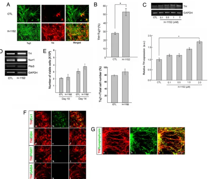
Fig. 2. Effect of treatment of H-1152, a kinase inhibitor, on differentiation of ESCs into TH-positive neurons. (A) Immunocytochemical analyses of TH-positive
neuronal generation from the H-1152-treated ESCs. ESCs were treated with H-1152 during days 5–9, which correspond to the neural precursor stage. Tuj1-positive cells (neurons) were stained with a green color, and TH-positive cells are shown in red. (Scale bar: 10m.) (B) Quantification of the ratios between the numbers of TH-positive and Tuj1-positive cells. TH-positive and Tuj1-positive cells were counted from 10 random fields per sample. Each group represents an average of three samples from independent experiment (*, P⬍ 0.05). Inhibitor concentration was 1M. (C) Dose-dependent effect of H-1152 measured by semiquantitative RT-PCR. TH gene expression went up as the concentration of H-1152 increased. The expression level of each gene was normalized to that of GAPDH. (D) Analyses of midbrain dopamine neuronal markers by semiquantitative RT-PCR. Treatment of ESCs with H-1152 resulted in an increase of dopamine neuronal markers (TH, Nurr1, and Pitx3) compared with the untreated control. The expression level of each gene was normalized to that of GAPDH. (E) Quantification of the number of total viable cells at days 10 and 14 (Left) and the ratios of Tuj1-positive cells among total cells at day 14 (Right). The number of viable cells was counted after staining the cells with trypan blue (n⫽ 3–4), and the proportion of neurons among total cells (Tuj1-positive cells per total cells) was determined after staining with Tuj1 antibody and DAPI. Each bar in Fig. 1E was from the counting from 10 random fields (n⫽ 3). Inhibitor concentration was 1M. (F) Immunocytochemical analyses of cells obtained after differentiation of H-1152-treated ESCs. The majority of TH-positive neurons coexpressed a midbrain dopamine neuronal marker, En1, but not DBH, PNMT, and GABA. (a, d, g, and j) Cells were stained with anti-TH. (b, e, j, and k) Cells were stained with anti-En1 (midbrain DA neuronal marker, b), anti-DBH (noradrenergic and adrenergic neuronal marker, e), anti-PNMT (adrenergic neuronal marker, h), and anti-GABA (k) antibodies. (c, f, i, and l) Merged images. (Scale bar: 10m.) (G) Expression of synaptophysin in TH-positive cells. (a and b) Differentiated cells were stained with antibodies against TH (a) and synaptophysin (b). (c) The coexpression of synaptophysin is an indication of synapse formation in the TH-positive cells. (Scale bar: 10m.)
CELL
were increased
⬇1.5- to 2.2-fold in H-1152-treated cells (Fig.
2D). There was no significant difference in the total number of
cells and neurons between H-1152-treated and untreated cells on
both days 10 and 14. For example, the total number of viable cells
and the percentage of neurons among total cells (Tuj1
⫹cells per
total cell) between H-1152-treated and untreated cells were
3.1
⫾ 1.0 ⫻ 10
6vs. 3.8
⫾ 1.1 ⫻ 10
6and 74.3
⫾ 2.43% vs. 81.2 ⫾
4.8%, respectively, on day 14, which was the end point of the
differentiation process (Fig. 2E). These results indicate that
H-1152 may regulate dopaminergic specification and
differen-tiation directly without affecting cell cycle or cell proliferation.
To confirm that the majority of TH-positive neurons
gener-ated in the presence of H-1152 retain characteristics of midbrain
dopamine neurons, we analyzed the TH-positive cell population
closely (Fig. 2F). First, we tried to determine whether the
TH-positive cell population contains other types of
catechol-amine neurons such as noradrenergic and adrenergic neurons.
To this end, a midbrain dopamine neuron-specific marker, En1,
and other catecholaminergic neuronal markers, such as
dopa-mine
-hydroxylase (DBH) and phenylethanolamine
N-methyltransferase (PNMT), were coimmunostained with TH.
Our results showed that most TH-positive cells expressed En1,
indicating that dopamine neurons are the major cell type in the
population (Fig. 2F a–c). This result is in line with the
obser-vation that only a few TH-positive cells expressed DBH, an
enzyme that converts dopamine into norepinephrine (Fig. 2F
d–f ), or PNMT, which catalyzes the formation of epinephrine
from norepinephrine (Fig. 2F g–i). These two enzymes are
known to be expressed in catecholaminergic neurons except
dopamine neurons. GABA expression was also not detected
among the TH-positive cells, indicating that the TH-positive
neurons are not the olfactory dopamine neurons (Fig. 2F j–l).
The TH-positive neurons generated in the presence of H-1152
also expressed synaptophysin, suggesting that the cells have the
capability to form synapses (Fig. 2G). Taken together, our results
suggest that the majority of TH-positive cells generated in the
presence of H-1152 are midbrain dopamine neurons that can
form synapses.
Conclusions and Discussions
Both observations described above, the enhanced differentiation
of rat MSCs to chondrocytes and of mouse ESCs to dopamine
neurons, support the notion that, although cell differentiation
may be the result of a complex orchestration of many signals
from multiple signaling pathways, even a single chemical
re-agent, a kinase inhibitor in this case, can alter the relative
balance of many signals enough to enhance differentiation of
stem cells to particular cell types. They also suggest that the
process may be optimized by a ‘‘mixture’’ of various multiple
kinase inhibitors. Interestingly, both inhibitors, H-89 and
H-1152, are two different derivatives of the same scaffold,
isoquinolinesulfonamide.
Because these compounds may interact with ‘‘off-target’’ kinases
as well as other unknown proteins, our observation should be
considered as a practical approach for finding chemical reagents for
inducing stem cell differentiation to a specific cell type even when
most of the signaling pathways are unknown. Thus, the approach
described or some variation of it may be applied to other stem cells,
including human stem cells, to find chemical molecules that can
trigger initiation, inhibition, or even reversion of the differentiation
process of stem cells or progenitor cells.
Materials and Methods
Isolation and Culture of MSCs. Isolation and primary culture of MSCs from the femoral and tibial bones of donor rats were performed. Bone marrow-derived mesenchymal stem cells were collected from the aspirates of the femurs and tibias of 4-week-old Sprague–Dawley male rats (⬇100 g) with 10 ml of MSC medium consisting of DMEM-low glucose supplemented with 10% FBS (Gibco)
and 1% antibiotic-penicillin and streptomycin solution (Gibco). Mononuclear cells recovered from the interface of Percoll-separated bone marrow were washed twice and resuspended in 10% FBS-DMEM, and plated at 1⫻ 106cells
per 100 cm2in flasks. Cultures were maintained at 37oC in a humidified
atmosphere containing 5% CO2. After 48 or 72 h, nonadherent cells were
discarded, and the adherent cells were thoroughly washed twice with PBS. Fresh complete medium was added and replaced every 3 or 4 days for⬇10 days. To further purify the MSCs, the Isolex Magnetic Cell Selection System (Baxter Healthcare) was used. Briefly, cells were incubated with Dynabeads M-450 coated with anti-CD34 mAb. A magnetic field was applied to the chamber, and the CD34⫹cell-bead complexes were separated magnetically from the remaining cell suspension with the CD34⫺fraction being further cultured. The cells were harvested after incubation with 0.25% trypsin and 1 mM EDTA (Gibco) for 5 min at 37°C, replated in 1⫻ 105per 100-cm2plates, and
again grown for⬇10 days.
ESCs Culture andin Vitro Differentiation. Undifferentiated mouse ESCs (J1)
were maintained on gelatin-coated dishes in DMEM (Gibco) supplemented with 2 mM glutamine (Gibco), 0.001%-mercaptoethanol (Sigma), 1⫻ non-essential amino acids (Gibco), 2,000 units/ml human recombinant leukemia inhibitory factor (LIF) (Chemicon International), 50 units/ml penicillin and 50 g/ml streptomycin (Gibco), and 10% FBS (Gibco). PA6 cells were purchased from Riken and maintained in␣-MEM (Gibco) supplemented with penicillin and streptomycin (Gibco), and 10% FBS (Gibco). To differentiate ESCs in vitro, PA6 cells were plated on gelatin-coated culture dishes to make a uniform feeder monolayer 1 day before the addition of J1, and then ESCs were added at a density of 1⫻ 103per well of a 24-well plate and four-well plate. ES
differentiation medium I [Glasgow minimum essential medium (G-MEM) (Gibco) supplemented with 10% knockout serum replacement (Gibco), 0.1 mM nonessential amino acid (Gibco), 1 mM sodium pyruvate (Sigma), 0.1 mM -mercaptoethanol (Sigma), and penicillin and streptomycin (PEST) (Gibco)] was used for 8 days and then replaced with embryonic stem differentiation medium II [G-MEM (Gibco) supplemented with 1⫻ N2 supplement (Gibco), 0.1 mM nonessential amino acid (Gibco), 1 mM sodium pyruvate (Sigma), 0.1 mM -mercaptoethanol (Sigma), and PEST (Gibco)] for an additional 6 days. The culture medium was changed on day 4 and every other day thereafter. Screening of Protein Kinase Inhibitors for Differentiation of MSCs and ESCs. We assembled a small library (41 compounds) of commercially available protein kinase inhibitors that are known to inhibit various members of protein kinase subfamilies (16). Of these, a subset of compounds was selected based on their low cytotoxicity. These compounds were screened for their activity to differ-entiate rat MSCs to chondrocytes and mouse ESCs to dopamine neurons. Alcian Blue Staining. Cells were first rinsed with PBS (Gibco) three times then fixed with 100% methanol (Sigma) for 10 min at⫺20°C. Staining was
accom-plished by applying a solution of 1% Alcian blue 8GX (Bio Basic) in 0.1 M HCl (pH 1.0) (Sigma) to the cells for 2 h at room temperature. To quantify the intensity of the staining, the stained culture plates were rinsed with PBS three times, and each well was extracted with 1 ml of 6 M guanidine-HCl (Sigma) overnight at room temperature. The optical density of extracted dye was measured at 650 nm.
Sandwich ELISA. The capture antibody was bound to the bottom of each well and then the plate was incubated overnight at 4°C. The plate was washed
twice with PBS (Gibco) and treated with 100l of 3% BSA (Sigma) /PBS for ⬇2–3 h at 37°C at room temperature. After washing the plate twice with PBS,
cell lysate was added to each well, and the plate was incubated for at least 2 h at room temperature in a humid atmosphere. The plate was washed four times with PBS containing 0.02% Tween-20 (Sigma). After addition of detector antibody, the plate was incubated for 2 h at room temperature in a humid atmosphere. The plate was then incubated with peroxidase-conjugated sec-ondary Ab for 1 h at 37°C. Finally, the plate was treated with 100l of
tetramethylbenzidine (Sigma) as substrate and 25l of 0.1 M H2SO4as stop
buffer, then detected immediately at 450 nm on an ELISA plate reader. Immunostaining. After 14 days of differentiation on PA6 feeder layer, ESCs were fixed with 4% formaldehyde for 30 min, washed with PBS, and perfo-rated with PBS containing 0.1% Triton X-100 for 10 min. Then coverslips were incubated with blocking buffer [PBS containing 5% normal donkey serum (NDS)] for 1 h. Cells were incubated at room temperature with primary antibodies diluted in PBS containing 5% NDS for 1 h. The following primary antibodies were used: mouse anti-neuronal class III-tubulin (1:1,000; Co-vance), rabbit anti-TH (1:300; Pel-Freeze), mouse anti-TH (1:100; Chemicon), mouse En1 (1:50; Developmental Studies Hybridoma Bank), sheep
DBH (1:250; Chemicon), rabbit PNMT (1:200; Chemicon), and rabbit anti-mammalian GABA antibodies (1:2,500; Sigma). The cells were washed with PBS and then incubated with fluorescent-labeled secondary antibodies [Alexa Fluor 488 (green) or Alexa Fluor 594 (red)-labeled mouse/rabbit IgG (1:1,000; Molecular Probes/Invitrogen)] in PBS with 5% NDS for 30 min at room tem-perature. The coverslips were rinsed three times for 5 min in PBS and mounted onto slides by using VECTASHIELD Hardset mounting medium with DAPI (Vector Laboratories). Images were obtained under a fluorescence microscope (Olympus IX71) and confocal microscope.
Immunoblot Analysis. Cells were washed once in PBS and lysed in a lysis buffer (Cell Signaling) containing 20 mM Tris (pH 7.5), 150 mM NaCl, 1 mM Na2EDTA,
1 mM EGTA, 1% Triton, 2.5 mM sodium pyrophosphate, 1 mM -glycerophos-phate, 1 mM Na3VO4, 1 mg/ml leupeptin, and 1 mM PMSF. Protein
concen-trations were determined by using the Bradford protein assay kit (BioRad). Proteins were separated in a 12% SDS-polyacrylamide gel and transferred to PVDF membrane (Millipore). After blocking the membrane with Tris-buffered saline-tween 20 (TBS-T, 0.1% Tween 20) containing 5% nonfat dried milk for 1 h at room temperature, the membrane was washed twice with TBS-T and incubated with primary antibodies for 1 h at room temperature or overnight at 4°C. The membrane was washed three times with TBS-T for 10 min, and then
incubated for 1 h at room temperature with HRP-conjugated secondary antibodies. After extensive washing, the bands were detected by ECL reagent (Santa Cruz Biotechnology). The band intensities were quantified by using the Photo-Image System (Molecular Dynamics).
RT-PCR Analysis. The expression levels of various genes were analyzed by RT-PCR. Total RNA was prepared by using the Ultraspec-II RNA system (Biotecx Laboratories) and easy-BLUE (Intron Biotechnology). Single-stranded cDNA was then synthesized from isolated total RNA by avian myeloblastosis virus (AMV) reverse transcriptase (Power cDNA Synthesis Kit; Intron Biotechnol-ogy). A 20-l reverse transcription reaction mixture containing 1 g of total RNA, 1⫻ reverse transcription buffer (10 mM Tris䡠HCl, pH 9.0, 50 mM KCl, 0.1% Triton X-100), 1 mM deoxynucleoside triphosphates (dNTPs) 0.5 unit of RNase inhibitor, 0.5g of oligo(dT)15, and 15 units of AMV reverse transcriptase was
incubated at 42°C for 60 min, heated to 99°C for 5 min, and then incubated at
0 –5°C for 5 min. PCR was performed by using a standard procedure with Taq
Polymerase (i-Max DNA Polymerase; Intron Biotechnology). The number of cycles varied from 25 to 40 cycles with denaturation at 94°C for 30 s, annealing
at 58°C to 65°C for 30 s, and elongation at 72°C for 30 s. Primers were as follows:
fibronectin, 5⬘-CCTTAAGCCTTCTGCTCTGG-3⬘, 5⬘-CGGCAAAAGAAAGCA-GAACT-3⬘ (300 bp); 1 integrin, GCCAGTGTCACCTGGAAAAT-3⬘, 5⬘-TCGTCCATTTTCTCCTGTCC-3⬘ (344 bp);␣5 integrin, 5⬘-CTTCGGTTCACTGTTC-CTC-3⬘, 5⬘-TGGCTTCAGGGCATTT-3⬘ (283 bp); N-cadherin, 5 ⬘-GCCACCATATGACTCCCTTTTAGT-3⬘, 5⬘-CAGAAAACTAATTCCAATCTGAAA-3⬘ (454 bp); TH, 5⬘-GAGAGGACAGCAT TCCACAG-3⬘, 5⬘-TCACGGGCAGACAGTA-GACC-3⬘ (100 bp); Nurr1, 5⬘-GCTAA ACAAAACTTGCATGC-3⬘, 5⬘-CTCATATCAT-GTGCCATACTAG-3⬘ (208 bp); and Pitx3, 5⬘-CTAGACCTCCCTCCATGGAG-3⬘, 5⬘-TCTTGAACCACACCCGCACG-3⬘ (348 bp). The GAPDH primers [5⬘-CTC-CCAACGTGTCTGTTGTG-3⬘, 5⬘-TGAGCTTGA CAAAGTGGTCG-3⬘ (450 bp) and 5⬘-ACCACAGTCCATGCCATCAC-3⬘, 5⬘-TCCACCACCCTGTTGCT GTA-3⬘ (450 bp)] were used as the internal standard. The signal intensity of the amplification product was normalized to its respective actin signal intensity.
Viable Cell Counting. Cultured cells were washed with PBS, detached from the dish with 1⫻trypsin/EDTA, and then collected in the medium. After spinning down, the cell pellet was resuspended in trypan blue solution for counting under a microscope.
Statistical Analysis. Results are expressed as mean⫾ SEM. Statistical analysis
was performed by Student’s t test. Relationships were considered statistically significant with P⬍ 0.05.
ACKNOWLEDGMENTS. This research was supported by Grant SC4001 from the Stem Cell Research Center of the 21st Century Frontier Research Program funded by the Ministry of Science and Technology (Republic of Korea), Korea Science and Engineering Foundation Grant M1064102000106N410200110 funded by the Ministry of Science and Technology, and Grant KGM1310713 from the Korea Research Council of Fundamental Science and Technology/ Korean Research Institute of Bioscience and Biotechnology Research Program of the Ministry of Science and Technology.
1. Jaiswal RK, et al. (2000) Adult human mesenchymal stem cell differentiation to the osteogenic or adipogenic lineage is regulated by mitogen-activated protein kinase.
J Biol Chem 275:9645–9652.
2. Castelo-Branco G, Rawal N, Arenas E (2004) GSK-3 inhibition/-catenin stabilization in ventral midbrain precursors increases differentiation into dopamine neurons. J Cell
Sci 117:5731–5737.
3. Hu X, Jin L, Feng L (2004) Erk1/2 but not PI3K pathway is required for neurotrophin 3-induced oligodendrocyte differentiation of postnatal neural stem cells. J Neurochem 90:1339 –1347.
4. Koyanagi M, et al. (2005) Noncanonical Wnt signaling enhances differentiation of human circulating progenitor cells to cardiomyogenic cells. J Biol Chem 280:16838 –16842. 5. Ding S, et al. (2003) Synthetic small molecules that control stem cell fate. Proc Natl Acad
Sci USA 100:7632–7637.
6. Manning G, et al. (2002) The protein kinase complement of the human genome.
Science 298:1912–1934.
7. Pittenger MF, et al. (1999) Multilineage potential of adult human mesenchymal stem cells. Science 284:143–147.
8. Leemhuis J, Boutillier S, Schmidt G, Meyer DK (2002) The protein kinase A inhibitor H89 acts on cell morphology by inhibiting Rho kinase. J Pharmacol Exp Ther 300:1000 –1007.
9. Davies SP, Reddy H, Caivano M, Cohen P (2000) Specificity and mechanism of action of some commonly used protein kinase inhibitors. Biochem J 351:95–105.
10. Kawasaki H, et al. (1998) A family of cAMP-binding proteins that directly activate Rap1.
Science 282:2275–2279.
11. Combest WL, Bloom TJ, Gilbert LI (1998) Polyamines differentially inhibit cyclic AMP-dependent protein kinase-mediated phosphorylation in the brain of the tobacco hornworm, Manduca sexta. J Neurochem 51:1581–1591.
12. Kawasaki H, et al. (2000) Induction of midbrain dopaminergic neurons from ES cells by stromal cell-derived inducing activity. Neuron 28:31– 40.
13. Kim DW, et al. (2006) Stromal cell-derived inducing activity, Nurr1, and signaling molecules synergistically induce dopaminergic neurons from mouse embryonic stem cells. Stem Cells 24:557–567.
14. Ikenoya M, et al. (2002) Inhibition of rho-kinase-induced myristoylated alanine-rich C kinase substrate (MARCKS) phosphorylation in human neuronal cells by H-1152, a novel and specific Rho-kinase inhibitor. J Neurochem 81:9 –16.
15. Sasaki Y, Suzuki M, Hidaka H (2002) The novel and specific Rho-kinase inhibitor (S)-(⫹)-2-methyl-1-[(4-methyl-5-isoquinoline)sulfonyl]-homopiperazine as a probing molecule for Rho-kinase-involved pathway. Pharmacol Ther 93:225–232.
CELL