See discussions, stats, and author profiles for this publication at: https://www.researchgate.net/publication/6837168
Inhibitory effects of tilianin on the expression
of inducible nitric oxide synthase in low density
lipoprotein receptor deficiency mice
Article in Experimental and Molecular Medicine · September 2006 Impact Factor: 3.45 · DOI: 10.1038/emm.2006.52 · Source: PubMed CITATIONS10
READS17
14 authors, including: Kim Misun 268 PUBLICATIONS 6,549 CITATIONS SEE PROFILE Sei-Ryang Oh Korea Research Institute of Bioscience and … 204 PUBLICATIONS 2,093 CITATIONS SEE PROFILEAll in-text references underlined in blue are linked to publications on ResearchGate, letting you access and read them immediately.
Available from: Sei-Ryang Oh Retrieved on: 19 May 2016
Ki-Hoan Nam
3, Jae-Hoon Choi
1,
Yun-Jeong Seo
1, Young-Mi Lee
1,
Yong-Sung Won
4, Mi-Ran Lee
1,
Mi-Ni Lee
1, Jong-Gil Park
1,
Young-Myeong Kim
2, Hyoung-Chin Kim
3,
Chul-Ho Lee
3, Hyeong-Kyu Lee
3,
Sei-Ryang Oh
3and Goo Taeg Oh
1,5 1Division of Molecular Life Sciences andCenter for Cell Signaling Research Ewha Womans University
Seoul 120-750, Korea
2Vascular System Research Center Kangwon National University Chuncheon 200-701, Korea
3Korea Research Institute of Bioscience and Biotechnology Daejeon 305-333, Korea
4Department of Surgery St. Vincent’s Hospital
The Catholic University of Korea Suwon 442-723, Korea
5Corresponding author: Tel, 82-2-3277-4128; Fax, 82-2-3277-3760; E-mail, gootaeg@ewha.ac.kr Accepted 24 July 2006
Abbreviations: HUVEC, human umbilical endothelial cell; Ldlr, low density lipoprotein receptor; NO, nitric oxide; NOS, nitric oxide synthase; VCAM-1, vascular cell adhesion molecule-1
Abstract
We investigated the effect of tilianin upon inducible nitric oxide synthesis in the plasma of low-density lipoprotein receptor knock-out (Ldlr-/-) mice fed with high cholesterol diet and in primary peritoneal macrophages of Ldlr-/- mice. High cholesterol diet induced nitric oxide production in the plasma of Ldlr-/- mice. Tilianin reduced the level of nitric oxide (NO) in plasma from Ldlr-/- mice induced by the high cholesterol diet. Tilianin also inhibited the NO production from the primary culture of peritoneal macrophages treated with lipopolysaccharide. The inhibition of NO production was caused by the suppression of inducible nitric oxide synthase (iNOS) gene expression in peritoneal macrophages isolated from Ldlr-/- mice. Moreover, tilianin inhibited the
transcriptional activation of iNOS promoter that has NF-κB binding element. Thus, these results provide the first evidence that tilianin inhibit iNOS expression and production of NO and may act as a potential anti-inflammatory agent.
Keywords: anti-inflammatory agents; hyperlipopro-teinemia; nitric oxide synthase type II; nitric oxide; tilianin
Introduction
NO is a gaseous free radical with very short half-life in biological system and can act both as a weak oxidant and as a reductant (Kanner et al., 1991). NO is synthesized in mammals by the nitric oxide synthases (NOS). There are three NOS isoforms: endothelial NOS (eNOS), neuronal NOS (nNOS), and inducible NOS (iNOS). Unlike eNOS and nNOS, iNOS is not constitutively expressed in macrophages and smooth muscle cells of inflammatory vascular tissues. In human atherosclerotic lesions, however, all three isoforms can be demonstrated (Wilcox et al., 1997). Especially, iNOS expression has been lo-calized to vascular smooth muscle cells and mo-nonuclear leukocytes in early and advanced athero-sclerosis (Esaki et al., 1997).
Murine iNOS expression has been observed in various types of cells, including macrophages (Hibbs et al., 1988), endothelial cells (Gross et al., 1991), smooth muscle cells (Beasley et al., 1991), and cardiac myocytes (Schulz et al., 1992). In human patients with infectious or inflammatory disease, iNOS is most readily observed in monocytes or macrophages (MacMicking et al., 1997). Therefore, in contrast to low level of NO produced by eNOS (Wever et al., 1998), excessive NO prouction by iNOS in macrophages is considered to be proa-therogenic (Buttery et al., 1996; MacMicking et al., 1997). NO interacts with superoxide to form the strong oxidant peroxynitrite (ONOO-) (Beckman et
al., 1990), which induces lipid peroxidation and nitro-sylation of tyrosine residue in human atherosclerotic lesions (Beckmann et al., 1994; Wilcox et al., 1997). In addition, it has been shown that suppression of NO by NOS inhibitor or iNOS gene deficiency (iNOS-/-) resulted in reduced atherosclerosis (Behr-Roussel et al., 2000; Detmers et al., 2000). Thus excessive NO has the potential to exacerbate
Inhibitory effects of tilianin on the expression of inducible nitric
oxide synthase in low density lipoprotein receptor deficiency mice
446 Exp. Mol. Med. Vol. 38(4), 445-452, 2006
atherosclerosis.
In our previous study, we have reported that tilianin, a major component of Agastache rugosa (Labiatae) significantly inhibits the tumor necrotic factor- (TNF- )-induced expression of vascular cell adhesion molecule-1 (VCAM-1) in human umbilical vein endothelial cells (HUVEC) as its potential anti- atherogenic mechanism (Hong et al., 2001; Nam et al., 2005). However, it has not been determined the effect of tilianin on NO production. Here we report that tilianin potently inhibited NO production by iNOS, another possible anti-atherogenic mechanism. We have demonstrated the inhibitory effects in in vivo as well as in vitro.
Materials and Methods
Animal modelAll animal experiments were approved by Insti-tutional Animal Care and Use Committees (IACUC) of Ewha Womans University and followed National Research Council Guidelines. Twenty-five low den-sity lipoprotein receptor-null (Ldlr-/-) male mice were divided into three groups (normal diet fed, n = 5, high-cholesterol diet (HCD) only; n = 10, HCD supplemented with tilianin; n = 10). The HCD contained 1.25% cholesterol, 6% fat and 0.5% cholic acid (CRF-1, Oriental Yeast Co., Ltd., Chiba, Japan). Tilianin was supplemented with 0.1% (wt/wt) of HCD. After 8 week experiment, plasma from the mice were collected and subjected to the mea-surement of total nitric oxide.
Histological finding and measurement of fatty streak lesion
The frozen sections of the aortic sinus were reacted with a rabbit anti-mouse iNOS antibody (Santa Cruz Biotechnology, CA) and a rabbit anti-nitrotyrosine antibody (Upstate Biotechnology, NY) at 1:200 dilutions, then the reactivities were detected using ABC kit (Vector Laboratory, CA). Fatty streak lesions were quantified by evaluating the lesion size in the aortic sinus, as previously described (Paigen et al., 1987). All hearts were sectioned using a cryostat at -20oC and six consecutive 9 m-thick sections were cut from the aorta where the valve cusp becomes visible. Atherosclerotic plaque was stained with Oil red O and counter-stained with Harris hematoxylin. The lesion area was then quantified by compu-ter-assisted morphometry (Image pro plus, MD) and the average lesion size was calculated for each animal.
Determination of NO synthesis
Nitric oxide (NO) production was measured by monitoring levels of nitrite plus nitrate (NOx) in
plasma using automatic NO analyzer described previously (Morita et al., 1994). Nitrite in culture media was measured spectrophotometrically as described (Sherman et al., 1993) using the Griess reagent.
Culture of peritoneal macrophages
Mice were injected with 2 ml of 4% thioglycolate broth. After 6 days, peritoneal macrophages were isolated from peritoneal lavage of mice. Peritoneal macrophages were cultured in Dulbecco’s modified eagle medium (DMEM) supplemented with 10% fetal bovine serum, 2 mM L-glutamine, penicillin (100 U/ml), and streptomycin (100 g/ml).
Immunoblot analysis
The isolated peritoneal macrophages were incubat-ed with tilianin (1, 10, 100 M) for 2 h and imme-diately activated by LPS (2 g/ml) at 37oC for 18 h.
The macrophages were harvested, washed with PBS containing 0.1 mM PMSF, and lysed by three cycles of freezing and thawing in liquid nitrogen. Cytosolic fraction was obtained by centrifugation at 12,000 g at 4oC for 10 min. The concentration of
protein was analyzed by the Lowery method. Sam-ples (50 g) were separated on 8% SDS-PAGE and transferred to nitrocellulose membrane (Schleicher & Schuell, Keene, NY). The detection of iNOS was performed with anti-iNOS antibody (Santa Cruz Biotechnology, CA) and ECL western blotting analy-sis system (Amersham, UK).
Analysis of iNOS mRNA
The expressions of iNOS was determined by semi- quantitative RT-PCR, using -actin as an internal
control. Primary peritoneal macrophages (6 × 10⁶
cells per 100 mm culture dish) were pre-treated with tilianin (1, 10, and 100 M) for 2 h, and stimulated with LPS (2 g/ml) for 18 h. Total RNA was extracted from the macrophages using a TRIZOL (Gibco BRL, MD) reagent. The forward primer for mouse iNOS (1) 5'-CTG CAG CAC TTG GAT CAG GAA CCT G-3', and the reverse primer (2) 5'- GGG AGT AGC CTG TGT GCA CCT GGA A -3' define an amplicon of 311 bp. The forward primer for -actin (1) 5'-TGG AAT CCT GTG GCA TCC ATG AAA C-3', and the reverse primer (2) 5'-TAA AAC GCA GCT CAG TAA CAG TCC G-3' define an amplicon of 349 bp. The densitometric analysis was performed with Image Analysis Software (Bio 1D Version 99, Vilber
Lour-mat, France) to determine the relative band density. The internal control was used to avoid saturation of PCR product.
Transient transfection assay
A spontaneously transfected mouse monocyte/ma-crophage cell (RAW264.7) was used because of their high transfection efficiency. Cells were plated in 6-well plates at 5 × 10⁵ cells/well in DMEM (Gibco BRL, MD) containing 10% fetal calf serum. After growing at 37oC for 20 h, 3 g of piNOS-Luc
plas-mids containing iNOS promoter (-1,029~ + 159) (Lo-wenstein et al., 1993) and luciferase reporter were transfected using FuGENE6 (Roche, Mannheim, Germany) according to the manufacturer’s direc-tions. For NF- B activation, pNF- B-Luc plasmid (5
×NF- B; Stratagene, CA) was transfected to
RAW264.7 cells. The control plasmid pCMVb (1 g)
(Clontech, CA) was co-transfected to monitor the transfection efficiency. After 20 h of transfection, medium was changed and tilianin (1, 10, and 100 M) was pretreated for 2 h before LPS (200 ng/ml) treatment. After additional 48 h cultures, cells were then harvested and luciferase activities were deter-mined using the Dual Luciferase assay (Promega, WI). The luciferase activity was normalized for trans-fection efficiency after determining the -galacto-sidase activity of the same sample. Each trans-fection was carried out in triplicate and experiments were repeated at least four times.
Electrophoretic mobility shift assay
Nuclear extracts were prepared from cells by lysing them in buffer A [10 mM HEPES (pH 7.5), 1.5 mM
MgCl2, 10 mM KCl, and 0.1% Nonidet P-40].
Sub-sequently, the solution was centrifuged to pellet the
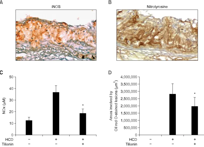
Figure 1. Localization of proatherogenic molecules in atherosclerotic lesions. iNOS (A, × 200) and nitrotyrosine (B, × 200) were detected by immunohistochemistry. Frozen sections of aortic segment were stained with polyclonal antibody for each molecule and detected using an ABC method. Section was then counterstained with Harris’ hematoxylin. Control staining without primary antibodies did not show any significant staining in atherosclerotic lesion (data not shown). (C) Effect of tilianin on the plasma NO level in HCD-fed Ldlr-/- mice. The plasma was obtained from mice fed normal diet only, HCD only, and HCD supplemented with tilianin (0.01% wt/wt diet). (D) Oil red O stained aortic valve lesion areas were quantified by computer-associated morphometry. All results are shown as mean ± SD. * indicates P < 0.01 compared with HCD only group.
448 Exp. Mol. Med. Vol. 38(4), 445-452, 2006
nuclei, and the collected nuclei was suspended in
buffer B [20 mM HEPES (pH 7.5), 1.5 mM MgCl2,
0.42 M NaCl, 0.2 mM EDTA, and 25% glycerol] and incubated on ice for 30 min. Following centrifugation, the nuclear extracts in the supernatant were har-vested and protein concentrations were determined by Bradford method (Bio-Rad Laboratories, Inc., CA). For Electrophoretic mobility shift assays, 5 g
of nuclear protein was reacted with [32P]- ATP-
labeled NF- B oligonucleotide (Promega, WI) in presence of 1 g of poly (dI-dC) (Sigma). To check for specificity, antibody against the p65 subunit of NF- B (Santa Cruz Biotechnology, CA) was added to the binding reaction. Nuclear extracts-oligonu-cleotide mixtures were then subjected to eletro-phoresis on 5% nondenaturing polyacrylamide gels. Gels were then dried and visualized by auto-radiography.
Statistics
The data are expressed as the mean ± SD. Statistical significance was determined by Student’s t test.
Results
Localization of iNOS, nitrotyrosine in atheromatous plaques
We confirmed the iNOS expression in atheromatous plaque of Ldlr-/- mice fed HCD by immunohistoche-mistry using antibodies specific for mouse iNOS. Control staining without primary antibodies showed no staining in atherosclerotic lesion (data not shown). Localization of iNOS protein was prominent in the atheromatous thickened intima (Figure 1A). No immunoreactivity for nitrotyrosine was detected in normal aortic sinus area (data not shown). Nitro-tyrosine, however, was easily detected in the corresponding lesion site in which prominent iNOS expression was detected (Figure 1B). These results indicate that iNOS expression in atheromatous plaques is associated with the formation of nitro-tyrosine.
Effects of tilianin on plasma levels of NO and lesion formation
We next examined the effect of tilianin on the plasma level of NO. Ldlr-/- mice fed with HCD resulted in increased plasma level of NOx, a stable
oxidized product of NO. In mice fed with HCD supplemented tilianin, however, the NOx level was
significantly decreased by about 50% (Figure 1C). In correspondence to the lowered NOx level, the
lesions induced by HCD were decreased by the treatment of tilianin, similar to our previous study (Nam et al., 2005) (Figure 1D).
Tilianin inhibits the NO production and iNOS expression under in vitro inflammatory condition
Since tilianin has anti-atherogenic effect, the de-crease of serum NO level may reflect the conse-quence of the reduction of inflammatory cell accu-mulation in the lesion area. Thus, we investigate whether tilianin directly affect the production of NO and the expression of iNOS gene in peritoneal macrophages stimulated with LPS (2 g/ml) in the presence of various concentrations of tilianin. LPS- induced increased NO production was decreased in concentration-dependent manner by tilianin (Figure 2). Moreover, increased expression of iNOS mRNA and protein in macrophages stimulated by LPS was markedly inhibited in a dose-dependent manner by the treatment of tilianin (Figure 3A, B and C, respectively). These results suggest that tilianin inhi-bits effectively the NO production by modulating iNOS expression under in vitro inflammatory con-dition.
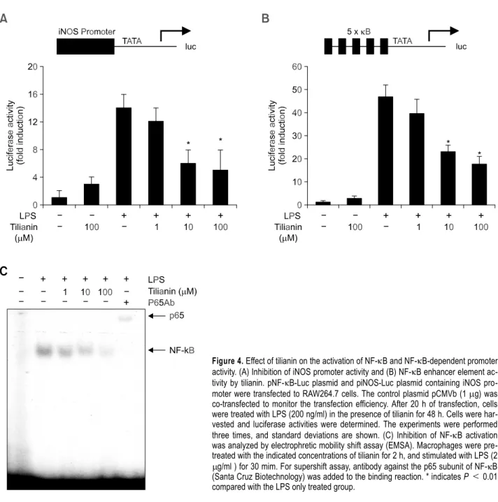
Tilianin inhibits iNOS promoter activity and NF-κB activation
We examined whether tilianin suppressed iNOS promoter activity. Transient transfection in the mouse
Figure 2. Effect of tilianin on NO production in LPS (2 g/ml) activated peritoneal macrophage. Inhibition of NO production in LPS activated peritoneal macrophage by tilianin. The primary peritoneal macro-phages from Ldlr-/- mice were incubated with tilianin (1, 10, 100 M) for 2 h and then activated with LPS (2 g/ml) for 18 h. The results are expressed as mean ± SD (n = 3). NO production was measured by Griess reaction. *,** indicates P < 0.05 and P < 0.01 compared with HCD only group, respectively.
macrophage cell line RAW264.7 cells was perform-ed with a murine iNOS promoter construct (piNOS- Luc) by lipofection. Treatment of the transfected cells with LPS and IFN- resulted in a 15-fold increase in luciferase activity. This increase was
suppressed in a dose-dependent manner by in-creasing concentrations of tilianin (Figure 4A). The expression of iNOS has been reported to be regulated by NF- B activation (Barnes and Karin, 1997). Therefore, it is possible that tilianin inhibits NO production in peritoneal macrophages by inhi-bition of NF- B activation. To investigate the roles of tilianin in NF- B dependent gene transcription, we conducted a transient transfection assay using a DNA construct containing SV40 promoter, 5 repeats of the consensus NF- B binding sequence, and the luciferase reporter gene. Stimulation of the trans-fectant with LPS resulted in an approximate 45-fold increase in luciferase activity, and this increase was inhibited dose-dependently by tilianin treatment (Figure 4B). These results indicate that tilianin inhibits NF- B activation and may subsequently suppress iNOS and inflammatory cytokines of which expressions are NF- B-dependent. Next, the effect of tilianin on NF- B activation in peritoneal ma-crophages was evaluated by electrophoretic mobility shift assay. The nuclear extract from LPS-stimulated primary macrophages showed an increase in NF- B- DNA binding activity, whereas any protein-oligo-nucleotide complex was not detected in unsti-mulated control cells. The binding activity was sup-pressed in a dose-dependent manner by the addi-tion of tilianin (Figure 4C). Specificity of the DNA- protein interaction for NF- B was demonstrated by the presence of antibody for the p65 subunit of NF- B in the complex using a supershift assay (Figure 4C).
Discussion
We investigated the effect of tilianin upon inducible nitric oxide production in vivo and in vitro. We de-monstrated that tilianin reduced the level of nitric oxide in plasma from Ldlr-/- mice fed with HCD and that tilianin suppressed the expression of iNOS gene in peritoneal macrophages isolated from Ldlr-/- mice. This compound also inhibited the transcriptional activation of iNOS promoter that has NF- B binding element. Thus, these results provide the first evidence that tilianin inhibit iNOS expression and NO production, suggesting a possible anti-athero-genic mechanism of tilianin in hyperlipidemic model mice.
Nitric oxide produced by iNOS inhibits the pro-liferation and induces apoptosis in vascular endo-thelial cells (Cornwell et al., 1994; Fukuo et al., 1996). It also produces the powerful oxidant pero-xynitrite (ONOO-) by interacting with the reactive oxygen superoxide (Beckman and Koppenol, 1996). Peroxynitrite has been strongly implicated as a Figure 3. Effect of tilianin on iNOS expression in LPS (2 g/ml)
acti-vated peritoneal macrophage. (A) Inhibition of iNOS expression in peri-toneal macrophage by tilianin. iNOS expression was determined by im-munoblot analysis as described in Methods. (B) Determination of iNOS mRNA expression levels by semi-quantitative RT-PCR. (C) A graph showing relative iNOS mRNA level in B. The primary peritoneal macro-phages from Ldlr-/- mice were incubated with tilianin (1, 10, 100 M) for 2 h and then activated with LPS (2 g/ml) for 18 h. The results are expressed as mean ± SD (n = 3). NO production was measured by Griess reaction. * indicates P < 0.05 compared with HCD only group, respectively.
450 Exp. Mol. Med. Vol. 38(4), 445-452, 2006
cytotoxic effector molecule contributing to cellular damage and promoting the formation of athero-sclerotic lesion (Wilcox et al., 1997). However, there are several conflicting papers regarding the effect of iNOS inhibition on atherosclerotic lesion formation using genetically engineered mice. When fed with normal diet, iNOS/ApoE double knockout mice did not have any different lesion compared to ApoE littermate controls (Knowles et al., 2000). Further-more, iNOS knockout mice fed with normal diet have hypertension, a two fold increase in plasma chole-sterol level, and aortic atherosclerotic lesion (Ihrig et al., 2001). On the contrary, when fed with athero-genic diet, the lesion size was decreased in iNOS/
ApoE double knockout mice compared to littermate ApoE-/- controls (Detmers et al., 2000; Kuhlencordt et al., 2001). These findings suggest that excessi-vely elevated plasma lipid level induced by athero-genic diet discloses the proatherathero-genic potential of iNOS. In the present study, we investigated the possibility that tilianin would prevent atherosclerosis induced by HCD. Our data showed that iNOS expression and nitrotyrosine formation were detect-ed in the atheromatous lesion, and tilianin effectively reduced NO synthesis in vitro or in vivo, suggesting that the inhibitory effect of tilianin on the production of NO and peroxynitrite is plausible mechanism responsible for its antiatherogenic activity.
Figure 4. Effect of tilianin on the activation of NF- B and NF- B-dependent promoter activity. (A) Inhibition of iNOS promoter activity and (B) NF- B enhancer element ac-tivity by tilianin. pNF- B-Luc plasmid and piNOS-Luc plasmid containing iNOS pro-moter were transfected to RAW264.7 cells. The control plasmid pCMVb (1 g) was co-transfected to monitor the transfection efficiency. After 20 h of transfection, cells were treated with LPS (200 ng/ml) in the presence of tilianin for 48 h. Cells were har-vested and luciferase activities were determined. The experiments were performed three times, and standard deviations are shown. (C) Inhibition of NF- B activation was analyzed by electrophretic mobility shift assay (EMSA). Macrophages were pre-treated with the indicated concentrations of tilianin for 2 h, and stimulated with LPS (2
g/ml ) for 30 mim. For supershift assay, antibody against the p65 subunit of NF- B (Santa Cruz Biotechnology) was added to the binding reaction. * indicates P < 0.01 compared with the LPS only treated group.
We further demonstrated that iNOS expression was significantly lowered by treatment of tilianin. Promoter region of iNOS contains NF- B binding sequence, and NF- B activation is required for iNOS gene expression (Lowenstein et al., 1993). We examined the role of tilianin in iNOS expression and its promoter activity in vitro. Transient transfection with reporter genes with NF- B binding elements to RAW264.7 cells showed clearly that tilianin inhibited significantly NF- B-dependent iNOS expression, which is responsible for reduced NO production. In conclusion, tilianin significantly inhibit NO production and iNOS expression through the inhibition of NF- B dependent transcription. Thus, the inhibition of NO production could be a novel possible mechanism of the anti-atherogenic potential of tilianin. And, it also implies that tilianin has potential as an anti-inflam-matory agent.
Acknowledgement
This work was supported by grants from the Vas-cular System Research Center of the Korean Science and Engineering Foundation, Molecular Cel-lular Biodiscovery Research Group (2004-01587), and Korea Research Foundation Grant (KRF-2005- 0828-1) funded by Korea Government.
References
Barnes PJ, Karin M. Nuclear factor-kappaB: a pivotal tran-scription factor in chronic inflammatory diseases. N Engl J Med 1997;336:1066-71
Beasley D, Schwartz JH, Brenner BM. Interleukin 1 induces prolonged L-arginine-dependent cyclic guanosine mono-phosphate and nitrite production in rat vascular smooth mus-cle cells. J Clin Invest 1991;87:602-8
Beckman JS, Beckman TW, Chen J, Marshall PA, Freeman BA. Apparent hydroxyl radical production by peroxynitrite: im-plications for endothelial injury from nitric oxide and superoxide. Proc Natl Acad Sci USA 1990;87:1620-4 Beckman JS, Koppenol WH. Nitric oxide, superoxide, and per-oxynitrite: the good, the bad, and ugly. Am J Physiol 1996; 271:C1424-37
Beckmann JS, Ye YZ, Anderson PG, Chen J, Accavitti MA, Tarpey MM, White CR. Extensive nitration of protein tyrosines in human atherosclerosis detected by immunohistochemistry. Biol Chem Hoppe Seyler 1994;375:81-8
Behr-Roussel D, Rupin A, Simonet S, Bonhomme E, Coumail-leau S, Cordi A, Serkiz B, Fabiani JN, Verbeuren TJ. Effect of chronic treatment with the inducible nitric oxide synthase in-hibitor N-iminoethyl-L-lysine or with L-arginine on progression of coronary and aortic atherosclerosis in hypercholesterole-mic rabbits. Circulation 2000;102:1033-8
Buttery LD, Springall DR, Chester AH, Evans TJ, Standfield EN, Parums DV, Yacoub MH, Polak JM. Inducible nitric oxide
synthase is present within human atherosclerotic lesions and promotes the formation and activity of peroxynitrite. Lab Invest 1996;75:77-85
Cornwell TL, Arnold E, Boerth NJ, Lincoln TM. Inhibition of smooth muscle cell growth by nitric oxide and activation of cAMP-dependent protein kinase by cGMP. Am J Physiol 1994;267:C1405-3
Detmers PA, Hernandez M, Mudgett J, Hassing H, Burton C, Mundt S, Chun S, Fletcher D, Card DJ, Lisnock J, Weikel R, Bergstrom JD, Shevell DE, Hermanowski-Vosatka A, Sparrow CP, Chao YS, Rader DJ, Wright SD, Pure E. Deficiency in in-ducible nitric oxide synthase results in reduced athero-sclerosis in apolipoprotein E-deficient mice. J Immunol 2000;165:3430-5
Esaki T, Hayashi T, Muto E, Yamada K, Kuzuya M, Iguchi A. Expression of inducible nitric oxide synthase in T lymphocytes and macrophages of cholesterol-fed rabbits. Atherosclerosis 1997;128:39-46
Fukuo K, Hata S, Suhara T, Nakahashi T, Shinto Y, Tsujimoto Y, Morimoto S, Ogihara T. Nitric oxide induces upregulation of Fas and apoptosis in vascular smooth muscle. Hypertension 1996;27:823-6
Gross SS, Jaffe EA, Levi R, Kilbourn RG. Cytokine-activated endothelial cells express an isotype of nitric oxide synthase which is tetrahydrobiopterin-dependent, calmodulin-indepen-dent and inhibited by arginine analogs with a rank-order of po-tency characteristic of activated macrophages. Biochem Biophys Res Commun 1991;178:823-9
Hibbs JB Jr, Taintor RR, Vavrin Z, Rachlin EM. Nitric oxide: a cytotoxic activated macrophage effector molecule. Biochem Biophys Res Commun 1988;57:87-94
Hong JJ, Choi JH, Oh SR, Lee HK, Park JH, Lee KY, Kim JJ, Jeong TS, Oh GT. Inhibition of cytokine-induced vascular cell adhesion molecule-1 expression; possible mechanism for an-ti-atherogenic effect of Agastache rugosa. FEBS Lett 2001; 495:142-7
Ihrig M, Dangler CA, Fox JG. Mice lacking inducible nitric oxide synthase develop spontaneous hypercholesterolaemia and aortic atheromas. Atherosclerosis 2001;156:103-7
Kanner J, Harel S, Granit R. Nitric oxide as an antioxidant. Arch Biochem Biophys 1991;289:130-6
Knowles JW, Reddick RL, Jennette JC, Shesely EG, Smithies O, Maeda N. Enhanced atherosclerosis and kidney dysfunc-tion in eNOS-/-Apoe-/- mice are ameliorated by enalapril treatment. J Clin Invest 2000;105:451-8
Kuhlencordt PJ, Chen J, Han F, Astern J, Huang PL. Genetic deficiency of inducible nitric oxide synthase reduces athero-sclerosis and lowers plasma lipid peroxides in apolipoprotein E-knockout mice. Circulation 2001;103: 3099-104
Lowenstein CJ, Alley EW, Raval P, Snowman AM, Snyder SH, Russell SW, Murphy WJ. Macrophage nitric oxide synthase gene: two upstream regions mediate induction by interferon gamma and lipopolysaccharide. Proc Natl Acad Sci USA 1993;90:9730-4
Luoma JS, Stralin P, Marklund SL, Hiltunen TP, Sarkioja T, Yla-Herttuala S. Expression of extracellular SOD and iNOS in
452 Exp. Mol. Med. Vol. 38(4), 445-452, 2006
macrophages and smooth muscle cells in human and rabbit atherosclerotic lesions: colocalization with epitopes charac-teristic of oxidized LDL and peroxynitrite-modified proteins. Arterioscler Thromb Vasc Biol 1998;18:157-67
MacMicking J, Xie QW, Nathan C. Nitric oxide and macro-phage function. Annu Rev Immunol 1997;15:323-50 Morita K, Ihnken K, Buckberg GD, Sherman MP, Young HH, Ignarro LJ. Role of controlled cardiac reoxygenation in re-ducing nitric oxide production and cardiac oxidant damage in cyanotic infantile hearts. J Clin Invest 1994;93:2658-66 Nam KW, Kim J, Hong JJ, Choi JH, Mar W, Cho MH, Kim YM, Oh SR, Lee HK, Nam KH, Oh GT. Inhibition of cytokine-induced I B kinase activation as a mechanism contributing to the an-ti-atherogenic activity of tilianin in hyperlipidemic mice. Athero-sclerosis 2005;180:27-35
Paigen B, Morrow A, Holmes PA, Mitchell D, Williams RA.
Quantitative assessment of atherosclerotic lesions in mice. Atherosclerosis 1987;68:231-40
Schulz R, Nava E, Moncada S. Induction and potential bio-logical relevance of a Ca(2+)-independent nitric oxide syn-thase in the myocardium. Br J Pharmacol 1992;105:575-80 Sherman MP, Aeberhard EE, Wong VZ, Griscavage JM, Ignarro LJ. Pyrrolidine dithiocarbamate inhibits induction of ni-tric oxide synthase activity in rat alveolar macrophages. Biochem Biophys Res Commun 1993;191:1301-8
Wever RM, Luscher TF, Cosentino F, Rabelink TJ. Atheroscle-rosis and the two faces of endothelial nitric oxide synthase. Circulation 1998;97:108-112
Wilcox JN, Subramanian RR, Sundell CL, Tracey WR, Pollock JS, Harrison DG, Marsden PA. Expression of multiple isoforms of nitric oxide synthase in normal and atherosclerotic vessels. Arterioscler Thromb Vasc Biol 1997;17:2479-88