저작자표시-비영리-변경금지 2.0 대한민국 이용자는 아래의 조건을 따르는 경우에 한하여 자유롭게 l 이 저작물을 복제, 배포, 전송, 전시, 공연 및 방송할 수 있습니다. 다음과 같은 조건을 따라야 합니다: l 귀하는, 이 저작물의 재이용이나 배포의 경우, 이 저작물에 적용된 이용허락조건 을 명확하게 나타내어야 합니다. l 저작권자로부터 별도의 허가를 받으면 이러한 조건들은 적용되지 않습니다. 저작권법에 따른 이용자의 권리는 위의 내용에 의하여 영향을 받지 않습니다. 이것은 이용허락규약(Legal Code)을 이해하기 쉽게 요약한 것입니다. Disclaimer 저작자표시. 귀하는 원저작자를 표시하여야 합니다. 비영리. 귀하는 이 저작물을 영리 목적으로 이용할 수 없습니다. 변경금지. 귀하는 이 저작물을 개작, 변형 또는 가공할 수 없습니다.
Prognostic Significance of Substaging according to the
Depth of Lamina Propria Invasion in Primary T1
Transitional Cell Carcinoma of the Bladder
by
Ji Yong Lee
Major in Medicine
Department of Medical Sciences
The Graduate School, Ajou University
Prognostic Significance of Substaging according to the
Depth of Lamina Propria Invasion in Primary T1
Transitional Cell Carcinoma of the Bladder
by
Ji Yong Lee
A Dissertation Submitted to The Graduate School of
Ajou University in Partial Fulfillment of the Requirements
for the Degree of
Master of Medicine
Supervised by
Se Joong Kim, M.D., Ph.D.
Major in Medicine
Department of Medical Sciences
The Graduate School, Ajou University
This certifies that the dissertation
of Ji Yong Lee is approved.
SUPERVISORY COMMITTEE
Se Joong Kim
Hyun Soo Ahn
Hee Jae Joo
The Graduate School, Ajou University
December, 20th, 2011
i
- ABSTRACT -
Prognostic Significance of Substaging according to the Depth of
Lamina Propria Invasion in Primary T1 Transitional Cell
Carcinoma of the Bladder
Purpose: The aim of this study was to evaluate the prognostic significance of depth of lamina propria invasion on the recurrence and progression in primary T1 transitional cell carcinoma (TCC) of the bladder.
Materials and Methods: We retrospectively reviewed the medical records of 183 patients with primary T1 TCC of the bladder who had undergone transurethral resection (TUR) at our institution. Substaging was defined according to the depth of lamina propria invasion: T1a, superficial invasion of lamina propria; T1b, invasion into the muscularis mucosa (MM); T1c, invasion beyond the MM but not to the muscularis propria. The prognostic significance of various clinicopathological variables for recurrence and progression was analyzed.
Results: Of the 183 patients, substaging was T1a in 119, T1b in 57, and T1c in 7 patients. The recurrence rate was 32.8% for T1a and 40.6% for T1b/c, but there was no significant difference between the two groups (p=0.338). The progression rate was significantly different between the two groups: 5.8% in T1a and 21.9% in T1b/c (p=0.003). The cancer-specific mortality rate was also significantly different: 4.2% in T1a and 14.0% in T1b/c (p=0.036). In the univariate analysis, microscopic tumor architecture was the only significant prognostic factor for recurrence (p=0.029). In the univariate and multivariate analysis
ii
concerning progression, depth of invasion (p=0.001, p=0.006) and concomitant carcinoma in situ (p=0.026, p=0.047) were significant prognostic factors.
Conclusions: Substaging according to the depth of lamina propria invasion in primary T1 TCC of the bladder was independent prognostic factor for progression. This suggests that substaging would be helpful for guiding decisions about adjuvant therapies and the surveillance interval.
Keywords: Lamina propria, Muscularis mucosa, Progression, Substaging, Urinary bladder
iii
TABLE OF CONTENTS
ABSTRACT ··· i
TABLE OF CONTENTS ··· iii
LIST OF FIGURES ··· iv
LIST OF TABLES ··· v
. Ⅰ INTRODUCTION ··· 1
. MATERIALS AND METHODS Ⅱ ··· 2 . RESULTS Ⅲ ··· 4 . DISCUSSION Ⅳ ··· 12 . CONCLUSION Ⅴ S ··· 15 REFERENCES ··· 16 국문요약 ··· 20
iv
LIST OF FIGURES
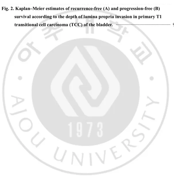
Fig. 1. Hematoxylin–eosin-stained paraffin sections of three distinct bladder cancers. ··· 3 Fig. 2. Kaplan–Meier estimates of recurrence-free (A) and progression-free (B)
survival according to the depth of lamina propria invasion in primary T1 transitional cell carcinoma (TCC) of the bladder. ··· 9
v
LIST OF TABLES
Table 1. Patient characteristics of 183 patients included in the study ··· 5 Table 2. Analysis of clinicopathological factors influencing recurrence ··· 7 Table 3. Analysis of clinicopathological factors influencing progression ··· 8 Table 4. Univariate and multivariate analysis of prognostic factors influencing recurrence
··· 10 Table 5. Univariate and multivariate analysis of prognostic factors influencing progression ··· 10 Table 6. Relationship between T1 substaging and other prognostic parameters ··· 11
- 1 -
.
Ⅰ
INTRODUCTION
Bladder cancer is the most common malignancy of the urinary tract (Hall et al, 2007; Babjuk et al, 2011). Approximately 75–85% of patients with bladder cancer are diagnosed as non-muscle invasive bladder cancer (NMIBC). In NMIBC, approximately 70% of patients present as pTa, 20% as pT1, and 10% as carcinoma in situ (CIS) (Rhijn et al, 2009). Generally, 30–80% of NMIBC will recur and 1–45% of cases will progress to muscle invasive bladder cancer within 5 years (Kirkali et al, 2005; Kiemeney et al, 1993; Sylvester et al, 2006). Of NMIBC, T1 transitional cell carcinoma (TCC) of the bladder with lamina propria invasion is known to have different degrees of aggressiveness with a progression rate varying from 12 to 54% (Herr 1991; Orsola et al, 2005). For T1 TCC of the bladder, it is important to select those patients who have a favorable prognosis and those who are at high risk for progression. Thus, many clinical (age, gender, multifocality, tumor size) and pathologic factors (concomitant CIS, tumor grade, architecture) for recurrence and progression have been studied extensively over the years to predict prognosis and guide management decisions (Rhijn et al, 2009; Sylvester et al, 2006).
Younes et al. were the first to use the muscularis mucosa (MM) in the transurethrally resected biopsy specimens to substage T1 TCC of the bladder. The MM is a scattered muscular fiber which is formed as a continuous or interrupted layer running along large blood vessel in the lamina propria between the epithelium and the detrusor muscle (Younes et al, 1990). From that time, there were several studies to know if the depth of lamina propria invasion is the valuable prognostic factor, using the MM as a landmark to substage. Several reports have demonstrated a worse prognosis for T1 tumors with deep lamina propia invasion, with progression rates ranging from 29 to 55% (Orsola et al, 2005; Younes et al, 1990; Hasui et al, 1994; Smits et al, 1998). However, Platz et al. stated that substaging of early invasive bladder cancer is technically difficult and does not have additional prognostic value with respect to survival (Platz et al, 1996).
In this study, we retrospectively reviewed our single center experience in patients with primary T1 TCC of the bladder and evaluated the feasibility and the prognostic significance of depth of lamina propria invasion on the recurrence and progression.
- 2 -
.
Ⅱ
METERIALS AND METHODS
Between January 1999 and December 2009, 651 patients underwent transurethral resection (TUR) procedures for bladder tumor at our institution. Among these patients, 184 patients were diagnosed as primary T1 TCC of the bladder and followed up for at least 12 months. Of 184 patients, 1 patient who had immediate radical cystectomy was excluded in this study. Thus, the medical records of the remaining 183 patients (149 men and 34 women) were retrospectively reviewed. Mean patient age was 63.5 years (range, 27-93 years). None of the patients included in this study had upper urinary tract malignancy at diagnosis. After TUR, all patients routinely received intravesical BCG instillation once per week for 6 weeks except 3 patients. First patient received Mitomycin-C due to the previous operation history of kidney transplantation, second patient had current active pulmonary tuberculosis, and third patient refused the BCG therapy.
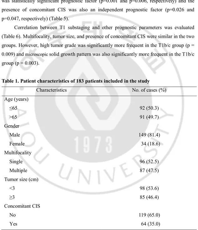
All TUR specimens from the routine histologic stains (hematoxylin-eosin) were reviewed again by one genitourinary pathologist. Substaging was defined according to the depth of lamina propria invasion using the Younes et al. classification: T1a, invasion of lamina propria superficial to the level of the MM; T1b, invasion to the level of the MM; T1c, invasion through the level of the MM but superficial to the muscularis propria (Fig. 1) (Younes et al, 1990). When the MM could not be found and the vascular plexus was identified, it served as a marker of MM level (Orsola et al, 2005; Soukup et al, 2008). Tumors were staged by using the 2002 TNM staging system (Greene et al, 2002) and were graded as high or low grade according to the 2004 World Health Organization/International Society of Urological Pathology (WHO/ISUP) grading system (Epstein et al, 1998). The microscopic tumor architecture was divided into papillary and solid. A solid growth pattern refers to a tumor that is composed of diffusely growing sheets of tumor cells without fibrovascular core formation. To classify a tumor as solid, at least 10% of the components need to be judged as solid. (Andius et al, 2007).
The median follow-up period was 36 months (mean, 43.5; range, 12 to 146 months). Patients were followed until death or last visit and follow-up included urinalysis, cystoscopy, and urine cytology every 3 months for the first 2 years, every 6 months for the subsequent 2 years, and then annually thereafter. Abdominopelvic computed tomography was performed
- 3 -
annually during follow-up or when clinically indicated.
Clinical outcome was evaluated in terms of tumor recurrence or progression. Recurrence was defined as the histological detection of TCC of the bladder through TUR or bladder biopsy after the first TUR. Progression was defined as the development of muscle-invasive disease or distant metastasis. The diagnosis of an advanced tumor of the upper urinary tract was not considered progression (Orsola et al, 2005; Andius et al, 2007).
Univariate and multivariate analyses were performed by using the log-rank test or the Cox’s proportional hazards regression model, respectively. The association between recurrence or progression and clinicopathological factors and relationship between substaging and other prognostic parameters were examined by using the chi-square test. The prognostic parameters assessed were clinical (gender, multifocality, tumor size) and
pathologic (concomitant CIS, tumor grade, microscopic tumor architecture, substaging)
factors. All statistical analyses were performed by using SPSS ver. 13.0 (SPSS Inc., Chicago, IL, USA). Values of p<0.05 were considered to be statistically significant in all of the analyses.
Fig. 1. Hematoxylin–eosin-stained paraffin sections of three distinct bladder cancers. A. Ta; no invasion into the muscularis mucosa (MM) showing MM and muscularis propria (MP). B. T1a; superficial invasion of lamina propria. C. T1b; invasion into the MM. (The tumor base limit is marked with an arrow).
- 4 -
.
Ⅲ
RESULTS
The clinicopathological characteristics of the 183 patients with primary T1 TCC of the bladder are summarized in Table 1. The number of tumors was 2 or more in 87 cases (47.5%), mean number of tumors were 2.1 (range, 1 to 9), and tumor size was 3 cm or more in 85
cases (46.4%). ConcomitantCIS was present with main tumor in the 64 cases (35.0%), high
grade of tumor was found in the 171 cases (93.4%), and microscopic tumor architecture was papillary growth pattern in the 175 cases (95.6%). Substaging was possible in all 183 cases. Substaging was as follows: T1a in 119 (65.0%), T1b in 57 (31.1%), and T1c in 7 (3.9%). And 8 patients (4.4%) underwent radical cystectomy after TUR due to frequent recurrence or progression.
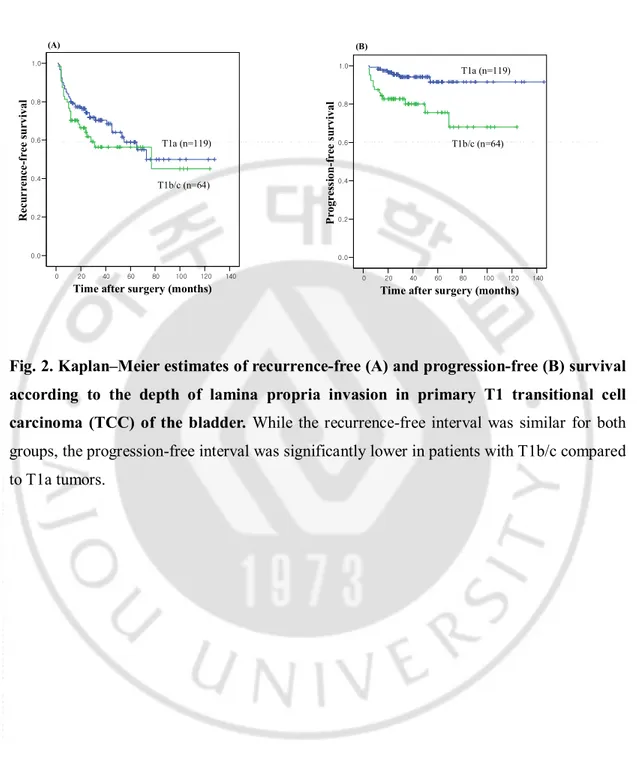
To evaluate the risks of recurrence and progression, T1b and T1c tumors were pooled together because only 7 cases have T1c tumors. The recurrence rate was 32.8% (39/119) for T1a and 40.6% (22/57 for T1b and 4/7 for T1c) for deep lamina propria invasive tumors including T1b and T1c (T1b/c) during the follow-up period, which was not significantly different (p=0.338) (Table 2). Overall recurrence rate was 35.5% (65/183). The average period until first recurrence was 16.3 months (range, 1 to 77) and the mean number of recurrence was 1.5 times (range, 1 to 4). The progression rate was significantly higher in patients with deep lamina propria invasion (p=0.003): 5.8% (7/119) in T1a and 21.9% (14/57 for T1b and 0/7 for T1c) in T1b/c (Table 3). Overall progression rate was 11.5% (21/183). And the average period until first progression was 18.3 months (range, 4 to 69). The cancer-specific mortality rate was also significantly higher in patients with deep lamina propria invasion (p=0.036): 4.2% (5/119) in T1a and 14.0% (8/64) in T1b/c. Eight patients died of unrelated causes, giving a overall mortality rate was 11.5% (21/183). Table 2 and 3 summarize analysis of clinicopathological factors influencing recurrence and progression. According to the Kaplan–Meier analysis, the progression-free interval was significantly lower in patients with T1b/c compared to T1a tumors (p=0.001). The recurrence-free interval, in contrast, was similar for both groups (p=0.235). Curves for progression-free and recurrence-free estimates are plotted in Fig. 2.
In the univariate analysis of prognostic factors influencing recurrence, microscopic tumor architecture was the only significant prognostic factor for recurrence (p=0.029).
- 5 -
However, it was not statistically significant in the multivariate analysis (p=0.075). And depth of invasion was not statistically significant prognostic factors influencing recurrence in the univariate and multivariate analysis (p=0.235, p=0.797, respectively) (Table 4).
In the univariate and multivariate analysis concerning progression, depth of invasion was statistically significant prognostic factor (p=0.001 and p=0.006, respectively) and the presence of concomitant CIS was also an independent prognostic factor (p=0.026 and p=0.047, respectively) (Table 5).
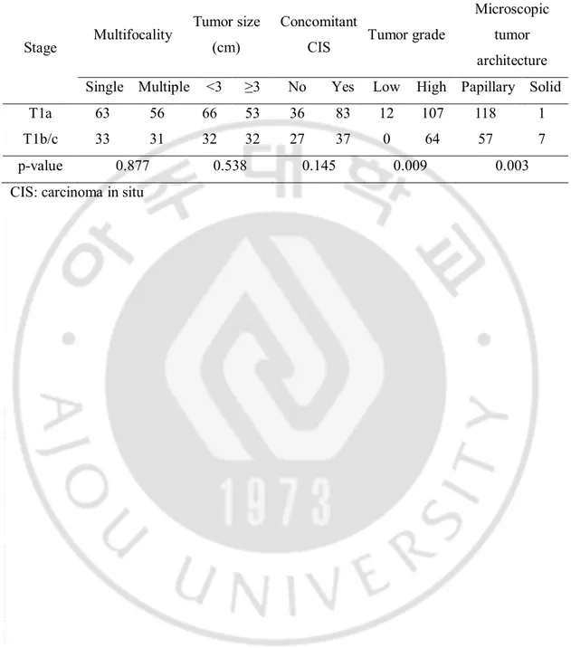
Correlation between T1 substaging and other prognostic parameters was evaluated (Table 6). Multifocality, tumor size, and presence of concomitant CIS were similar in the two groups. However, high tumor grade was significantly more frequent in the T1b/c group (p =
0.009) and microscopic solid growth pattern was also significantly more frequent in the T1b/c
group (p = 0.003).
Table 1. Patient characteristics of 183 patients included in the study
Characteristics No. of cases (%)
Age (years) ≤65 92 (50.3) >65 91 (49.7) Gender Male 149 (81.4) Female 34 (18.6) Multifocality Single Multiple 96 (52.5) 87 (47.5) Tumor size (cm) <3 ≥3 98 (53.6) 85 (46.4) Concomitant CIS No Yes 119 (65.0) 64 (35.0)
- 6 - Tumor grade Low High 12 (6.6) 171 (93.4) Microscopic tumor architecture
Papillary Solid 175 (95.6) 8 (4.4) Depth of invasion T1a T1b T1c 119 (65.0) 57 (31.1) 7 (3.9)
Radical cystectomy after TUR 8 (4.4)
Recurrence 65 (35.5)
Progression 21 (11.4)
Death
Death of disease Death of other disease
13 (7.1) 8 (4.3) CIS: carcinoma in situ, TUR: transurethral resection
- 7 -
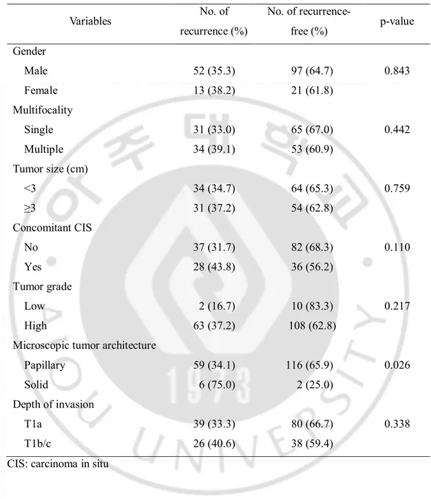
Table 2. Analysis of clinicopathological factors influencing recurrence
CIS: carcinoma in situ
Variables No. of recurrence (%) No. of recurrence-free (%) p-value Gender Male Female 52 (35.3) 13 (38.2) 97 (64.7) 21 (61.8) 0.843 Multifocality Single Multiple 31 (33.0) 34 (39.1) 65 (67.0) 53 (60.9) 0.442 Tumor size (cm) <3 ≥3 34 (34.7) 31 (37.2) 64 (65.3) 54 (62.8) 0.759 Concomitant CIS No Yes 37 (31.7) 28 (43.8) 82 (68.3) 36 (56.2) 0.110 Tumor grade Low High 2 (16.7) 63 (37.2) 10 (83.3) 108 (62.8) 0.217 Microscopic tumor architecture
Papillary Solid 59 (34.1) 6 (75.0) 116 (65.9) 2 (25.0) 0.026 Depth of invasion T1a T1b/c 39 (33.3) 26 (40.6) 80 (66.7) 38 (59.4) 0.338
- 8 -
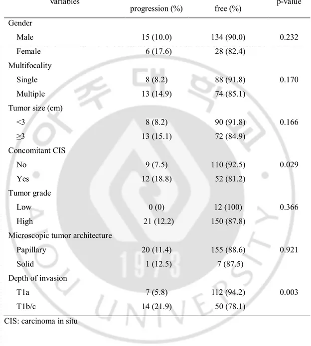
Table 3. Analysis of clinicopathological factors influencing progression
CIS: carcinoma in situ
Variables No. of progression (%) No. of progression-free (%) p-value Gender Male Female 15 (10.0) 6 (17.6) 134 (90.0) 28 (82.4) 0.232 Multifocality Single Multiple 8 (8.2) 13 (14.9) 88 (91.8) 74 (85.1) 0.170 Tumor size (cm) <3 ≥3 8 (8.2) 13 (15.1) 90 (91.8) 72 (84.9) 0.166 Concomitant CIS No Yes 9 (7.5) 12 (18.8) 110 (92.5) 52 (81.2) 0.029 Tumor grade Low High 0 (0) 21 (12.2) 12 (100) 150 (87.8) 0.366 Microscopic tumor architecture
Papillary Solid 20 (11.4) 1 (12.5) 155 (88.6) 7 (87.5) 0.921 Depth of invasion T1a T1b/c 7 (5.8) 14 (21.9) 112 (94.2) 50 (78.1) 0.003
- 9 -
0 20 40 60 80 100 120 140 Time after surgery (months) 0.0 0.2 0.4 0.6 0.8 1.0 R ec ur re nc e-fr ee s u rv iv al T1a (n=119) T1b/c (n=64) (A) 0 20 40 60 80 100 120 140 Time after surgery (months) 0.0 0.2 0.4 0.6 0.8 1.0 P ro gr es si on -f re e su rv iv al T1a (n=119) T1b/c (n=64) (B)
Fig. 2. Kaplan–Meier estimates of recurrence-free (A) and progression-free (B) survival according to the depth of lamina propria invasion in primary T1 transitional cell carcinoma (TCC) of the bladder. While the recurrence-free interval was similar for both groups, the progression-free interval was significantly lower in patients with T1b/c compared to T1a tumors.
- 10 -
Table 4. Univariate and multivariate analysis of prognostic factors influencing recurrence
Variables Univariate Multivariate
p-value Hazards ratio (95% CI) p-value
Gender (Male vs. Female) 0.910 1.087 (0.582-2.029) 0.793
Multifocality (Single vs. Multiple) 0.485 1.048 (0.633-1.736) 0.855
Tumor size (<3 cm vs. ≥3 cm) 0.688 1.271 (0.766-2.107) 0.353
Concomitant CIS (No vs. Yes) 0.078 1.473 (0.870-2.492) 0.141
Tumor grade (Low vs. High) 0.141 0.444 (0.105-1.876) 0.269
Microscopic tumor architecture
(Papillary vs. Solid) 0.029 2.282 (0.920-5.659) 0.075
Depth of invasion (T1a vs. T1b/c) 0.235 1.072 (0.631-1.821) 0.797
CIS: carcinoma in situ
Table 5. Univariate and multivariate analysis of prognostic factors influencing progression
Variables Univariate Multivariate
p-value Hazards ratio (95% CI) p-value
Gender (Male vs. Female) 0.240 1.760 (0.672-4.612) 0.250
Multifocality (Single vs. Multiple) 0.165 1.577 (0.643-3.869) 0.320
Tumor size (<3 cm vs. ≥3 cm) 0.148 2.251 (0.909-5.572) 0.079
Concomitant CIS (No vs. Yes) 0.026 2.561 (1.013-6.473) 0.047
Tumor grade (Low vs. High) 0.197 0 0.985
Microscopic tumor architecture
(Papillary vs. Solid) 0.894 0.402 (0.049-3.304) 0.396
Depth of invasion (T1a vs. T1b/c) 0.001 2.009 (1.024-3.942) 0.006
- 11 -
Table 6. Relationship between T1 substaging and other prognostic parameters
Stage Multifocality
Tumor size (cm)
Concomitant
CIS Tumor grade
Microscopic tumor architecture
Single Multiple <3 ≥3 No Yes Low High Papillary Solid
T1a 63 56 66 53 36 83 12 107 118 1
T1b/c 33 31 32 32 27 37 0 64 57 7
p-value 0.877 0.538 0.145 0.009 0.003
- 12 -
.
Ⅳ
DISCUSSION
The presence of MM in the lamina propia was first reported by Dixon and Gosling from the biopsy specimens of 25 patents and the radical cystectomy specimens of 4 patents in the 1983 (Dixon et al, 1983). The use of MM to substage T1 TCC of the bladder was first described by Younes et al (Younes et al, 1990). And then, several studies about the MM were conducted to identify prognostic significance. The present study shows that the depth of lamina propria invasion is a significant predictor of progression in patients with primary T1 TCC of the bladder, with 21.9% progression rate for T1 tumors deeply invading the lamina propria (T1b/c) as opposed to 5.8% progression rate for T1 tumors without deep lamina propria involvement (T1a). In previous other studies, progression rates for T1 tumors deeply invading the lamina propria (T1b/c) have been shown to be at least 29% and up to 55%, which is similar to our study (Smits et al, 1998; Kondylis et al, 2000). In contrast, progression rates for T1a, i.e. those with limited invasion, range at least 6.7% and up to 11%, which is also similar to the 5.8% in our study (Orsola et al, 2005; Cheng et al, 1999). In the previous Korean study, Kim et al. reported similar results that the progression rate of T1b/c was significantly higher than that of T1a (41.4% and 3.8%, respectively) (Kim et al, 1995). Shin et al. reported that progression rates of T1a, T1b, and T1c were 0%, 10%, and 20.6% respectively (p=0.005) (Shin et al, 1999). Kim et al. also reported statistically significant results that the 2-year risk of progression of T1a, T1b, and T1c was 5%, 19%, and 33% respectively (Kim et al, 2000). Regarding cancer-specific mortality, it was also significantly different between the two groups: 4.2% in T1a and 14.0% in T1b/c in this study. Among different series, survival rates range at least 58% and up to 95% for T1a and between 11% and 82% for T1b/c, which is similar to our study (Hasui et al, 1994; Holmang et al, 1997).
From the above findings, even though depth of lamina propria invasion has consistently remained the most important factor related to progression in primary T1 TCC of the bladder, both the usefulness and the effectiveness of T1 substaging have been debated by pathologists. Criticisms are difficulty for identification of MM and interpathologist variation and disagreement. That is thought to be uneven distribution, misorientation, thermal injury of resection fragments, or necrosis (Younes et al, 1990; Hasui et al, 1994). However, Orsola et al. contended that they could be overcome through practice and reported that the
- 13 -
identification rate of MM progressed from an initial 60% to an 87% rate at present (Orsola et al, 2005). Similarly, in previous publications the rate of substaging has increased from the initial 65–72% to up to 100% in more recent reports (Bernardini et al, 2001; Hermann et al, 1998). In difficult cases where specimen orientation and artifact could create a major hindrance, Mhawech et al. reported that immunohistochemistry (IHC) using desmin and keratin in comparison to hematoxylin-eosin was superior in lowering the rate of imprecision and appeared to be a useful tool in substaging (Mhawech et al, 2002). And they also reported that the combination of p21 and p16 may have value in distinguishing T1b tumors from T1a tumors from the study of molecular markers (Mhawech et al, 2004). Therefore, the accuracy of T1 substaging could be improved through practice and experience of pathologist, IHC, and expression of molecular markers.
And in the previous study some authors claimed that T1b and T1c might be pooled together since T1b shows an almost clinically similar behavior to T1c (Hasui et al, 1994; Holmang et al, 1997; Bernardini et al, 2001). Thus, it could be reasonable to categorize tumors with deep lamina propria involvement invading to or through the level of MM or vascular plexus in the lamina propria as a one category. In our study, T1b and T1c were pooled together and T1b/c was used to evaluate the prognostic significance.
Concomitant CIS has been reported to have a negative effect on the prognosis and is a well-known risk factor for recurrence and progression (Rodriguez-Alonso et al, 2002; Millan-Rodriguez et al, 2000). Orsola et al. showed that depth of lamina propria involvement and associated CIS remained prognostic factors in patients receiving BCG (Orsola et al, 2005). Similarly, Bernardini et al. reported that tumors invading the MM associated with CIS had a risk of progression increased by a factor of 7.5 (Bernardini et al, 2001). And Smits et al. reported a 3-year progression rate of 39% in patients with CIS compared with 9% among patients without CIS (Smits et al, 1998). The present study also showed that the presence of concomitant CIS was a significant prognostic factor for progression with increased the hazards ratio on multivariate analysis.
This study has several limitations. First, this study was conducted in the single center. Second, treatment was not identical for all patients, because TUR was not conducted by a single operator. Third, interpathologist variation and disagreement could not be evaluated, because all of TUR specimens were reviewed and diagnosed by one pathologist. Fourth, it
- 14 -
was retrospective in nature and the size of the study population was small. Fifth, second resections were not routinely performed. Nevertheless, we believe that the results of our study support that patients with deep lamina propria invasion (T1b/c) have a significantly higher progression rate, especially those with concomitant CIS which is also an independent predictor of progression. In addition, designing multicenter and randomized prospective protocols including biological markers might contribute to determine the optimal treatment for deep lamina propria invasive T1 bladder cancer.
- 15 -
.
Ⅴ
CONCLUSIONS
The results of this study demonstrated that substaging according to the depth of lamina propria invasion in primary T1 TCC of the bladder and the presence of concomitant CIS were independent prognostic factors for progression and the patients with deep lamina propria invasion have significantly higher cancer-specific mortality rate. These findings will be helpful for guiding decisions about adjuvant therapies such as more radical treatment and the surveillance interval after TUR.
- 16 -
REFERENCES
1. Hall MC, Chang SS, Dalbagni G, Pruthi RS, Seigne JD, Skinner EC. Guideline for the management of nonmuscle invasive bladder cancer (stages Ta, T1, and Tis): 2007 update. J Urol 178: 2314-2330, 2007
2. Babjuk M, Oosterlinck W, Sylvester R, Kaasinen E, Böhle A, Palou-Redorta J. EAU Guidelines on Non–Muscle-Invasive Urothelial Carcinoma of the Bladder, the 2011 Update. Eur Urol 59: 997-1008, 2011
3. Rhijn BV, Burger M, Lotan Y, Solsona E, Stief CG, Sylvester RJ. Recurrence and pogression of disease in non–muscle-invasive bladder cancer: from epidemiology to treatment strategy. Eur Urol 56: 407-582, 2009
4. Kirkali Z, Chan T, Manoharan M, Algaba F, Busch C, Cheng L. Bladder cancer: epidemiology, staging and grading, and diagnosis. Urology 66(Suppl 6A): 4-34, 2005 5. Kiemeney LA, Witjes JA, Verbeek AL, Heijbroek RP, Debruyne FM. The clinical
epidemiology of superficial bladder cancer. Dutch South-East Cooperative Urological Group. Br J Cancer 67: 806-812, 1993
6. Sylvester RJ, van der Meijden APM, Oosterlinck W, Witjes JA, Bouffioux C, Denis L, Newling DW, Karlheinz Kurth K. Predicting recurrence and progression in individual patients with stage Ta T1 bladder cancer using EORTC risk tables: a combined analysis of 2596 patients from seven EORTC trials. Eur Urol 49: 4660-4677, 2006
7. Herr HW. Progression of stage T1 bladder tumors after intravesical bacillus Calmette-Guerin. J Urol 145: 40-43, 1991
8. Orsola A, Trias I, Raventos CX, Espanol I, Cecchinia L, Bucar S, Salinas D, Orsola I. Initial high-grade T1 urothelial cell carcinoma: feasibility and prognostic significance of lamina propria invasion microstaging (T1a/b/c) in BCG-treated and BCG-non-treated patients. Eur Urol 48: 231-238, 2005
- 17 -
9. Younes M, Sussman J, True LD. The usefulness of the level of the muscularis mucosae in the staging of invasive transitional cell carcinoma of the urinary bladder. Cancer 66: 543-548, 1990
10. Hasui Y, Osada Y, Kitada S, Nishi S. Significance of invasion to the muscularis mucosae on the progression of superficial bladder cancer. Urology 43: 782-786, 1994
11. Holmang S, Hedelin H, Anderstrom C, Holmberg E, Johansson SL. The importance of the depth of invasion in stage T1 bladder carcinoma: a prospective cohort study. J Urol 157: 800-803, 1997
12. Smits G, Schaafsma E, Kiemeney L, Caris C, Debruyne F, Witjes JA. Microstaging of pT1 transitional cell carcinoma of the bladder: identification of subgroups with distinct risks of progression. Urology 52: 1009-1013, 1998
13. Platz CE, Cohen MB, Jones MP, Olson DB, Lynch CF. Is microstaging of early invasive cancer of the urinary bladder possible or useful? Mod Pathol 9: 1035-1039, 1996 14. Soukup V, Babjuk M, Duskova J, Pesl M, Szakaczova M, Zamecnik L, DvorAcek J.
Does the expression of fascin-1 and tumor subclassification help to assess the risk of recurrence and progression in t1 urothelial urinary bladder carcinoma? Urol Int 80: 413-418, 2008
15. Greene FL, Page DL, Fleming ID, Fritz AG, Balch CM, Haller DG, Morrow M. AJCC cancer staging manual. 6th ed. New York: Springer-Verlag 329-331, 2002
16. Epstein JI, Amin MB, Reuter VR, Mostofi FK. The World Health Organization/International Society of Urological Pathology consensus classification of urothelial (transitional cell) neoplasms of the urinary bladder. Bladder Consensus Conference Committee. Am J Surg Pathol 22: 1435-1448, 1998
17. Andius P, Johansson SL, Holmang S. Prognostic factors in stage T1 bladder cancer: tumor pattern (solid or papillary) and vascular invasion more important than depth of invasion. Urology 70: 758-762, 2007
- 18 -
18. Dixon JS, Gosling JA. Histology and fine structure of the muscularis mucosae of the human urinary bladder. J Anat 136: 265-271, 1983
19. Rodriguez-Alonso A, Pita-Fernandez S, Gonzalez-Carrero J, Nogueira-March JL. Multivariate analysis of survival, recurrence, progression and development of metastasis in T1 and T2a transitional cell bladder carcinoma. Cancer 94: 1677-1684, 2002
20. Millan-Rodriguez F, Chechile-Toniolo G, Salvador-Bayarri J, Palou J, Vicente-Rodriguez J. Multivariate analysis of the prognostic factors of primary superficial bladder cancer. J
Urol 163: 73-78, 2000
21. Kondylis FI, Demirci S, Ladaga L, Kolm P, Schellhammer PF. Outcomes after intravesical bacillus Calmette-Guerin are not affected by substaging of high grade T1 transitional cell carcinoma. J Urol 163: 1120-1123, 2000
22. Cheng L, Neumann RM, Weaver AL, Spotts BE, Bostwick DG. Predicting cancer progression in patients with stage T1 bladder carcinoma. J Clin Oncol 17: 3182-3187, 1999
23. Kim HS, Ryu DS, Kwon GY, Kim CI, Lee SC. Significance of invasion to the muscularis mucosae on the progression of superficial bladder cancer. Korean J Urol 36: 272-276, 1995
24. Shin SM, Chung JY, Noh CH. Predictive Value of Microstaging in the recurrence and the progression of T1 superficial bladder cancer. Korean J Urol 40: 1459-1464, 1999 25. Kim YJ, Moon KH, Kim KR, Kim CS. Significance of T1 Substaging on the Recurrence
and Progression of Bladder Cancer. Korean J Urol 41: 1323-1328, 2000
26. Bernardini S, Billerey C, Martin M, Adessi GL, Wallerand H, Bittard H. The predictive value of muscularis mucosae invasion and p53 overexpression on progression of stage T1 bladder carcinoma. J Urol 165: 42-46, 2001
- 19 -
and the prevalence of p53 nuclear accumulation on survival in stage T1 transitional cell bladder cancer. J Urol 159: 91-94, 1998
28. Mhawech P, Iselin C, Pelte MF. Value of immunohistochemistry in staging T1 urothelial bladder carcinoma. Eur Urol 42: 459-463, 2002
29. Mhawech P, Greloz V, Oppikofer C, Szalay-Quinodoz I, Herrmann F. Expression of cell cycle proteins in T1a and T1b urothelial bladder carcinoma and their value in predicting tumor progression. Cancer 100: 2367-2375, 2004
- 20 - - 국문요약 -
일차성 T1 방광이행세포암에서 고유판 침범 깊이에 따른
소분류의 예후적 중요성
아주대학교 대학원 의학과
이 지 용
(지도교수: 김 세 중)
목적: 일차성 T1 방광이행세포암 환자들에서 고유판 침범 깊이에 따른 소분류가 방광암의 재발과 진행의 예후 인자로 작용하는지 알아보고자 하였다. 대상 및 방법: 1999년 1월부터 2009년 12월까지 본원에서 처음으로 경요도방 광종양절제술을 시행 받은 651명의 환자 중 일차성 T1 방광이행세포암으로 진 단되고 12개월 이상 추적 관찰이 가능하였던 183명 (남자 149명, 여자 34명, 평균 연령 63.5세)의 환자를 대상으로 후향적으로 조사하였다. 평균 추적관찰 기간은 43.5개월 (12-146)이었다. 병리조직은 1명의 병리학자에 의해 재검토 되었다. T1 소분류는 암세포가 고유판을 침범하였으나 점막근층까지 침범하지 않았으면 T1a, 점막근층까지 침범하였으면 T1b, 점막근층을 침범하였으나 고유 근을 침범하지 않았으면 T1c라고 정의하였다. 결과: 183명의 대상 환자 중 종양이 2개 이상인 경우는 87례 (47.5%), 종양의 크기가 3 cm 이상인 경우는 85례 (46.4%), 상피내암이 동반된 경우는 64례- 21 - (35.0%), 분화도가 나쁜 경우는 171례 (93.4%), 현미경적 유두상 종양이 175 례 (95.6%)였다. T1a는 119례 (65.0%), T1b는 57례 (31.1%), T1c는 7례 (3.9%)였다. 추적관찰 기간 동안에 T1a에서 39례 (32.8%), T1b/c에서 26례 (40.6%)에서 재발되었고, 소분류와 재발 사이에 유의한 연관성은 없었다 (p=0.338). 방광 고유근을 침범하거나 타장기로 전이가 발생한 경우를 진행으 로 정의하였을 때 T1a에서 7례 (5.8%), T1b/c에서 14례 (21.9%)에서 진행되 었고, 소분류와 진행 사이에 유의한 연관성이 있었다 (p=0.003). 추적관찰 기간 동안 T1a에서 5명, T1b/c에서 8명이 방광암으로 사망하였고 소분류와 유의한 연관성이 있었다 (p=0.036). 방광암 재발에 대한 단변량 분석에서 현미경적 종 양 형태 (p=0.029)가 유의한 인자였고, 방광암 진행에 대한 단변량 및 다변량 분석에서 상피내암 동반 여부 (p=0.026, p=0.047)와 T1 소분류 (p=0.001, p=0.006)가 유의한 인자였다. 결론: T1 방광이행세포암에서 고유판 침범 깊이에 따른 소분류는 방광암의 진행 을 예측하는 독립적인 예후인자였다. 따라서 T1 병기의 소분류는 이들 환자들의 추적관찰 계획 수립과 치료 방침 결정에 도움을 줄 것으로 생각한다. 핵심어: 고유판, 점막 근층, 진행, 소분류, 방광 종양