저작자표시 2.0 대한민국 이용자는 아래의 조건을 따르는 경우에 한하여 자유롭게 l 이 저작물을 복제, 배포, 전송, 전시, 공연 및 방송할 수 있습니다. l 이차적 저작물을 작성할 수 있습니다. l 이 저작물을 영리 목적으로 이용할 수 있습니다. 다음과 같은 조건을 따라야 합니다: l 귀하는, 이 저작물의 재이용이나 배포의 경우, 이 저작물에 적용된 이용허락조건 을 명확하게 나타내어야 합니다. l 저작권자로부터 별도의 허가를 받으면 이러한 조건들은 적용되지 않습니다. 저작권법에 따른 이용자의 권리는 위의 내용에 의하여 영향을 받지 않습니다. 이것은 이용허락규약(Legal Code)을 이해하기 쉽게 요약한 것입니다. Disclaimer 저작자표시. 귀하는 원저작자를 표시하여야 합니다.
Therapeutic effect ofintra-arterial delivery
of neural-induced mesenchymal stem cells
in the ischemic stroke
By Gyu hee Kim
Department of Biomedical Science
The Graduate School, Ajou University
Therapeutic effect ofintra-arterial delivery
of neural-induced mesenchymal stem cells
in the ischemic stroke
Supervised by
Haeyoung Suh-KimPh.D
A Dissertation Submitted to The Graduate School of
Ajou University in Partial Fulfillment of the Requirements
for the Degree of Ph.D in Medical Science
February, 2017
Department of Biomedical Science
The Graduate School, Ajou University
i
-ABSTRACT-Therapeutic Effects of Intra-Arterial Delivery of neural-induced
mesenchymal stem cells in The Ischemic Brain
Mesenchymal stem cells (MSCs) have been shown to improve a variety of neurological dysfunction by their paracrine effects. Neurogenin-1 (Ngn1) is a proneural gene that directs neuronal differentiation of progenitor cells during development. In recently study, intracranial injection of Ngn1-expessing MSCs showed the remarked improvement of motor dysfunction in stroke model compared to MSC and PBS treated group. However, intracranial injection is not feasible method to use in clinical field. Therefore, we conducted the study to investigate that intra-arterial injection of Ngn1-expressing MSCs can improve motor deficit in ischemic rat model.Cerebral ischemic stroke is a serious public health concern. It causes considerable death and disability. Only limited treatment options are available in the acute phase of stroke.Fifteen Sprague-Dawley rats were subinjected to transient middle cerebral artery occlusion(tMCAo) of 2 hours with the suture occlusion model. Magnetic resonance image (MRI), including diffusion-weighted imaging (DWI) and T2-weighted imaging was performed at 2, 7 and 28days after withdrawal of the suture. Motor function evaluation including ratarod test and adhesive removal test was performed at 1, 7, 14 and 28 days. Amimals were divided into 3 subgroups. Each group received 1×10⁶ MSC-Ngn1 cells, 1×10⁶ MSC-lacZ cells and normal saline respectively. The distribution and phenotype of injected stem cells were compared among the groups. Rat injected with MSC-Ngn1 showed the tendency of motor dysfunction improvement compared to MSC-LacZ and control groups. The induction of neural stem cell number was greater in MSC-Ngn1 injected rat than in other groups. Intra-arterial injection of MSC-Ngn1 cell in stroke model showed the remarkable improvement of motor dysfunction. Intra-arterial injection can be the feasible method of stem cell transplant.
ii
iii
TABLE OF CONTENTS
ABSTRACT ... i
TABLE OF CONTENTS...iii
LIST OF FIGURES ... v
Part. I. Comparison of MSC-Neurogenin1 administration modality in MCAo rat model I. INTRODUCTION ... 2
II. MATERIALS AND METHODS ... 4
A. MSCs/Ngn1 preparation ... 4
B. Induction of ischemic models... 5
C. Transplantation ... 8
D. Behavioral testing ... 10
E. Infarct volume measurement ... 11
F. In vivo tracking of superparamagnetic iron oxide (SPIO)-labeled cells ... 12
G. Histological analysis ... 13
H. Statistical analysis ... 14
III.RESULTS... 15
A. Therapeutic effects of MSCs/Ngn1 according to administration modality ... 15
B. Serial changes in infarct volume according to administration modality ... 17
C. MSCs/Ngn1 distribution according to administration modality ... 19
iv
IV.DISCUSSION... 23
Part. II. Therapeutic Effects of Intra-Arterial Delivery of neural-inducedmesenchymal stem cells in The Ischemic Brain I. INTRODUCTION ... 28
II. MATERIALS AND METHODS ... 29
A. Cell preparation and culture ... 29
B. Transient middle cerebral artery occlusion (MCAo) model ... 30
C. Cell administration ... 31
D. Assessment of neurologic deficit ... 32
E. Measurement of infarct volume... 33
F. SPIO labeling of cells ... 34
G. In Vivo MRI cell tracing ... 35
H. Cell Migration Assays ... 36
I. RNA isolation and RT-qPCR ... 37
J. Immunohistochemistry ... 39
K. Statistical Analysis ... 40
III.RESULTS... 41
A. IA transplantation of MSC-ngn1 improve functional recovery ... 41
B. MRI Tracking of cells after IA TP ... 44
C. Cell migration mechanism ... 50
v
E. Tissue integrity was preserved in the MSC-Ngn1 treated brain ... 57
F. Transdifferentiation of MSC-Ngn1 Cells In Vivo ... 59
IV. DISCUSSION ... 61
V. CONCLUSION ... 64
VI. REFERENCE ... 65
국문요약 ... 70
LIST OF FIGURES
Fig. 1. Cerebral infarct in rat model ... 6Fig. 2. Cerebral infarct in rat model ... 9
Fig. 3. Effects of functional recovery in the ischemic stroke ………...16
Fig. 4. Comparison of serial infarct volume in MRI ... 18
Fig. 5. MSCs/Ngn1 tracing by using SPIO labeling. (A) MSCs/Ngn1 labeled with ferridex and protamine sulfate ... 20
Fig. 6.Transdifferentiation of MSCs/Ngn1 at 4 weeks after intra-arterial grafting. ... 22
Fig. 7.Effects of transplantation on functional recovery in the ischemic stroke ... 42
Fig. 8. Effects of transplantation on infarct volume in the MCAo animal model ... 43
Fig. 9. In vivo tracing of MSC and MSCs/Ngn1 after IA TP in ischemic brain ... 45
vi
Fig. 11. localization of the grafted cells in the ischemic brain at 1day and 4days after
IA TP. ... 47
Fig. 12. Intra-arterial transplanted cells at 4h after injection in the ischemic brain ... 48
Fig. 13. Grafted cells at 25day after IA injection in the ischemic brain .. ... 49
Fig. 14.The expressions of chemokine molecules and cell migration under ischemic condition ... 51
Fig. 15. The expressions of TGF-β in MSC and MSC/Ngn1 ... 52
Fig.16.Effects of grafted cells on cell death.. ... 54
Fig. 17.Effects of grafted cells on inflammation response ... 55
Fig. 18. GFAP expression of reactive astrocytes in the ischemic brains ... 56
Fig. 19. tissue integrity in ischemic brain after IA TP ... 58
Fig. 20. Trans differentiation of MSCs/Ngn1 after intra-arterial administration. ... 60
LIST OF TABLES
Table 1. Experimental animal groups ··· 71
PART. 1. Comparison of MSC-Neurogenin1 administration
modality in MCAo rat model
2
I. INTRODUCTION
Mesenchymal stem cells (MSCs) exist in adult bone marrow and occupy <0.01% of all nucleated bone marrow cells [1]. Even though the main MSC differentiation is of mesodermal lineage, several experimental studies have revealed that these cells can differentiate into neuronal and glial ectodermal cells [2-4]. Animal and human trials have suggested that MSC administration exerts beneficial effects in ischemic heart disease, stroke, and even progressive neurodegenerative diseases, regardless of their autologous or allogeneic origin [5-7].
Neurogenins (Ngn) are a family of basic helix-loop-helix (bHLH) transcription factors involved in neuronal or glial differentiation, and their subtypes are classified into Ngn 1, 2, or 3 [8]. Among Ngn subtypes, Ngn1 facilitates neuronal differentiation as an activator for downstream transcription factors such as NeuroD [9]. A previous study demonstrated that intracerebral (IC) administration of Ngn1-expressing MSCs (MSCs/Ngn1) produced restorative augmentation of behavioral functions and induced the expression of neuron-specific proteins and voltage-gated Ca2+ and Na+ channels, suggesting survival of the grafted MSCs [10].
Animal experiments have shown that intracerebral (IC) administration is a reliable route for stem cell transplantation [11-13]. IC cell grafting is avoided in the clinical field due to its hazardous complications, such as seizure, chronic subdural hematoma, cortical vein occlusion, and postprocedural clinical deterioration [14-16]. The intravenous (IV) administration, on the other hand, the majority of MSCs are entrapped within the lung and liver before they are targeted to the brain, which potentially reduces their therapeutic potential [5, 17]. Theoretically, intra-arterial (IA) cell grafting is more feasible in clinical applications because of more predictable engrafting (vs. IV approach) and less invasiveness (vs. IC approach). An animal study reported that IA administration of bone marrow stromal cells can facilitate axonal sprouting and remyelination in the cortical ischemic boundary zone and improve neurological
3
function [18]. Recent animal studies showed that IA administration is a safe and more effective method than IV administration for treating cerebral ischemia [19, 20]. Nonetheless, there are no comparative studies on the engrafting effectiveness of IA versus IC stem cell administration. Therefore, the purpose of this study was to investigate behavioral changes, infarct volume, cell distribution, and stem cell identification in IA and IC MSC/Ngn1 groups in a rat model of ischemic stroke..
4
II. MATERIALS AND METHODS
A. MSCs/Ngn1 preparation
MSCs were isolated from human bone marrow with approval of the Institutional Review Board of Ajou University Medical Center (Suwon, South Korea). Human MSCs were transduced to prepare the MSCs/Ngn1 as previously described [10]. Briefly, MSCs were transduced with Ngn1-expressing retrovirus for 8 hours in the presence of 4 μg/mL polybrene (Sigma-Aldrich, St Louis, MO, USA) and 10 ng/mL basic fibroblast growth factor (Dong-A Pharmaceutical Co., Youngin, Korea). The transduced cells were enriched for 2 weeks in the presence of 2 μg/mL puromycin (Sigma-Aldrich) and maintained by subculturing every 5 to 7 days in the growth medium, which was composed of Dulbecco’s modified Eagle’s medium (Welgene, Daegu, Korea) supplemented with 10% fetal bovine serum (HyClone, Logan, UT, USA), 100 units/mL penicillin, and 100 µg/mL streptomycin (Gibco, Grand Island, NY, USA)..
5
B. Induction of ischemic modelsAll animal protocols were approved by the Institutional Animal Care and Use Committee of Ajou University Medical School (Suwon, South Korea). A transient focal ischemia model of 120-minute MCAo with an intraluminal filament was performed in male Sprague Dawley rats (approximately 250 g) according to a modified procedure originally described by Longa et al [21]. Briefly, anesthesia was induced with 5% isoflurane in 70% N2O and 30% O2 and maintained at 3% isoflurane. A 4-0 monofilament nylon suture with a rounded tip was introduced into the external carotid artery (ECA) lumen and gently advanced into the internal carotid artery (ICA) until it blocked the bifurcating origin of the MCA and suture was removed two hours after MCA occlusion. A double screen-out method was employed to ensure consistent ischemic injuries between rats. First, test subjects with negligible or moderate ischemic symptoms on any type of behavior tests on day 1 were excluded. Second, rats with a small infarct only involving the striatum or cortex were excluded after magnetic resonance imaging (MRI) analysis at day 2 (Fig. 1). Twenty two animals showing similar degree of neurologic deficit on behavior test and similar infarct pattern and volume on MRI were selected and randomly assigned to the following groups: (I) to receive normal saline (NS) via the IA route (n=5), (II) to receive NS via the IC route (n=5), (III) to receive MSCs/Ngn1 via the IA route (n=5), and (IV) to receive MSCs/Ngn1 via the IC route (n=5). (Table 1)
6
Fig. 1.Cerebral infarct in rat modelInfarction of rat brainresulted from the occlusion of middle cerebral artery (MCAo) wasdocumented by T2 and diffusion weight image (DWI) of brain magneticresonance image (MRI).
7
Table 1. Experimental animal groupsGroup labeling Cell Transplantation† Assay point End Figures
I
-
IC, NS (n=5)
MRI Behavior test Cresyl violet stain Immunohistochemistry 28d Fig. 2 & 4 II IA, NS (n=5) III IC, MSCs/Ngn1 (n=5) IV IA, MSCs/Ngn1 (n=5) V SPIO
IC, MSCs/Ngn1 (n=1) MRI cell tracking Prussian blue stain
Immunohistochemistry 7d Fig. 3 VI IA, MSCs/Ngn1 (n=1)
*Middle cerebral artery occlusion (MCAo) was validated by behavior test on day 1 and magnetic resonance imaging (MRI) on day 2.
†Transplantation was carried out on day 3.
Abbreviation: IC= intra-cerebral, IA=intra-arterial, NS=normal saline, MSCs/Ngn1=Ngn1 expressing mesenchymal stem cell
8
C. TransplantationThree days after MCAo, the animals were anesthetized with 5% isoflurane in 70% N2O and 30% O2 using an induction chamber and maintained at 3% isoflurane using a face mask. After the skull was opened, MSCs/Ngn1 (1.0 106 cells) or NS in a total fluid volume of 10 μL were IC transplanted into the striatum (anteroposterior [AP], 0.5; mediolateral [ML], 2.5; dorsoventral [DV], 5.0) and cortex (AP, -0.5; ML, 2.0; DV, 2.5) in the ipsilateral hemisphere using a stereotactic apparatus (David Kopf Instruments, Tujunga, CA) (Fig. 1A). IA administration was performed as previously described [22] with modifications. Briefly, the CCA was ligated, and a catheter (25G) tip was inserted to the common carotid artery (CCA) bifurcation through the ECA. After blood flow from the CCA to ICA was recovered by CCA occlusion release, stem cells (1 106) in 1.2 mL saline were directly injected into the CCA bifurcation over the course of 5 minutes (Fig. 1B).
9
Fig. 2.Cerebral infarct in rat modelInfarction of rat brainresulted from the occlusion of middle cerebral artery (MCAo) wasdocumented by T2 and diffusion weight image (DWI) of brain magneticresonance image (MRI).
10
D. Behavioral testingTests were performed to measure motor and sensory behaviors. The battery consisted of the Rotarod test and adhesive removal test. All animals were trained for 7 days before MCAo induction. Only the animals capable of remaining on the Rotarod cylinder for more than 300 seconds and removing adhesive dots within 10 seconds were used for experiments. In the Rotarod test, the Rotarod cylinder (Ugobasile, Comerio, Italy) was accelerated from 4 to 40 rpm within 5 minutes, and the amount of time each animal remained on the Rotarod was measured with a cut-off time of 300 seconds. The data are presented as the percentage of the mean duration from three trials with respect to the performance before MCAo. For adhesive removal tests, square dots of adhesive-backed paper (100 mm2) were used as bilateral tactile stimuli occupying the distal-radial region on the wrist of each forelimb. The time taken for each animal to remove each dot was recorded, and animals were given 3 trials with a cut-off time of 300 seconds. The data are presented as the mean time to remove the dots. We performed behavior tests at 1, 7, 14, and 28 days after MCAo.
11
E. Infarct volume measurementMRI scanning was performed using a 3.0-Tesla whole-body MRI scanner (Achieva 3.0T X-Series Qasar Dual, Philips Healthcare, Amsterdam, Netherlands) equipped with a gradient system capable of 35 milliteslas/m at 3, 7, 14, 28 days after MCAo. A fast-spin echo imaging sequence was used to acquire T2-weighted anatomical images of the rat brain in vivo, using the following parameters: repetition time, 4,000 milliseconds; effective echo time, 96 milliseconds; field of view, 55 x 55 mm2; image matrix, 256 x 256; slice thickness, 1.5 mm; flip angle, 90°; number of excitations, 2; pixel size, 0.21 x 0.21 mm2. A 300-mmdiameter quadrature 16-rung birdcage coil arrangement was used for RF excitation, and a 40-mm-diameter saddle coil was used for signal detection. A total of 15 slices were scanned to cover the entire brain. For each slice, the ischemic area from each T2-weighted image was manually marked and calculated using the software program Osiris (University of Geneva, Geneva, Switzerland).Relative infarct volume (RIV) was normalized as described by Neumann-Haefelin et al [23] using the equation RIV = (LT – (RT – RI)) d, where LT and RT represented the areas of the left and right hemisphere, respectively, in square millimeters; RI was the infarcted area in square millimeters; and d was the slice thickness (1.5 mm). RIV was expressed as a percentage of the right hemispheric volume.
12
F. In vivo tracking of superparamagnetic iron oxide (SPIO)-labeled cells MSCs/Ngn1 were labeled using ferridex (Ferridex IV, TAEJOON Pharm, Korea) and protamine sulfate (Sigma-Aldrich) according to procedure described by Arbab et al [24]. A Prussian blue staining method [24] was used to identify ferridex incorporation into the MSCs/Ngn1. We transplanted the SPIO-labeled MSCs/Ngn1 via IC and IA administration as described above. Distribution and migration of MSCs/Ngn1 were serially monitored by MRI using the SPIO-labeled MSCs/Ngn1 after 4 h and 1, 4, and 7 days. To identify labeled MSCs/Ngn1 in the ischemic brain, animals were sacrificed at 7 days after transplantation. Transplanted cells were detected by Prussian blue staining and immunohistochemistry using anti-human mitochondria (hMT, 1:100, Milllipore, Temecula, CA, USA) or anti-CD68 (ED1, 1:200, Abcam, Cambridge, MA, USA) antibodies
13
G. Histological analysisAt 28 days after transplantation, the anesthetized rats were transcardially perfused with 0.9% saline followed by 4% paraformaldehyde (PFA). Brains were removed, postfixed in 4% PFA overnight, and then cut through the areas of interest after embedding in paraffin. Serial 6-μm-thick paraffin sections were deparaffinized and then placed in boiled citrate buffer (pH 6.0) for 10 minutes. After blocking in 1% bovine serum albumin (BSA) and 5% normal serum, the sections were incubated with antibodies against MAP2 (microtubule-associated protein 2, 1:100, Sigma-Aldrich) which is a marker for mature neuron, ED1 (1:200, Abcam) to detect phagocytic activity, and hMT (1:100, Milllipore) which is a marker for transplanted human stem cell at 4°C overnight. Then, the sections were incubated with Alexa Fluor 488- or 594-conjugated anti-IgG secondary antibodies and counterstained with bis-benzamide (Molecular Probes, Eugene OR, USA) to visualize the entire population of cells. Fluorescent images were acquired using a Zeiss LSM510 confocal microscope (Carl Zeiss, Oberkochen, Germany).
14
H. Statistical analysisTwo-group comparisons were done with independent t-tests. Multiple comparisons for each treatment group were analyzed using one-way analysis of variance (ANOVA) with independent variables of treatment groups and days of testing, followed by Tukey’s post hoc tests. All statistical analyses were performed using SPSS version 12.0 (SPSS; Chicago, IL, USA), and results were for multiple comparisons.
15
III. RESULTS
A. Therapeutic effects of MSCs/Ngn1 according to administration
modality
The effectiveness of MSCs/Ngn1 administration in a rat stroke model was evaluated followed by comparison of efficacy between IA and IC administration. Control animals that received IA or IC normal saline spontaneously recovered to a limited degree for 7 days and then no further improvement was observed until the end of the study at 28 days. The IC MSCs/Ngn1-transplanted animals showed significantly better performance in Rotarod testing than the IC control group at 28 days (Tukey’s post hoc test, p<0.0001 at 28 days only). The IA MSCs/Ngn1-transplanted animals showed significantly better Rotarod performance than the IA control group from 7 to 28 days (Tukey’s post hoc test, all p<0.05). The adhesive removal test revealed greater improvement for the IA MSCs/Ngn1 group compared to the IA control (Tukey’s post hoc test, p<0.05 for 21 and 28 days). Interestingly, the IC MSCs/Ngn1 group showed a decrease in functional score on the Rotarod test at 7 days compared to other groups (including controls); this was the peri-procedural period after IC stereotactic administration. Functional recovery was higher in IA MSCs/Ngn1 animals than in the IC MSCs/Ngn1 group for both the Rotarod and adhesive removal tests. At 28 days, the IA MSCs/Ngn1 administration group achieved significantly higher functional scores in the Rotarod test (independent test, p=0.003) and adhesive removal test (independent t-test, p=0.009), compared to the IC MSCs/Ngn1 transplantation group (Fig. 2A, 2B).
16
Fig.3.Effects of functional recovery in the ischemic stroke.
Behavioral improvements of the animals transplanted with normal saline or MSCs/Ngn1 stem cells via intra-cerebral or intra-arterial route were evaluated by (A) Rotarod test or (B) adhesive removal test. MSC/Ngn1 stem cell showed the therapeutic effect compared with control and intra-arterial route had better functional recovery compared with intra-cerebral route. The data were collected from 5 animals per group and are presented as mean values ± S.D. Statistical significant differences between the groups were determined by analysis of variance (*, p < .05 compared with IA-control group; #, p < .05 compared with IC-MSCs/Ngn1 group). Arrows indicate transplantation time of cells
17
B. Serial changes in infarct volume according to administration
modality
The RIV of brains were monitored using MRI analysis over the 28-day experimental period (Fig. 2C) and performed histologic assessments of ischemic rat brain at 28 days (Fig. 2D). No differences were seen in the initial infarct volume ratios between the four groups (p=0.966). Although the IC-MSCs/Ngn1 administration group showed a tendency toward a reduced infarct volume ratio compared to the IA control (p=0.061) and IC control (p=0.223), the IA MSCs/Ngn1 administration group showed a significant reduction of infarct volume compared with IA controls (p=0.044) and a trend toward reduction compared with IC controls (p=0.174) (Table 1) at 28 days.
18
Fig.4.Comparison of serial infarct volume in MRI.(A) Representative MR imaging from 2 to 28 days after ischemia. (B) Cresyl violet staining at 28 days after ischemia. MSCs/Ngn1 transplanted groups showed more reduced infarct volume on MR images and histology. MSCs/Ngn1 transplanted groups showed more reduced infarct volume on MR images and histology. (C) Quantitative analysis of infarct volume was evaluated using brain magnetic resonance image.
19
C. MSCs/Ngn1 distribution according to administration modality
SPIO labeling was evaluated in MSCs/Ngn1 stained with Prussian blue after ferridex and protamine sulfate incubation for 12 hours (Fig. 3A). The distribution patterns of transplanted MSCs/Ngn1 were discretely different. IA-transplanted SPIO-labeled MSCs/Ngn1 were widely distributed in the ischemic area, whereas IC-transplanted SPIO-labeled MSCs/Ngn1 on MRI were localized to the stereotactically injected site after 4 hours. Serial MRI imaging studies at 1, 4, and 7 days showed that each distribution pattern was not dramatically changed from the initial pattern on MRI (Fig. 3B). To identify the SPIO signal on MR, animals were sacrificed at 7days. IA MSCs/Ngn1-transplanted ischemic rat brain specimens were stained with Prussian blue to identify SPIO-labeled cells. Identification of MSCs/Ngn1 and phagocytic immune cells were performed with immunohistochemistry with a hMT antibody and ED1 antibody, respectively (Fig. 3C). Some SPIO remained within the injected MSCs/Ngn1, and other SPIO stained the immune cells, suggesting that immune cells phagocytosed MSCs/Ngn1.20
Fig.5.MSCs/Ngn1 tracing by using SPIO labeling. (A) MSCs/Ngn1 labeled with ferridex and protamine sulfate.
Prussian blue staining for the identification of incorporation of ferridex into the MSCs/Ngn1. (B) Distribution of MSCs/Ngn1 was monitored by SPIO labeling on MRI. Intra-arterial transplanted SPIO labeled MSCs/Ngn1 was widely distributed in the ischemic area. Each distribution pattern remained during the serial MR imaging studies at 1 day, 4 day, and 7 day. (C) Prussian blue staining at 7 days of the ischemic brain transplanted with intra-arterial MSCs/Ngn1 cells. Immunostaining for identifying transplanted SPIO labeled MSCs/Ngn1 cell and phagocytic activity (microglia/macrophages) by using anti hMT (human Mitochondria) and ED1 antibody respectively were performed in ischemic boundary (I) and core (II) region. SPIO correlated with hMT (red arrowheads) and ED1 (black arrowheads). Scale bars = 20 um.
21
D. MSCs/Ngn1 transdifferentiation in a rat stroke model
To assess transdifferentiation of the IA-transplanted MSCs/Ngn1, we identified hMTimmunoreactive cells in the ischemic core and border zone at 28 days. Most hMT positive cells express MAP2 (a neuronal marker) but do not express GFAP (an astrocyte marker).
22
Fig.6. Transdifferentiation of MSCs/Ngn1 at 4 weeks after intra-arterial grafting.
(A) Neuronal transdifferentiated MSCs/Ngn1 cells were identified by the anti-MAP2 (Microtubule-Associated Proteins) antibody (neuronal marker) and hMT (human mitochondrial) antibody. Arrowheads denote the donor-derived cells co-localized with cell type specific antigens in enlarged orthographic images. (B) None of the MSCs/Ngn1 that identified with anti-hMT antibody (arrows) co-stained with antibody recognizing glial fibrillary acidic protein (GFAP). Scale bars = 20 um.
23
IV. DISCUSSION
The experiment demonstrated that MSCs/Ngn1 have therapeutic properties in an acute ischemic stroke model and provide more benefits when they were administered intra-arterially. SPIO-labeled MSCs/Ngn1 delivered via the IA route was more evenly distributed within the infarct area on MRI, whereas they remained at the injection site in the IC group. Moreover, the neuronal differentiation of IA-transplanted MSCs/Ngn1 in the ischemic area was observed.
Stem cell therapy has emerged as a promising strategy for treating neurological diseases [25, 26]. MSCs are of particular clinical interest because they are easily isolated from bone marrow, adipose tissue, and umbilical cord blood and can be expanded through as many as 50 population doublings in around 10 weeks [27]. In the previous studies, MSCs were shown to ameliorate neurologic dysfunction in patients with acute ischemic stroke [5, 17, 28]. The previous study reported that IC administration of Ngn1-expressing MSCs was more beneficial than parentally delivered MSCs in an acute animal stroke model [10]. A possible explanation is the paracrine effects of MSCs/Ngn1 on surrounding tissues – that is, compared to parental MSCs, MSCs/Ngn1 can induce the neurogenesis of resident stem cells in the subventricular zone (SVZ); reduce delayed neuronal cell death in penumbra regions; ameliorate inflammation by activating resident microglia and stimulating infiltration of leukocytes into the microvessels; and reduce ischemic cerebral penumbra followed by ischemic insult. Another plausible reason is prolonged survival of MSCs/Ngn1 in the ischemic brain area and a much higher proportion of transdifferentiation into functional neuronal cells in ischemic regions. Therefore, the comparison between IA and IC routes were done using MSCs/Ngn1.
The behavior tests showed significantly better functional recovery in the IA-MSCs/Ngn1 group compared to the IC-IA-MSCs/Ngn1 group. This finding indicates that the route of administration of stem cells, which have a beneficial effect for neuronal recovery, can be another important factor in stem cell therapy for stroke treatment.
24
Injected MSCs/Ngn1 stem cells were evenly distributed in IA-MSCs/Ngn1 group on serial MRIs using SPIO-labeled MSCs/Ngn1. However, almost all MSCs/Ngn1 remained around the injected area in the IC-MSCs/Ngn1 group. Theoretically, the paracrine effects of IC-engrafted MSCs would be limited because their locations are bounded in the vicinity of the injected area. On the other hand, evenly distributed MSCs/Ngn1 administered via IA might facilitate greater paracrine and neuronal transdifferentiation effects by stimulating and revitalizing areas adjacent to ischemic tissue. This result demonstrates that the restorative effect of stem cells is conveyed differently into the ischemic lesion by different distribution pattern according to administration route and even distribution is more beneficial for recovery of the neurologic deficit in ischemic stroke.
The experiment confirmed the neuronal differentiation of administered MSCs/Ngn1 cells via IA. The previous report indicated that greater than 45% of IC-transplanted MSCs/Ngn1 differentiate into morphologically mature neurons and express functional neuronal proteins such as vesicular glutamate transporter 2 (VGLUT2), NF200, and Tau [10]. However, whether the injected stem cells via IA can be moved into the ischemic lesion and can be differentiated into neuronal cell or not is questionable. This neuronal differentiation of mesenchymal stem cell showed the capability of neuronal induction by genetic engineering through transfection of the Ngn1 gene. In addition to restorative paracrine effect, neuronal replacement by differentiation into neuronal cell with mature function may be another mechanism to recover the injured neuronal function by stem cell therapy with MSCs/Ngn1.
Although IA-MSCs/Ngn1 group showed the significant reduction of infarct volume on MRI compared with IA controls, IA administration of MSCs/Ngn1 did not significantly reduced RIV on MRI compared to IC-MSCs/Ngn1 group in this study. Because we performed behavior test at 1 day and brain MRI study at 2 day serially to verify and include MCAo rat model with the similar neurologic impairment and similar infarct volume into our study, we cannot avoid the delay of stem cell administration and MSCs/Ngn1 were administrated on day 3 from stroke onset in this study. The late
25
treatment with MSCs/Ngn1 may affect the results such as low differences in size of ischemic lesion volume between IA and IC groups.
It is known that IA delivery of MSCs enables efficient cell administration to the infarct area and results in significant functional recovery after ischemic stroke in a rat model [29, 30]. However, the major issue of IA administration is microembolization due to aggregation of the cells in the blood vessels [31]. The major determinants of the safety of IA stem cell administration are cell size, velocity of injection, and cell dose [22, 32] Janowski et al. [22] showed that administration of MSCs with infusion velocity over >1 mL/minute often resulted in stroke whereas a lower velocity (0.2 mL/minute) was safe. They also showed that stroke lesions occurred frequently when injecting 2 x10⁶ MSCs, but not after lowering the dose to 1x 10⁶ cells. In this study, we administered low dose of MSCs/Ngn1 (1x10⁶ cells/1.2mL) with lower infusion velocity (0.24 mL/minute) to avoid microembolism. Here, we did not find any newly developed microembolic infarcts on MRI after MSCs/Ngn1 administration via the IA route, but further assessment of the optimum cell administration procedure is required to evaluate the adverse effects. To minimize the risk of distant embolism in human patients undergoing an endovascular approach, a skilled interventionist and well-equipped clinical setting are essential for ensuring the safety of IA administration of MSCs or intensified MSCs. Safe IC administration in human patients is technique sensitive and require general anesthesia. Moreover, the direct deposit of grafting cells via the IC route may inevitably results in damage to the brain parenchyma. For these reasons, previous human trials with the IC route reported both serious and non-serious adverse effects [14-16]. Although IV injection is a clinically relevant and minimally invasive technique, only a small amount of injected cells reach the targeted brain area due to the first pass effect in the lungs and liver [19]. Because the IA route increases the chances of MSC migration and distribution, it facilitates efficient engrafting of stem cells to the target brain areas [33].
The present study compared the therapeutic effect after IC and IA administrations of stem cells. We also showed the hemispheric distributions of injected stem cells on serial
26
MRI and observed neuronal differentiation on immunohistochemical analyses. The result requires cautious interpretation because of the lack of comparisons with surrogate biomarkers for paracrine effects of injected MSCs.
Although we did not found newly formed microembolic stroke on MRI after IA delivery of stem cells, we cannot exclude the possibility of microembolsim. It is possible that the microembolisms caused by MSCs infusion might be masked by the large ischemic lesions on MRI. Therefore, the continuous stirring technique should be implemented to prevent distant embolism due to the tendency of MSCs to aggregate. Further human stem cells trials using the IA approach are required to investigate the clinical benefits in acute ischemic stroke patients.
27
PART. II. Therapeutic Effects of Intra-Arterial Delivery of
neural-induced mesenchymal stem cells in The Ischemic Brain
28
I. INTRODUCTION
Cerebral ischemic stroke is a serious public health concern. It causes considerable death and disability. Only limited treatment options are available in the acute phase of stroke. Cell-based therapy is a promising experimental approach to enhance poststroke recovery. Mesenchymal stem cells (MSCs) have been shown to have successful results in experimental animal models of ischemic stroke when administered systematically, reducing the severity of cerebral ischemic injury and promoting neurological function . Neurogenin1 (Ngn1) is a member of the basic helix-loophelix (bHLH) transcription factor family. It is expressed in early neuronal progenitor cells during development of the nervous system. Forced expression of Ngn1 has been shown to increase the number of neuronal cells in Xenopus embryos and in uncommitted pluripotent stem cells, such as embryonic carcinoma P19 cells, by activation of downstream proneuralbHLH transcription factors, including NeuroD. Once NeuroD is expressed, the cells exit the cell cycle and terminally differentiate into neurons. Meanwhile, Ngn1 suppresses astroglial differentiation by sequestering CREB binding protein/p300 and Smad1 away from signal transducers and activators of transcription-containing complexes that are required for glial fibrillary acidic protein (GFAP) expression. Several delivery routes are available, but the intravenous transplantation technique is the most commonly used in both preclinical and clinical trials. However, the current treatment strategies are far from optimal. For example, most of the infused cells are rapidly trapped in the lung, followed by their relocation to the internal organs. The pulmonary circulation can be circumvented by giving an intra-arterial infusion to increase the cell homing to the ischemic hemisphere, which has been claimed to enhance therapeutic outcome.
29
II. MATERIALS AND METHODS
A. Cell preparation and culture
All experimental protocols using MSCs were approved by the Institutional Review Board of the Ajou University Medical Center (Suwon, South Korea). Human MSCs were isolated from bone marrow aspirates and cultured in vitro, as previously described. Cultured MSCs were positive for surface antigens STRO-1, CD29, CD49a, CD73, CD90, and CD105 and were negative for HLA-DR, CD45, CD34, CD11b, and CD117 and displayed the capacity for multi-lineage differentiation into adipocytes, osteoblasts, and chondrocytes (Sung Park et al., 2013). MSCs at passages 7 was used in this study. MSCs-Ngn1 were prepared by transducing MSCs with a retroviral vector encoding Ngn1, as previously described and were maintained in growth medium. Neural induction by Ngn1 was verified by monitoring the terminal differentiation of MSCs-Ngn1 into neuronal cells in vitro.
30
B. Transient middle cerebral artery occlusion (MCAo) model
Transient focal ischemia was induced by intraluminal filament occlusion of the middle cerebral artery (MCAo) according to a modified procedure originally described by Longa et al . In brief, male Sprague-Dawley rats were anesthetized with 5% isoflurane in 70% of N2O and 30% of O2 and maintained at 3% isoflurane. The right common carotid artery (CCA), external carotid artery (ECA), and internal carotid artery (ICA) were exposed. A 4-0 monofilament nylon suture with a rounded tip was advanced from the ECA lumen into the ICA until it blocked the bifurcating origin of the MCA. Two hours after MCAo, animals were reanesthetized and reperfusion was performed by withdrawal of the suture until the tip cleared the lumen of the ECA.
31
C. Cell administrationThree days after MCAo, the animals were anesthetized by 5% isoflurane in 70% of N2O and 30% of O2 using an induction chamber and maintained at 3% isoflurane using a face mask. The CCA was ligated, followed by insertion of a catheter (25G) tip to the CCA bifurcation through the ECA. After the blood flow from the CCA to ICA was recovered by the release of the CCA occlusion. To prevent cell aggregation, the cells (1 × 10⁶) was shacked in 0.5mL saline prior to administration into the CCA bifurcation
32
D. Assessment of neurologic deficitBehavioral tests were carried out in a blinded manner and all animals were trained for 1 week. The Rotarod and Adhesive-removal test were used to assess motor coordination, spontaneous activity, and sensory motor function of animals at 1, 7, 14, 21 and 28 days after the onset of injury. In the rotarod performance test, the Rotarod cylinder was accelerated from 4 to 40 rpm within 5 minutes, and the cutoff time was 300 seconds. For adhesive removal tests, two square dots of adhesive-backed paper dots (100 mm2) were used as bilateral tactile stimuli occupying the distal-radial region on the wrist of each forelimb.
33
E. Measurement of infarct volumeTo measure the infarct volume, MR imaging was conducted with 3.0 Tesla whole body MRI scanner (Achieva 3.0T X-Series Qasar Dual, Philips Healthcare) equipped with shielded gradients capable of producing 35 milliteslas/m at 2, 7, 14, 28 days after injury. Diffusion- and T2-weighted images were acquired at each imaging time point monitoring the infarct evolution. Coronal brain images were obtained with a rapid acquisition with relaxation enhancement T2-weighted imaging acquired with the following parameters: repetition time (TR) = 3000ms, echo time (TE) = 120ms, average = 8, FOV = 60mm × 60mm, Acquisition Matrix = 240 × 220; 14 slices with 2-mm slice thickness, Flip Angle: 90°. Diffusion-weighted images were obtained using repetition time (TR) = 3116s, echo time (TE) = 73ms, average = 8, FOV = 60mm × 60mm, Acquisition Matrix = 120 × 119; 14 slices with 2-mm slice thickness, Flip Angle: 90°. For each slice, the ischemic area from each T2-weighted image was marked manually and calculated using the software program Osiris (University of Geneva).
Relative infarct volume (RIV) was normalized as described by Neumann-Haefelin et al [23] using the equation RIV = (LT – (RT – RI)) × d, where LT and RT represented the areas of the left and right hemisphere, respectively, in square millimeters; RI was the infarcted area in square millimeters; and d was the slice thickness (1.5 mm). RIV was expressed as a percentage of the right hemispheric volume.
34
F. SPIO labeling of cellsCells were seeded into a 100mm dish at a density of 1 × 10⁶/plate and cultured with Dulbecco’s modified Eagle’s medium [DMEM] supplemented with 10% fetal bovine serum [FBS] and 10 ng/ml basic fibroblast growth factor [bFGF] for 1 day. After aspirating the growth medium, labeling medium comprising growth medium and 50g Fe/ml of SPIO (Sigma, Cat. # 51238) was added to plates and incubated for 16 hrs at 37℃ in a 5% CO2 humidified atmosphere. Then the cells were washed three times with PBS after removing the labeling medium. The labeled cells were detached with trypsin and stopped the reaction using culture medium (10%FBS-DMEM). Then the cells were centrifuged at 500g for 5 min and washed with PBS removing trypsin. Before intra-arterial administration to the ischemic brain, the viability of the SPIO-labeled cells was evaluated by trypan blue exclusion assay and over the 95% of the cells remained viable after labeling with SPIO.
35
G. In Vivo MRI cell tracingTo confirm that labeled cells could be detected by MRI, MSC and MSC-Ngn1 cells labeled with SPIO for 16 hrs were injected (1 × 10⁶ cells in 0.5ml of saline) through the ICA. The cells were traced in the ischemic brain at 30min, 4hrs, 1day, 4day and 25days after injection through MRI.MR imaging was performed using a 4.7-T animal MRI system (Biospec 47/40; Bruker, Karlsruhe, Germany). The animal was placed on a non-magnetic holder equipped with a nose cone for administration of anesthetic gases at 3% Isoflurane. During the imaging procedure, anesthesia was maintained with 2% isoflurane in 70% of N2O and 30% of O2. T2-weighted images were obtained using repetition time (TR) = 5000 ms, echo time (TE) = 90 ms, average = 4, Acquisition Matrix=256 × 256, ; 15 slices with 1-mm slice thickness, Flip Angle: 180° T2*-weighted multislice images were acquired with the following parameters: TR = 561 ms, TE = 20 ms, average = 4, Acquisition Matrix=256 × 256, 15 slices with 1-mm slice thickness, Flip Angle: 30°. The images were analysed with ParaVision Acquisition 5.1\ParaVision 5.1.
36
H. Cell Migration AssaysMigration assays were performed in Transwell cell culture chambers with polycarbonate filters (six-well, 8-μm pore; Corning Costar, Cambridge, MA, USA). Cells were placed in the upper compartment of the chamber at a density of 1×10⁴ cells
in 100㎕ of assay media (DMEM with 2% FBS). The lower chamber was loaded with 500㎕ DMEM, 100 μg/ml of brain ischemic extract (IE). After incubation of the transwell apparatus for 4 h at 37 °C in 5% CO2, the cells adhering to the upper side of the membrane were removed with a cotton swab, and transmigrated cells at the bottom side of the membrane were visualized by Hoechst dye staining. The average cell number from each well was calculated.
37
I.RNA isolation and RT-qPCRTotal RNA extraction from the cells was isolated using RNAzol B (Tel-Test Inc., Friendswood, TX) according to the manufacturer's protocol. 1 μg of RNA was reverse transcribed in a 20μl reaction using a 1st Strand cDNA synthesis kit (Roche Diagnostics). qPCR was performed using SYBR green master mix (4368708; Applied Biosystems) in a Applied Biosystems StepOnePlus™ Real-Time PCR system (4376592; Applied Bio systems) and analyzed using the ΔΔCt method according to the following equation: ΔCt (test) = Ct (target, test) − Ct (ref, test); ΔCt (calibrator) = Ct (target, calibrator) − Ct (ref, calibrator); ΔΔCt = ΔCt (test) − ΔCt (calibrator); 2−ΔΔCt = relative quantitative value. The PCR primers are listed in Table 1. This assay was repeated at three times.
38
Table 2. Primers for quantitative real-time PCR39
J. ImmunohistochemistryRats were anesthetized with ketamine and perfused transcardially with ice-cold 0.9% saline followed by 4% PFA at indicated time after transplantation. Brains were removed, post fixed in 4% PFA overnight, and then embedded in paraffin. Serial 5um-thick paraffin sections were deparaffinized and then placed in boiled citrate buffer (pH 6.0) for 10 minutes. After blocking in 10% normal serum, the sections were incubated with antibodies against NeuN (1:100) (chemicon), human mitochondria (1:100) (millipore); GFP (1:500) (abcam) Rat endothelial cells antigen-1 (RECA 1:50) (Lifespan); at 4°C overnight. Then, the sections were incubated with Alexa Fluor 488- or 594-conjugated anti-IgG secondary. Fluorescent images were acquired using a Zeiss LSM510 confocal microscope (Carl Zeiss). To increase the confidence in the colocalization analysis care was taken to collect potentially overlapping emissions separately using the “multitrack” function. For orthogonal projections, a stack of 20–30 confocal images that were 0.25 um apart were typically collected and analyzed.
40
K. Statistical AnalysisResults were analyzed using one-way or repeated measures analysis of variance with independent variables of treatment groups and days of testing, followed by Scheffe´’s post hoc test for multiple comparisons at each treatment group. The level of statistical significance was set at p < .05. All values are presented as the mean ± SD.
41
III. RESULTS
A. Intra-arterial transplantation of MSC-ngn1 improve functional
recovery
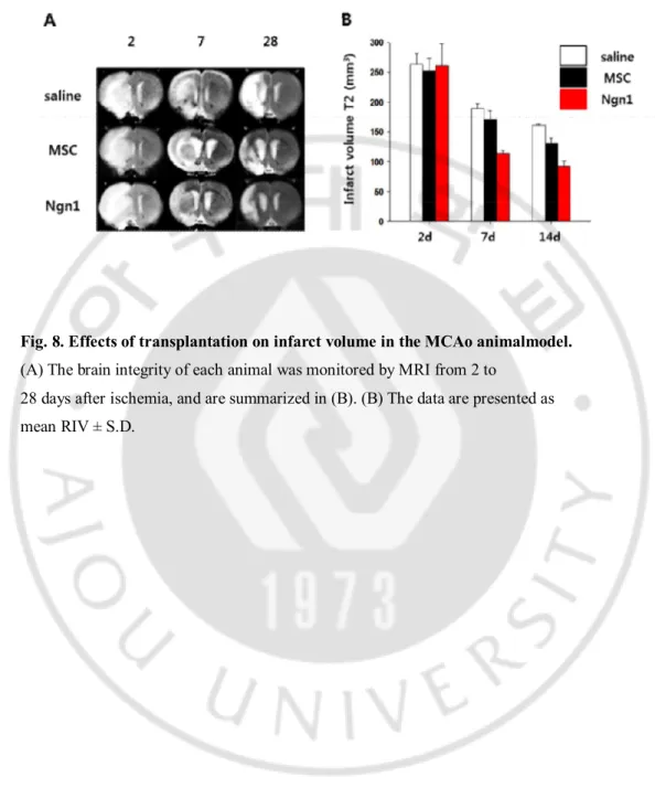
We analyzed motor functions with Rotarod and adhesive removal test (Fig.1 A, B) from 1 day after ischemic stroke. Compared with the saline and MSC group, MSC-Ngn1 group showed significant recovery at day 7, which continued until day 28 after stroke in the both behavior tests. The infarct volumes of the ischemic animals were measured by MRI (Fig.1 C, D) over the 4-week period monitoring the physical integrity of the brains. Animals grafted with MSC and MSC-Ngn1 cells showed higher recoveries than controls over the 4weeks. MSC cells reduced the infarct to a lesser degree compared with MSC-Ngn1 cells. These data indicated that the animals with MSC-Ngn1 cells exhibited the highest functional recovery among the tested animals.
42
Fig.7 . Effects of transplantation on functional recovery in the ischemic stroke.
Performance in the (A) rotarod and (B) adhesive removal from 1 to 28 days after ischemia. The data were collected from 5 animals per group and are presented as mean values ± S.D. Statistically significant differences between the groups were determined by ANOVA (*, p < 0.05, **, p < 0.01, ***, p < 0.001 compared to the control saline group; #, p < 0.05, ##, p < 0.01, ###, p < 0.001 compared to the MSC-LacZgroup).
43
Fig. 8. Effects of transplantation on infarct volume in the MCAo animalmodel.
(A) The brain integrity of each animal was monitored by MRI from 2 to
28 days after ischemia, and are summarized in (B). (B) The data are presented as mean RIV ± S.D.
44
B. MRI Tracking of cells after IA TP
We traced SPIO-labeled MSC and MSC-Ngn1 cells in the ischemic brain after intra-arterial administration (Figure 2). The cells could be detected as soon as 30min after IA injection scattering discretely throughout the ipsilateral side of the brain (Fig.2 A). While MSCs and MSC-ngn1 were shown similar amount and distribution pattern in the ipsilateral side until 4h after injection, a few MSC were seen from 1day comparing with the patterns of MSC-ngn1 cells. These data indicate that MSC-ngn1 were remained stable in the ischemic brain until 25 days after administration compared with MSCs. We also check the localization the cells labeled GFP at 1day and 4day after IA injection. Most of the transplanted cells were localized in the blood vessels at 1day (Fig.2 B) and some cells were located near the blood vessels at 4days after IA injection (Fig.2 C). The SPIO-labeled cells could be detected through PB-staining and the signals were merged with hMT and ED1 indicating that some cells survived exhibiting human specific marker and the others were already died by macrophages at 25day after IA injections (Fig.2 D). These patterns were detected both MSCs and MSC-Ngn1 cells (Fig.2 C, D).
45
Fig.9 . In vivo tracing of MSC and MSCs/Ngn1 after IA TP in ischemic brain.
Migration of MSC and MSCs/Ngn1 was monitored by the MRI using the SPIO labeling ofcells. SPIO-labeled cells were visualized in T2*-weighted images at 30min, 4hrs, 1day, 4days and 25ays after intra –arterial injection. The ischemic lesions were located on the right side of the brain.
46
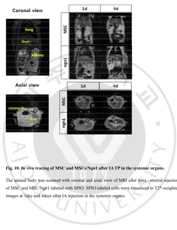
Fig. 10. In vivo tracing of MSC and MSCs/Ngn1 after IA TP in the systemic organs.
The animal body was scanned with coronal and axial view of MRI after intra –arterial injection of MSC and MSC/Ngn1 labeled with SPIO. SPIO-labeled cells were visualized in T2*-weighted images at 1day and 4days after IA injection in the systemic organs.
47
Fig.11 . localization of the grafted cells in the ischemic brain at 1day and 4daysafter IA TP.
Confocal images of MSC/Ngn1 and blood vessels at 1day (A) and 4days (B) after intra-artery administration. MSC-Ngn1 and blood vessels were identified by the GFP (green) and the RECA (red) respectively. Scale bars = 20 μm.
48
Fig. 12. Intra-arterial transplanted cells at 4h after injection in the ischemic brain.
Fluorescence scanning images show MSC/Ngn1(green) and immune cells (red) at 4h after IA in the ischemic brain. The MSC/Ngn1 and immune cells were identified by GFP(green), ED1(red) and Iba1(red) antibody.
49
Fig.13 . Grafted cells at 25day after IA injection in the ischemic brain.
Prussian blue staining at 25 days of the ischemic brain transplanted with intra-arterial MSCs/Ngn1 cells. Immunostaining for identifying transplanted SPIO labeled MSCs/Ngn1 cell and phagocytic activity (microglia/macrophages) by using anti hMT (human Mitochondria) and ED1 antibody respectively were performed in ischemic region. SPIO correlated with hMT (red arrowheads) and ED1 (black arrowheads). Scale bars = 20 μm.
50
C. Cell migration mechanism
To compare the ability of cell migration under ischemic condition between MSC and Ngn1, we performed the migration assay using transwell chamber. MSC or MSC-Ngn1 cells was seeded in the upper Transwell chamber, while ischemic extract was added to the lower chamber. MSCs exhibited barely migration to the lower chamber, while MSC-Ngn1 showed high migratory activity to the ischemic extract. Verifying the differences of cell migration mechanism between MSC and MSC-Ngn1 after IA administration, we investigated the expression of chemoattractant receptors which involved in transmigration from blood vessels to ischemic brain, including CCR1, CCR2, CXCR4, CD44, MMP2, VLA-4 and c-Met in vitro. All of gene expressions in MSC/Ngn1 was higher than in MSCs. Especially CCR1, CCR2 and CXCR4 considering chemoattractant receptors were highly expressed in MSC/Ngn1. These data suggest that the better migration ability of MSC/Ngn1 in the ischemic brain results from the higher expressions level of receptors concerning cell adhesion, chemoattractant and transmigration.
51
Fig.14 . The expressions of chemokine molecules and cell migration under ischemic condition.
Relative expressions of cell receptors relating to the cell adhesion, chemoattraction and transmigration between MSC and MSC/Ngn1(A). All of the receptors were higher expressed in MSC/Ngn1 than MSC. Transwell migration assays were used to assess the migratory activity of MSCs and MSCs-Ngn1 toward ischemic extract (B). The presence of IE in the lower chamber evoked the migration of cells, and chemotactic migration were enhanced in MSC/Ngn1.Abbreviations: CCR1, C-C chemokine receptor type 1; CCR2, C-C motif chemokine receptor 2; CXCR4, C-X-C motif chemokine receptor 4; VLA-4, Very Late Antigen-4 (Integrin alphaAntigen-4beta1); MMP2, matrix metallopeptidase 2; c-Met, Met,hepatocyte growth factor receptor (HGFR).
52
Fig.15.The expressions of TGF-β in MSC and MSC/Ngn1.
Relative expressions of TGF-beta suppressing immune propagation between MSC and MSC/Ngn1. TGF-β was similarly expressed in both of cells.
53
D. Paracrine effect of MSC-ngn1 cells on ischemic brain
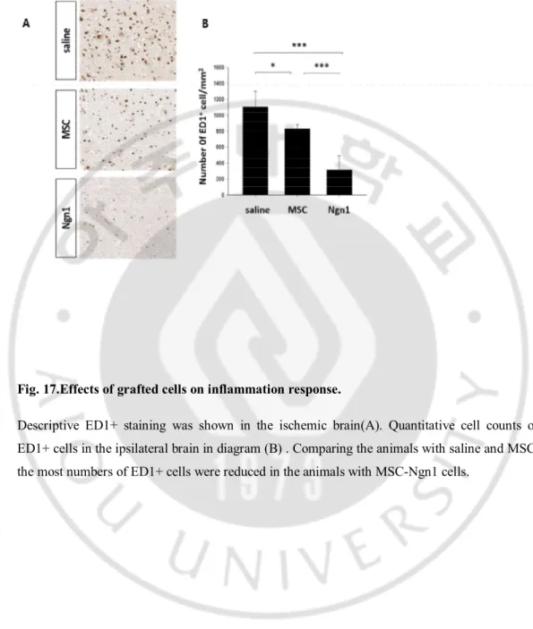
We evaluated the differences in inflammatory response 4 days after administrations. Active microglia, monocytes, and macrophages could be recognized by ED1 indicating ongoing inflammation. Comparing the animals with saline and MSC, the most numbers of ED1+ cells were reduced in the animals with MSC-Ngn1 cells. To confirm the anti-apoptotic effect in the ipsilateral regions, we performed TUNEL assays on the ischemic rat brains. The enormous apoptosis was shown in the control animals 1 week after injury. Intra-arterial injection of MSC and MSC-Ngn1 cells reduced the number of TUNEL+ cells considerably in the ischemic brain. Notably the higher reduction of TUNEL+ cells was shown in the animals with MSC-Ngn1 than MSC. These data indicate that MSCs and MSC-Ngn1 have paracrine effect on the ischemic brain 4 days after intra-arterial administration.
54
Fig. 16.Effects of grafted cells on cell death.Representative histological images of TUNEL assay (A). Quantitative analysis of TUNEL+ cells in the ipsilateral side of ischemic stroke is illustrated (B). The higher reduction of TUNEL+ cells was shown in the ischemic brain with MSC-Ngn1 than MSC.
55
Fig. 17.Effects of grafted cells on inflammation response.
Descriptive ED1+ staining was shown in the ischemic brain(A). Quantitative cell counts of ED1+ cells in the ipsilateral brain in diagram (B) . Comparing the animals with saline and MSC, the most numbers of ED1+ cells were reduced in the animals with MSC-Ngn1 cells.
56
Fig. 18.GFAP expression of reactive astrocytes in the ischemic brains.
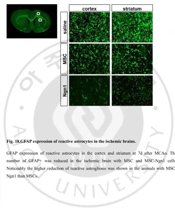
GFAP expression of reactive astrocytes in the cortex and striatum at 7d after MCAo. The number of GFAP+ was reduced in the ischemic brain with MSC and MSC-Ngn1 cells. Noticeably the higher reduction of reactive astrogliosis was shown in the animals with MSC-Ngn1 than MSCs.
57
E. Tissue integrity was preserved in the MSC-Ngn1 treated brain
Ischemic brain at the level of the striatum were stained with anti-NeuN antibody to visualize the neuronal loss 4 weeks after the ischemic injury. In the saline group, most of NeuN+ cells in the cortex and striatum were lost. Some NeuN+ cells in the ischemic striatum were found in the MSC group indicating degeneration of striatum was less severe. Loss of NeuN+ cells was intensely reduced by transplantation of MSC-Ngn1 cells comparing the other groups. This data indicated that the higher recovery of behavioral impairment detected in the MSC-Ngn1 group is related to the higher tissue integrity of the host brain.58
Fig.19.Tissue integrity in ischemic brain after IA TP.Coronal sections of brain at the level of the striatum were immunostained with anti-NeuN antibody to visualize the ischemic neuronal loss 4 weeks after MCAo. Loss of NeuN-positive cells in the ipsilateral striatum was protected by transplantation of MSC-Ngn1 (C,F,I) compared to MSC-LacZ (B,E,H) or saline-treated control (A,D,G). Higher magnification of areas marked in (A–C). (G–I): Higher magnification of areas marked in (D–F).
59
F. Transdifferentiation of MSC-Ngn1 Cells In Vivo
we sought evidence that the functional recovery in animals with MSC-Ngn1 cells was due, at least in part, to neuronal differentiation of the transplanted cells. Evaluation of neuronal differentiation of grafted cells in vivo was performed. We identified MSC-Ngn1 cells were identified with anti-human mitochondrial antibody at 25days after transplantation. MSC-Ngn1 cells could be found in both cortical core and IBZ, where they differentiated into NeuN+ cells.
60
Fig.20.Trans differentiation of MSCs/Ngn1 after intra-arterial administration.
Confocal images of the transplanted cells at 4 weeks after ischemia. MSC-Ngn1 cells were identified by the anti-NeuN antibody and human mitochondrial antibody. Arrowheads denote the donor-derived cells colocalized with cell type specific antigens in enlarged orthographic images.
61
IV. DISCUSSION
Stem cell therapy has emerged as an exciting candidate for the treatment of neurological diseases. Among the various types of stem cells, MSCs are of particular clinical interest because they can be isolated from bone marrow, adipose tissues, and umbilical cord blood. MSCs were shown to improve the neurological dysfunctions in stroke patients. The present study presents the scientific basis for a requirement of neural induction of MSCs for better treatment of neurological dysfunctions.
We analyzed motor functions with Rotarod and adhesive removal test (Figure.1) from 1 day after ischemic stroke. Compared with the saline and MSC group, MSC-Ngn1 group showed significant recovery at day 7, which continued until day 28 after stroke in the both behavior tests. The infarct volumes of the ischemic animals were measured by MRI over the 4-week period monitoring the physical integrity of the brains. Animals grafted with MSC and MSC-Ngn1 cells showed higher recoveries than controls over the 4weeks. MSC cells reduced the infarct to a lesser degree compared with MSC-Ngn1 cells. These data indicated that the animals with MSC-Ngn1 cells exhibited the highest functional recovery among the tested animals.
From 4days after IA administration, MSC-Ngn1 group showed significant functional recovery comparing the other groups. That is not because of neuronal induction but the paracrine effects from the cells. We previously confirmed that MSC and MSC/Ngn1 have similar paracrine effects in the ischemic stroke. To check whether the MSC and MSC/Ngn1 differently distribute in the ischemic brain or not, we traced SPIO-labeled MSC and MSC-Ngn1 cells in the ischemic brain after intra-arterial administration (Figure 2). The cells could be detected as soon as 30min after IA injection scattering discretely throughout the ipsilateral side of the brain. While MSCs and MSC-ngn1 were shown similar amount and distribution pattern in the ipsilateral side until 4h after injection, a few MSC were seen from 1day comparing with the patterns of MSC-ngn1 cells. These data indicate that MSC-ngn1 were remained stable in the ischemic brain until 25 days after administration compared with MSCs. We also check the localization
62
the cells labeled GFP at 1day and 4day after IA injection. Most of the transplanted cells were localized in the blood vessels at 1day and some cells were located near the blood vessels at 4days after IA injection. The SPIO-labeled cells could be detected through PB-staining and the signals were merged with hMT and ED1 indicating that some cells survived exhibiting human specific marker and the others were already died by macrophages at 25day after IA injections. These patterns were detected both MSCs and MSC-Ngn1 cells.
To compare the ability of cell migration under ischemic condition between MSC and Ngn1, we performed the migration assay using transwell chamber. MSC or MSC-Ngn1 cells was seeded in the upper Transwell chamber, while ischemic extract was added to the lower chamber. MSCs exhibited barely migration to the lower chamber, while MSC-Ngn1 showed high migratory activity to the ischemic extract. Verifying the differences of cell migration mechanism between MSC and MSC-Ngn1 after IA administration, we investigated the expression of chemoattractant receptors which involved in transmigration from blood vessels to ischemic brain, including CCR1, CCR2, CXCR4, CD44, MMP2, VLA-4 and c-Met in vitro. All of gene expressions in MSC/Ngn1 was higher than in MSCs. Especially CCR1, CCR2 and CXCR4 considering chemoattractant receptors were highly expressed in MSC/Ngn1. These data suggest that the better migration ability of MSC/Ngn1 in the ischemic brain results from the higher expressions level of receptors concerning cell adhesion, chemoattractant and transmigration.
It is known that MSCs produce bioactive cytokines that attenuate immune responds after ischemic injury and protect delayed cell death in the detrimental environment of damaged tissues. Though the MSC and MSC/Ngn1 exert similar paracrine effect in the ischemic brain, they differently remain in the ipsilateral side of the brain after IA administration. So we hypothesize that different amount of remaining cells cause different paracrine effect after IA transplantation. We evaluated the differences in inflammatory response 4 days after administrations. Active microglia, monocytes, and macrophages could be recognized by ED1 indicating ongoing inflammation.
63
Comparing the animals with saline and MSC, the most numbers of ED1+ cells were reduced in the animals with MSC-Ngn1 cells. To confirm the anti-apoptotic effect in the ipsilateral regions, we performed TUNEL assays on the ischemic rat brains. The enormous apoptosis was shown in the control animals 1 week after injury. Intra-arterial injection of MSC and MSC-Ngn1 cells reduced the number of TUNEL+ cells considerably in the ischemic brain. Notably the higher reduction of TUNEL+ cells was shown in the animals with MSC-Ngn1 than MSC. These data indicate that MSCs and MSC-Ngn1 have paracrine effect on the ischemic brain 4 days after intra-arterial administration.
Ischemic brain at the level of the striatum were stained with anti-NeuN antibody to visualize the neuronal loss 4 weeks after the ischemic injury. In the saline group, most of NeuN+ cells in the cortex and striatum were lost. Some NeuN+ cells in the ischemic striatum were found in the MSC group indicating degeneration of striatum was less severe. Loss of NeuN+ cells was intensely reduced by transplantation of MSC-Ngn1 cells comparing the other groups. This data indicated that the higher recovery of behavioral impairment detected in the MSC-Ngn1 group is related to the higher tissue integrity of the host brain.
we sought evidence that the functional recovery in animals with MSC-Ngn1 cells was due, at least in part, to neuronal differentiation of the transplanted cells. Evaluation of neuronal differentiation of grafted cells in vivo was performed. We identified MSC-Ngn1 cells were identified with anti-human mitochondrial antibody at 25days after transplantation. MSC-Ngn1 cells could be found in both cortical core and IBZ, where they differentiated into NeuN+ cells.
64
V. CONCLUSION
This study in an acute ischemic stroke model suggests that MSCs/Ngn1 stem cells have promising therapeutic effectiveness. IA administration would be a feasible grafting modality compared to IC administration, based on the behavior test results and MRI analysis.
65
VI.REFERENCE
1. Pittenger MF, Mackay AM, Beck SC, Jaiswal RK, Douglas R, Mosca JD, et al. Multilineage potential of adult human mesenchymal stem cells. Science. 1999, 284, 143-147.
2. Woodbury D, Schwarz EJ, Prockop DJ, Black IB. Adult rat and human bone marrow stromal cells differentiate into neurons. J. Neurosci Res. 2000, 61, 364-370.
3. Deng W, Obrocka M, Fischer I, Prockop DJ. In vitro differentiation of human marrow stromal cells into early progenitors of neural cells by conditions that increase intracellular cyclic AMP. Biochem. Biophys. Res. Commun. 2001, 282, 148-152.
4. Phinney DG, Prockop DJ. Concise review: mesenchymal stem/multipotent stromal cells: the state of transdifferentiation and modes of tissue repair--current views. Stem Cells. 2007, 25, 2896-2902.
5. Bang OY, Lee JS, Lee PH, Lee G. Autologous mesenchymal stem cell transplantation in stroke patients. Ann. Neurol. 2005, 57, 874-882. 6. Chen J, Li Y, Wang L, Zhang Z, Lu D, Lu M, et al. Therapeutic benefit of
intravenous administration of bone marrow stromal cells after cerebral ischemia in rats. Stroke. 2001, 32, 1005-1011.
7. Pittenger MF, Martin BJ. Mesenchymal stem cells and their potential as cardiac therapeutics. Circ. Res. 2004, 95, 9-20.
8. Ma Q, Kintner C, Anderson DJ. Identification of neurogenin, a vertebrate neuronal determination gene. Cell. 1996, 87, 43-52.