저작자표시-비영리-변경금지 2.0 대한민국 이용자는 아래의 조건을 따르는 경우에 한하여 자유롭게 l 이 저작물을 복제, 배포, 전송, 전시, 공연 및 방송할 수 있습니다. 다음과 같은 조건을 따라야 합니다: l 귀하는, 이 저작물의 재이용이나 배포의 경우, 이 저작물에 적용된 이용허락조건 을 명확하게 나타내어야 합니다. l 저작권자로부터 별도의 허가를 받으면 이러한 조건들은 적용되지 않습니다. 저작권법에 따른 이용자의 권리는 위의 내용에 의하여 영향을 받지 않습니다. 이것은 이용허락규약(Legal Code)을 이해하기 쉽게 요약한 것입니다. Disclaimer 저작자표시. 귀하는 원저작자를 표시하여야 합니다. 비영리. 귀하는 이 저작물을 영리 목적으로 이용할 수 없습니다. 변경금지. 귀하는 이 저작물을 개작, 변형 또는 가공할 수 없습니다.
Sustained-release Recombinant
Human Growth Hormone improved
body composition and QoL in adults
over 50 years with somatopause
Jeong Kyung Park
Department of Medicine
Sustained-release Recombinant
Human Growth Hormone improved
body composition and QoL in adults
over 50 years with somatopause
Directed by Professor Eun Jig Lee
The Master's Thesis
submitted to the Department of Medicine,
the Graduate School of Yonsei University
in partial fulfillment of the requirements for the degree
of Master of Medical Science
Jeong Kyung Park
June 2015
This certifies that the Master's Thesis of
Jeong Kyung Park is approved.
---Thesis Supervisor : Eun Jig Lee
---Thesis Committee Member#1 : Kyung Soo Park
---Thesis Committee Member#2 : Yoon-Sok Chung
---The Graduate School
Yonsei University
ACKNOWLEDGEMENTS
It took me a long time to complete my thesis. I’d like to thank
all those who have believed in and supported me.
I would like to special thank my supervisor, Prof. Eun Jig Lee,
for his guidance and support throughout this study. His
creative ideas and initiatives have inspired my learning. I feel
very fortunate to have had a chance to work with him. I
appreciate Prof. Yoon-Sok Chung and Prof. Kyung Soo Park
for giving me valuable advice for this study. Although I cannot
list the names of everyone who helped my studies in this
limited space, I do remember and appreciate every single help
and advice I have received.
Finally, I am also grateful for my husband and parents for
their support and unlimited love throughout my life.
<TABLE OF CONTENTS>
ABSTRACT ···1
I. INTRODUCTION ···3
II. MATERIALS AND METHODS···5
1. Study design ···5
2. Study population···5
3. Study protocol ···6
4. Hormone assays ···7
5. Assessment of body composition ···7
6. Assessment of quality of life ···8
7. Statistical analyses ···8
III. RESULTS ···10
1. Baseline characteristics···10
2. Serum IGF-1 concentration ···12
3. Body size···13
4. Body composition···13
5. Abdominal fat distribution···15
6. Mid-thigh composition···15
7. Quality of life ···15
8. Assessment of safety and tolerability···17
IV. DISCUSSION ···19
V. CONCLUSION ···24
REFERENCES ···25
ABSTRACT(IN KOREAN) ···31
LIST OF FIGURES
Figure 1. Serum level of IGF-1 (ng/ml) following administration
of SR-rhGH··· 13
Figure 2. Estrogen substitute and SR-rhGH··· 14
Figure 3. Changes over 26 weeks in BAP and CTX··· 18
LIST OF TABLES
Table 1. Demographic characteristics of the study subjects ·· 10
Table 2. Effect of the treatment on vital sign, body
composition, and QoL ··· 11
Table 3. Effect of the growth hormone administration on serum
ABSTRACT
Sustained-release Recombinant Human Growth Hormone improved
body composition and QoL in adults over 50 years with somatopause
Jeong Kyung Park
Department of Medicine
The Graduate School, Yonsei University
(Directed by Professor Eun Jig Lee)
Context: The elderly experiencing somatopause and the resultant metabolic
impairment can obtain partial recovery from administration of recombinant human GH (rhGH). However, aged adults suffer inconvenience from daily injection of existing rhGH.
Objectives: To evaluate the effects, safety, and compliance of weekly
administered low dose of sustained-release recombinant human GH (SR-rhGH) in aged adults with somatopause.
Design: This is a 26-week prospective, single-arm, multicenter phase IV trial in
adults.
Intervention/Participants: A total of 38 subjects, aged ≥ 50 years with
somatopause (serum IGF-1 < 150 ng/ml) were enrolled and each received 2 mg of SR-rhGH for 26 weeks.
Results: Mean baseline IGF-1 level of 123.4 ± 41.6 ng/ml increased to 174.8 ±
59.6 ng/ml after administration of SR-rhGH at 4 weeks and it was maintained for the remainder of the study period. At 26 weeks, average lean body mass increased by 0.45 kg, waist circumference reduced by 1.06 cm, and Quality of Life was improved significantly (P<0.01 in each index). There was a simultaneous increase in serum levels of biochemical markers of bone resorption and formation. Estrogen substitute in women attenuated the beneficial effects of SR-rhGH on body composition and metabolic indices.
There was no significant change in the body fat distribution or fat mass. Adverse events included pruritus (10.5%), arthralgias (5.3%), and edema (5.3%), but their symptoms were well tolerable.
Conclusions: Body composition and Quality of Life can be restored in part by
the replacement of low dose SR-rhGH for 26 weeks in patients with somatopause without significant adverse effects.
---Key words:
growth hormone replacement, somatopauseSustained-release Recombinant Human Growth Hormone improved
body composition and QoL in adults over 50 years with somatopause
Jeong Kyung Park
Department of Medicine
The Graduate School, Yonsei University
(Directed by Professor Eun Jig Lee)
I. INTRODUCTION
Normal aging is paralleled by a progressive decline in growth hormone (GH) secretion.1 Adolescents have the highest level of GH, followed by a
progressive decline in GH secretion with advancing age; each decade of increasing age attenuating the GH production rate by 14%.2,3 Therefore, most
elderly adults are confronted with somatopause. A decreased GH secretion provides central fat accumulation, sarcopenia, dyslipidemia, increased cardiovascular mortality and diminished quality of life (QoL).4,5 Especially,
sarcopenia, a complex syndrome that is associated with decreased muscle mass and increased fat mass, is increasingly recognized by physicians to be associated with higher risks for multiple adverse outcomes in the elderly. Sarcopenia is considered a major contributor in the pathway leading to an elder patient’s frailty.6For these elderly populations, a number of clinical trials of GH
beneficial effects on lipid profile, body fat distribution, bone metabolism, and well-being.8,10,11 Nevertheless, several concerns remain regarding diabetes
mellitus (DM), peripheral edema, and arthralgias.12 The technical difficulty of
GH administration is also an important problem for elderly patients.
A sustained release formulation of recombinant human GH (SR-rhGH, Declage® LG Life Sciences, Ltd., Seoul, Korea) using sodium hyaluronate
microparticles, was developed aiming at once-a-week injection.13 This new
formulation was expected to lead to better patient compliance with comparable efficacy and adverse effects. In the current study, we administered low dose of SR-rhGH for 26 weeks to patients aged > 50 years with somatopause and investigated the safety and the effects of the formulation on body composition and quality of life. We also assessed the role of gender differences and the influence of sex hormones.
II. MATERIALS AND METHODS 1. Study Design
This study was a prospective, single-arm, multicenter phase IV trial. The protocol was reviewed and approved by an independent Institutional Review Board at each participating center, and all subjects provided written, informed consent prior to registration for this study.
2. Study Population
Men and women aged ≥ 50 years with documented somatopause (serum IGF-1 levels of 150 ng/ml or lower) were eligible for this study. If subjects required glucocorticoid, thyroxine, or sex hormone replacements, doses of all hormone replacement was adjusted to maintain a state of stabilization for at least 4 weeks before GH administration. No subjects had received GH treatment within the past 3 months. Subjects were excluded if they had impaired glucose tolerance or diabetes mellitus, acromegaly, intracranial hypertension, active malignancy, or concurrent antitumor therapy, critical illness, history of cardiac or abdominal surgery, cognitive impairment, or GH deficiency following an organic cause such as pituitary tumor, head trauma, or other pituitary disorders. Patients with renal impairment (defined as serum creatinine > 1.6 mg/dl) or liver disease (defined as alanine aminotransferase or aspartate aminotransferase levels greater than three times the upper limit of normal) were also excluded.
3. Study Protocol
This study included a screening visit (-week 4), baseline visit, and 3 visits over the 26 week study period. After a 4-week screening period, participants received 6 IU (2 mg) of SR-rhGH subcutaneously weekly for 26 weeks. The dosage of administered SR-rhGH was adjusted upon physician’s judgment. At every visit, vital signs, height, weight, and waist circumference were investigated and symptoms and signs of adverse effects were carefully monitored. Serum IGF-1 was assessed to evaluate the effectiveness of the drug at baseline and at 4, 13, and 26 weeks. Body composition, quality of life, and laboratory assessment [lipid profile, bone alkaline phosphatase (B-ALP), C-telopeptide (CTX), free thyroxine (fT4), thyroid-stimulating hormone (TSH)] were determined at baseline and at 26 weeks. Laboratory tests including complete blood count, total protein, albumin, liver function tests, blood urea nitrogen (BUN), creatinine, total bilirubin, uric acid, calcium, phosphate, sodium, potassium, HbA1c, fasting plasma glucose (FBS), and insulin were measured at baseline, at 13 weeks, and at 26 weeks. The homeostasis model assessment (HOMA-IR) was used to determine insulin resistance14 and
calculated as: [fasting glucose (mg/dl) x fasting insulin (μU/ml)] / 405. Participants were instructed not to change their diet and exercise patterns during the treatment period. Caloric intake was assessed by daily self-recording of episodes by the subjects in a nutrition diary at baseline and at 4, 13, and 26
weeks. At every visit, researchers inquired about compliance and the empty vial was returned for counting. Compliance was calculated at 26 weeks as the percentage of the number of actual injection per the number of scheduled injection.
4. Hormone Assays
All blood samples were taken after an overnight fast of 10 hours. Serum IGF-1 concentrations were measured with a commercially available standard enzyme-linked immunosorbent assay kit, according to the manufacturer’s protocols (Immunodiagnostic Systems, Fountain Hills, AZ, USA). The coefficient of inter-assay variation was 7.4-9.1% and intra-assay variation was 2.6-4.4% in the concentration range of 180-360 ng/ml. TSH and fT4 were determined by chemiluminescence immunoassay (Siemens Healthcare Diagnostics, Cergy Pontoise, France).
5. Assessment of Body Composition
Body weight (kg), height (cm), body mass index (BMI, calculated as weight in kilograms divided by height in meters squared), and waist circumference (WC, measured as the minimum circumference around level of umbilicus) were determined.
Measurements of fat mass (FM) and lean body mass (LBM) were obtained using whole body dual X-ray absorptiometry (QDR 4500A, Hologic
Inc., Bedford, MA, USA). Head, trunk, both arms and legs were included for calculation of FM and LBM. Computed tomography (CT) scans (SOMATOM Sensation 64, SIEMENS, Germany) were used to estimate levels of visceral, subcutaneous, and total adipose tissue (VAT, SAT, and TAT, respectively). At least five-slice CT scans were performed at the level of second to fifth lumbar vertebra (L2-L5) and at the mid-thigh lesion. Abdominal VAT and SAT were estimated as area (cm2) at level of L2-3. TAT measurements were interpreted as
the sum of VAT and SAT measurements.
6. Assessment of Quality of life
Quality of life was assessed using the ‘Quality of Life-Assessment of Growth Hormone Deficiency in Adults’ questionnaire (QoL-AGHDA), which has been shown to have good reliability, and reproducibility while constructing validity across a range of languages.15The score of QoL-AGHDA, ranging from
0 to 25, was assessed at baseline and wk26. A high score on the measure indicates poor quality of life.
7. Statistical analyses
Statistical analyses were performed using the SPSS software package for Windows (Version 16.0; SPSS Inc., Chicago, IL, USA). The efficacy of SR-rhGH was analyzed using serum IGF-1, body composition, lipid profile, quality of life comparing between baseline values and those at week 26 and the
significance was determined by paired t-test, if the data were normally distributed, and by Wilcoxon’s signed rank test if they were not. The safety of the SR-rhGH was estimated using vital signs, laboratory assessments comparing between baseline and week 26 by paired t-test or Wilcoxon’s signed rank test as appropriate. The data were expressed as the mean ± standard deviation. A P-value of > 0.05 was regarded as significant.
III. RESULTS
1. Baseline Characteristics
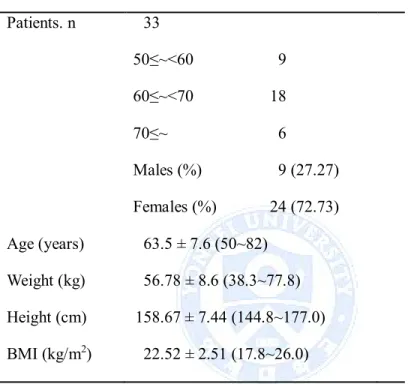
Table 1 Demographic characteristics of the study subjects
Patients. n 33 50≤~<60 9 60≤~<70 18 70≤~ 6 Males (%) 9 (27.27) Females (%) 24 (72.73) Age (years) 63.5 ± 7.6 (50~82) Weight (kg) 56.78 ± 8.6 (38.3~77.8) Height (cm) 158.67 ± 7.44 (144.8~177.0) BMI (kg/m2) 22.52 ± 2.51 (17.8~26.0)
BMI, body mass index
In total, 38 patients received at least one dose of SR-rhGH and were included in safety analyses. Five subjects dropped out early for the following reasons; lost to follow-up (one), withdrawal the consent (two), violation the protocol (two). Thirty three subjects were analyzed, the mean age was 64 ± 8 yr (50-82), and there were 24 (72.8%) females and 9 males (27.8%) (Table 1). Women were postmenopausal and six had been taking sex hormone replacement therapy (HRT). None of the male subjects had been taking testosterone replacement
therapy. The mean weight of all subjects was 56.8 ± 8.6 kg (38.3-77.8 kg), and the mean height was 158.7 ± 7.4 cm (144.8-177.0 cm). Calculated BMI was 22.5 ± 2.5 kg/m2(17.8~26.0). As shown in Table 2, at baseline, subjects overall
exhibited high VAT/SAT ratio, indicating central obesity in spite of relatively low BMI. In comparison to women, men had a higher LBM (women, 45.6 ± 6.2 kg; men, 35.2 ± 3.2 kg; P<0.0001) and lower body fat mass (women, 32.7 ± 5.4 kg; men, 19.3 ± 8.7 kg; P=0.001). Women on estrogen substitution had higher LBM as those without sex hormone (37.9 ± 2.8 kg, 34.2 ± 2.9 kg respectively, P=0.014). There were no significant differences in baseline levels of IGF-1, BMI, and WC between men and women, and between women who received concurrent HRT and those did not.
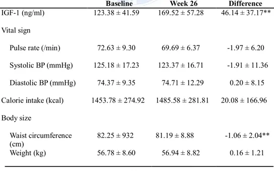
Table 2 Effect of the treatment on vital sign, body composition, and QoL Baseline Week 26 Difference
IGF-1 (ng/ml) 123.38 ± 41.59 169.52 ± 57.28 46.14 ± 37.17**
Vital sign
Pulse rate (/min) 72.63 ± 9.30 69.69 ± 6.37 -1.97 ± 6.20
Systolic BP (mmHg) 125.18 ± 17.23 123.37 ± 16.71 -1.91 ± 11.36
Diastolic BP (mmHg) 74.37 ± 9.35 74.71 ± 12.29 0.20 ± 8.15
Calorie intake (kcal) 1453.78 ± 274.92 1485.58 ± 281.81 20.08 ± 166.96 Body size
Waist circumference
(cm) 82.25 ± 932 81.19 ± 8.88 -1.06 ± 2.04**
BMI (kg/m2) 22.52 ± 2.51 22.60 ± 2.64 0.08 ± 0.46 Body composition
% Body fat (%) 29.06 ± 8.79 28.82 ± 9.04 -0.25 ± 1.59
FM (kg) 16.38 ± 5.64 16.40 ± 5.86 0.01 ± 0.91
LBM (kg) 37.99 ± 6.28 38.44 ± 6.38 0.45 ± 1.10*
Abdominal fat distribution
Visceral fat (cm2) 110.16 ± 69.85 107.15 ± 71.45 -3.01 ± 15.30
Subcutaneous fat (cm2) 140.33 ± 67.61 135.66 ± 64.23 -4.67 ± 19.12 Total abdominal fat
(cm2) 250.49 ± 114.91 242.81 ± 115.36 -7.69 ± 27.96 VAT/SAT ratio 0.87 ± 0.53 0.96 ± 0.77 0.09 ± 0.68 Mid-thigh Subcutaneous fat (cm2) 54.65 ± 29.14 54.08 ± 28.75 -0.58 ± 10.03 Muscle (cm2) 83.26 ± 22.78 82.01 ± 22.55 -1.75 ± 8.60 AGHDA QoL 10.06 ± 5.41 7.33 ± 5.34 -2.73 ± 4.91**
* : P-value is less than 0.05 ** : P-value is less than 0.01
FM, Fat mass; LBM, lean body mass; VAT, visceral fat; SAT, subcutaneous fat
2. Serum IGF-1 concentration
After administration of SR-rhGH, IGF-1 levels increased from 123.4 ± 41.6 to 174.8 ± 59.6 ng/ml (P<0.0001) within 4 weeks and were maintained throughout the next 22 weeks (Figure 1). There was a greater response in females than in males. The differences in IGF-1 levels from baseline to week 26 were 37.2 ± 34.1 ng/ml in men and 49.5 ± 38.4 ng/ml in women respectively. In
women on HRT showed greater IGF-1 increments than in sex hormone naïve women at week 26 but this difference did not reached statistical significance (P=0.263).
3. Body Size
There was no significant change in intake calories from baseline to end of this study (Table 2). In body size analysis, the waist circumference reduced significantly after SR-rhGH administration (P=0.0021). However, in subgroup analysis, this difference was not observed in patient who received HRT. BMI and body weight were similar before and after treatment.
Fat mass showed no changes during treatment period and % body fat was slightly decreased without significance, whereas LBM increased significantly at 26 weeks (Table 2, P=0.0096). There were no significant differences between men and women in body composition. Concomitant use of sex hormone in women did not influence LBM, fat mass, and % body fat.
5. Abdominal Fat Distribution
As described in Table 2, TAT, VAT, and SAT showed a trend of reduction, whereas VAT/SAT ratio tended to increase without significance. Abdominal fat distribution showed exciting results in subgroup analysis (Figure 2); women who received HRT demonstrated an increased VAT and SAT (P=0.0313, P=0.0449, respectively). TAT values within this subgroup also showed a tendency to increase. However, fat distribution of patients who did not received HRT, by contrast, showed a decrease, even in VAT/SAT ratio.
6. Mid-thigh Composition
Before and after treatment, the changes of subcutaneous fat and muscle of mid-thigh were not significant. In subgroup analysis, the total area of subcutaneous fat and muscle of mid-thigh did not change significantly over the course of the SR-rhGH administration.
7. Quality of Life
In general, AGHDA score (Table 2) improved significantly (P=0.0032) after SR-rhGH administration, but this difference was not significant in men (P=0.1076). Basal mean AGHDA score was higher in females than in males (female; 10.88 ± 5.79, male; 7.89 ± 3.69). It was also observed that women who were treated with sex hormones had higher AGHDA score (12.67 ± 6.77) at baseline, and did not show an improvement after SR-rhGH treatment.
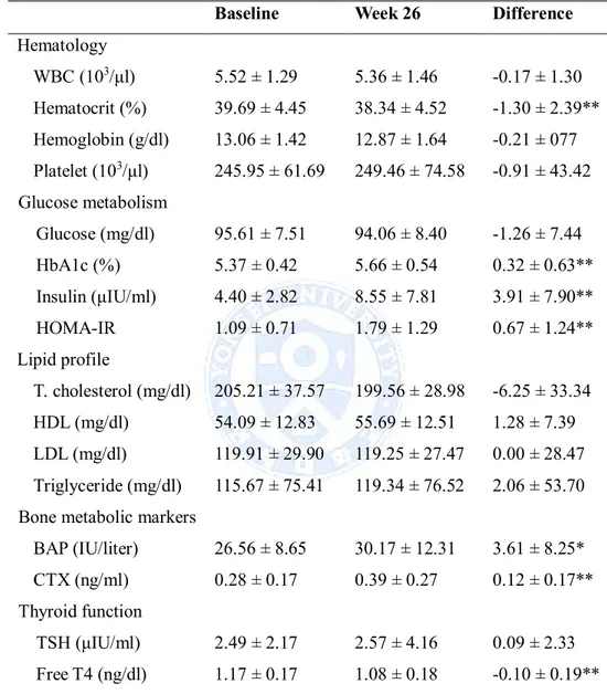
Table 3 Effect of the growth hormone administration on serum profile
Baseline Week 26 Difference
Hematology WBC (103/μl) 5.52 ± 1.29 5.36 ± 1.46 -0.17 ± 1.30 Hematocrit (%) 39.69 ± 4.45 38.34 ± 4.52 -1.30 ± 2.39** Hemoglobin (g/dl) 13.06 ± 1.42 12.87 ± 1.64 -0.21 ± 077 Platelet (103/μl) 245.95 ± 61.69 249.46 ± 74.58 -0.91 ± 43.42 Glucose metabolism Glucose (mg/dl) 95.61 ± 7.51 94.06 ± 8.40 -1.26 ± 7.44 HbA1c (%) 5.37 ± 0.42 5.66 ± 0.54 0.32 ± 0.63** Insulin (μIU/ml) 4.40 ± 2.82 8.55 ± 7.81 3.91 ± 7.90** HOMA-IR 1.09 ± 0.71 1.79 ± 1.29 0.67 ± 1.24** Lipid profile T. cholesterol (mg/dl) 205.21 ± 37.57 199.56 ± 28.98 -6.25 ± 33.34 HDL (mg/dl) 54.09 ± 12.83 55.69 ± 12.51 1.28 ± 7.39 LDL (mg/dl) 119.91 ± 29.90 119.25 ± 27.47 0.00 ± 28.47 Triglyceride (mg/dl) 115.67 ± 75.41 119.34 ± 76.52 2.06 ± 53.70 Bone metabolic markers
BAP (IU/liter) 26.56 ± 8.65 30.17 ± 12.31 3.61 ± 8.25*
CTX (ng/ml) 0.28 ± 0.17 0.39 ± 0.27 0.12 ± 0.17**
Thyroid function
TSH (μIU/ml) 2.49 ± 2.17 2.57 ± 4.16 0.09 ± 2.33
Free T4 (ng/dl) 1.17 ± 0.17 1.08 ± 0.18 -0.10 ± 0.19** * : P-value is less than 0.05
** : P-value is less than 0.01
8. Assessment of Safety and Tolerability
One patient treated with SR-rhGH required a dose decrease to 1.5 mg at visit 3, due to high blood pressure. The mean systolic and diastolic blood pressures were unchanged throughout the study (Table 2). The 26-week SR-rhGH treatment reduced mean value of hematocrit compared with baseline (Table 3, p=0.0028). Though FBS showed no difference, HbA1c and serum insulin levels were increased significantly by 0.30 ± 0.65% (P=0.0001) and 2.91 ± 5.67 μIU/ml (P=0.0004), respectively (Table 3). Corresponding increase in calculated HOMA-IR was observed (by 0.92 ± 1.83, P=0.0009). Mean total cholesterol was decreased by 6.25 ± 33.34 mg/dl compared to its initial level, but this difference was not significant. The values of serum electrolytes, ALT, AST, ALP, BUN, and creatinine did not change significantly (data not shown). Whereas no significant difference in serum calcium concentration was observed between baseline and at week 26, serum phosphorous concentration increased by 0.24 ± 0.62 mg/dl after 13-week of SR-rhGH administration (P=0.0227), and there was no difference at 26 weeks compared with baseline. The bone metabolic markers, BAP, a bone formation marker, and CTX, a bone resorption marker, were increased significantly (P=0.0171, P=0.00004, respectively). In subgroup analysis for bone metabolic markers (Figure 3), there were no significant differences in males and females receiving sex hormones. Only women who were not treated with sex hormone showed a significant increase. In the thyroid function test, serum free T4 decreased by 0.08 ± 0.19 ng/dl
(P=0.0066) at 26 weeks, as compared to the level at baseline, without significant elevation of TSH.
The most common drug-related adverse events were pruritus on injection site in 4 patients. Two patients exhibited arthralgias and in one, symptoms persisted until the end of the study. Two experienced edema at injection site, and another two cases complained of body pruritus after injection of SR-rhGH but the symptoms disappeared within the testing period. Most events were mild to moderate in severity. SR-rhGH administration was not associated with significant adverse effects. Returned vial counts suggested that compliance was excellent, with 99.7% of vials injected into participants.
IV. DISCUSSION
There has been a controversy surrounding replacement of rhGH in the elderly experiencing somatopause and the resultant functional and metabolic impairment including sarcopenia. The first evidence for GH replacement in the elderly was reported by Rudman and colleagues in 1990.8 They demonstrated
improvement in LBM, adipose-tissue mass, and bone mineral density (BMD) after administration of rhGH in men over 60 years old. Subsequently, further studies in the elderly have been conducted, yielding controversial results.16-21
In the current study, we examined the efficacy and safety in administration of SR-rhGH for the first time. During 26 weeks of SR-rhGH administration, serum IGF-1 levels increased to the normal range within 4 weeks and they were maintained throughout the treatment period (Figure 1). This pattern was consistent with an earlier study.8 We detected that the IGF-1
level was higher in females than in males at baseline, compatible with a previous report.22 Serum IGF-1 increased further in females than in males
during SR-rhGH administration, which conflicted with earlier reports12,23that
the effect being more pronounced in males than in females. This result may be due partly to the small number of men studied. It seems be also associated with that baseline mean age was higher in men than in women (65.0 vs. 62.9 years respectively) although the difference was not significant.
A reduction in IGF-1 concentrations during oral estrogen replacement has been observed in postmenopausal women.24There has been some sequential
evidence that estrogen administration lowers IGF-1 concentrations in women.22,25However, baseline IGF-1 level was higher in women with HRT than
those in women without HRT, although it was statistically insignificant. This opposite result possibly related to high LBM value at baseline in women received estrogen. Moreover, women on HRT were younger than those without HRT (61.5 vs. 63.4 years). Span et al.23 confirmed that serum IGF-1 initially
increased equally in both groups of estrogen-substituted and nonsubstituted women. The difference between two groups was statistically significant only after 18 months of rhGH supplements. This delayed blunting effect on IGF-1 might explain why IGF-1 increment was not lower in women received estrogen during our 26-week study.
According to the literature, participants who received GH increased their LBM and decreased their fat mass.8,20,21 Our study consolidates previous
reports in elevation of LBM, and decrease of WC after SR-rhGH administration. However, these effects of SR-rhGH were insignificant compared with previous studies that supplied GH therapy with concurrent lifestyle intervention.26-28
Using CT scans for body fat analysis, we did not detect a significant change in VAT, SAT and TAT. Though participants of this study had lower baseline BMI than those of previous studies, the mean VAT/SAT ratio of cases was 0.87, suggesting central obesity. A concurrent exercise intervention study might have led to better responses to the SR-rhGH. Furthermore, previous report showed
dose-dependent changes in body composition.29 In our study, relatively low
dose of SR-rhGH supplied compared to existing trials8,30 might also influence
insignificant effects on body composition.
We reproduced similar results to previous reports31,32 on biochemical
markers of bone metabolism. The administration of SR-rhGH for 26 weeks significantly increased serum levels of biochemical markers of bone resorption and formation. Joseph and colleagues32 confirmed a simultaneous increase in
bone resorption and formation with the increase in bone formation markers becoming significantly higher than resorption only by 6 months. It suggests there may be delayed increase in BMD after prolonged GH administration.
There was an additional benefit related to QoL but the difference was not significant as found in previous studies, which administrated GH for more than 12 months.33,34 However, there was an obvious improvement of QoL
through the 26-weeks administration of SR-rhGH, especially in women without estrogen substitution. It is expected that long-term trial in postmenopausal females would provide better outcomes in assessment of QoL.
In subgroup analysis of VAT and SAT, females with HRT showed a significant increase in visceral and subcutaneous fat, whereas nonsubstituted women showed a decrease at 26-week (Figure 2). Waist circumference also decreased significantly after administration of SR-rhGH, but not in females with HRT. Munzer and colleagues suggested that HRT attenuated the rhGH-mediated effects through their placebo-controlled study.30 Holloway et al.21 also found a
blunted effect of rhGH on body composition and metabolic indices in postmenopausal women receiving estrogen as compared with women without estrogen replacement. Our results also confirmed that estrogen significantly attenuated the response to SR-rhGH despite IGF-1 increments. It is suggested that estrogens inhibit GH effect not only by reducing IGF-1, but also by other way such as increasing resistance.
Previous studies have shown that participants treated with GH experienced higher rates of soft tissue edema, arthralgias, and carpal tunnel syndrome than those not receiving GH.12,30 In this study, two (5.3%) patients
experienced edema and another two (5.3%) patients presented arthralgias. In general, the rates of adverse events were not significantly greater among subjects who administrated once-weekly injection of SR-rhGH compared with those receiving 3 times/week injection of rhGH.17
There was a slight but significant decrease in hematocrit concentration at 13 weeks (P=0.0152) and at 26 weeks (P=0.0028) compared with the level at baseline. It has been reported that GH deficiency is associated with reducing total body water (TBW), which is mainly due to a decrease in extracellular water.35 The published literature has demonstrated that GH increases TBW and
plasma volume using radionuclide dilution method.36This may have contributed
to the slight decrease in hematocrit values shown in our results. However, increase in body weight was not observed.
significant elevation of TSH. This result supports Jorgensen’s theory that growth hormone increases the serum free T3 and decreases free T4, suggesting either suppression of D3 activity or increased T4 to T3 conversion.37Losa et al.
confirmed this theory through a clinical study which free T4 level decreased at 6 months after GH treatment in adult growth hormone deficiency patients.38
Showing in our study, although a decrement of free T4 was slight and remained within the normal range, it is recommendable to monitor thyroid function during administration of rhGH.
In recent meta-analysis of GH replacement in the GH-deficient adults, circulating glucose and insulin levels were found to be significantly increased after receiving GH,39 whereas it is debatable whether insulin sensitivity is
decreased or unchanged during prolonged GH replacement. We demonstrated that HbA1c, plasma insulin, and HOMA-IR were significantly increased at 26 weeks, namely, administration of SR-rhGH for 26 weeks decreased insulin sensitivity. Placebo-controlled studies are needed to compensate for the physiological decrease in insulin sensitivity with increasing age.
Apart from the single-arm study design, a limitation of this study was the small sample size. Although we found beneficial effects of SR-rhGH in LBM, WC, and QoL, the findings may not apply to the male population. Additional studies are required to verify these results in larger populations.
V. CONCLUSION
Our study suggests that body composition and Quality of Life can be restored in part by the replacement of SR-rhGH for 26 weeks in patients with somatopause. A once-weekly regimen of SR-rhGH was well tolerated without significant adverse effects. Further placebo-controlled studies examining long-term effects will be invaluable in characterizing the efficacy of SR-rhGH on elder’s sarcopenia.
REFERENCES
1. Rudman D, Kutner MH, Rogers CM, Lubin MF, Fleming GA, Bain RP.
Impaired growth hormone secretion in the adult population: relation to age and adiposity. J Clin Invest 1981:67:1361-9.
2. Iranmanesh A, Lizarralde G, Veldhuis JD. Age and relative adiposity are specific negative determinants of the frequency and amplitude of growth hormone (GH) secretory bursts and the half-life of endogenous GH in healthy men. J Clin Endocrinol Metab 1991:73:1081-8.
3. Zadik Z, Chalew SA, McCarter RJ, Jr., Meistas M, Kowarski AA. The
influence of age on the 24-hour integrated concentration of growth
hormone in normal individuals. J Clin Endocrinol Metab
1985:60:513-6.
4. Lanfranco F, Gianotti L, Giordano R, Pellegrino M, Maccario M, Arvat E. Ageing, growth hormone and physical performance. J Endocrinol Invest 2003:26:861-72.
5. Toogood AA, O'Neill PA, Shalet SM. Beyond the somatopause:
growth hormone deficiency in adults over the age of 60 years. J Clin Endocrinol Metab 1996:81:460-5.
6. Evans WJ, Paolisso G, Abbatecola AM, Corsonello A, Bustacchini S,
Strollo F, Lattanzio F Frailty and muscle metabolism dysregulation in the elderly. Biogerontology 2010:11(5):527-36.
7. Lamberts SW. Hormone replacement therapy for somatopause:
risk-benefit analysis and precedents in the treatment of menopause. Growth Horm IGF Res 2000:10 Suppl A: S52-3.
8. Rudman D, Feller AG, Nagraj HS, Gergans GA, Lalitha PY, Goldberg
AF et al. Effects of human growth hormone in men over 60 years old. N Engl J Med 1990:323:1-6.
9. Veldhuis JD, Patrie JM, Frick K, Weltman JY, Weltman AL. Administration of recombinant human GHRH-1,44-amide for 3 months reduces abdominal visceral fat mass and increases physical performance measures in postmenopausal women. Eur J Endocrinol 2005:153:669-77.
10. Monson JP, Abs R, Bengtsson BA, Bennmarker H, Feldt-Rasmussen
U, Hernberg-Stahl E, Thoren M, Westberg B, Wilton P, Wuster C. Growth hormone deficiency and replacement in elderly hypopituitary adults. KIMS Study Group and the KIMS International Board. Pharmacia and Upjohn International Metabolic Database. Clin Endocrinol (Oxf) 2000:53:281-9.
11. Fernholm R, Bramnert M, Hagg E, Hilding A, Baylink DJ, Mohan S,
Thoren M. Growth hormone replacement therapy improves body composition and increases bone metabolism in elderly patients with pituitary disease. J Clin Endocrinol Metab 2000:85:4104-12.
12. Blackman MR, Sorkin JD, Munzer T, Bellantoni MF,
Busby-Whitehead J, Stevens TE et al. Growth hormone and sex steroid administration in healthy aged women and men: a randomized controlled trial. JAMA 2002:288:2282-92.
13. Kim SJ, Hahn SK, Kim MJ, Kim DH, Lee YP. Development of a novel
sustained release formulation of recombinant human growth hormone using sodium hyaluronate microparticles. J Control Release 2005:104:323-35.
14. Matthews DR, Hosker JP, Rudenski AS, Naylor BA, Treacher DF,
Turner RC. Homeostasis model assessment: insulin resistance and beta-cell function from fasting plasma glucose and insulin concentrations in man. Diabetologia 1985:28:412-9.
Wiren L. The QoL-AGHDA: an instrument for the assessment of quality of life in adults with growth hormone deficiency. Qual Life Res 1999:8:373-3.
16. Giordano R, Bonelli L, Marinazzo E, Ghigo E, Arvat E. Growth hormone treatment in human ageing: benefits and risks. Hormones (Athens) 2008:7:133-9.
17. Liu H, Bravata DM, Olkin I, Nayak S, Roberts B, Garber AM, Hoffman AR. Systematic review: the safety and efficacy of growth hormone in the healthy elderly. Ann Intern Med 2007:146:104-15.
18. Franco C, Brandberg J, Lonn L, Andersson B, Bengtsson BA,
Johannsson G. Growth hormone treatment reduces abdominal visceral fat in postmenopausal women with abdominal obesity: a 12-month placebo-controlled trial. J Clin Endocrinol Metab 2005:90:1466-74.
19. Johannsson G, Marin P, Lonn L, Ottosson M, Stenlof K, Bjorntorp P, Sjostrom L, Bengtsson BA. Growth hormone treatment of abdominally obese men reduces abdominal fat mass, improves glucose and lipoprotein metabolism, and reduces diastolic blood pressure. J Clin Endocrinol Metab 1997:82:727-34.
20. Papadakis MA, Grady D, Black D, Tierney MJ, Gooding GA,
Schambelan M, Grunfeld C. Growth hormone replacement in healthy older men improves body composition but not functional ability. Ann Intern Med 1996:124:708-16.
21. Holloway L, Butterfield G, Hintz RL, Gesundheit N, Marcus R. Effects of recombinant human growth hormone on metabolic indices, body composition, and bone turnover in healthy elderly women. J Clin Endocrinol Metab 1994:79:470-9.
amplitude-specific divergence in the pulsatile mode of growth hormone (GH) secretion underlies the gender difference in mean GH concentrations in men and premenopausal women. J Clin Endocrinol Metab 1996:81:2460-7.
23. Span JP, Pieters GF, Sweep CG, Hermus AR, Smals AG. Gender difference in insulin-like growth factor I response to growth hormone (GH) treatment in GH-deficient adults: role of sex hormone replacement. J Clin Endocrinol Metab 2000:85:1121-5.
24. Dawson-Hughes B, Stern D, Goldman J, Reichlin S. Regulation of growth hormone and somatomedin-C secretion in postmenopausal women: effect of physiological estrogen replacement. J Clin Endocrinol Metab 1986:63:424-32.
25. Bellantoni MF, Vittone J, Campfield AT, Bass KM, Harman SM, Blackman MR. Effects of oral versus transdermal estrogen on the growth hormone/insulin-like growth factor I axis in younger and older postmenopausal women: a clinical research center study. J Clin Endocrinol Metab 1996:81:2848-53.
26. Hameed M, Lange KH, Andersen JL, Schjerling P, Kjaer M, Harridge
SD, Goldspink G. The effect of recombinant human growth hormone and resistance training on IGF-I mRNA expression in the muscles of elderly men. J Physiol 2004:555:231-40.
27. Taaffe DR, Thompson JL, Butterfield GE, Hoffman AR, Marcus R.
Recombinant human growth hormone, but not insulin-like growth factor-I, enhances central fat loss in postmenopausal women undergoing a diet and exercise program. Horm Metab Res 2001:33:156-62.
28. Thompson JL, Butterfield GE, Gylfadottir UK, Yesavage J, Marcus R, Hintz RL, Pearman A, Hoffman AR. Effects of human growth
hormone, insulin-like growth factor I, and diet and exercise on body composition of obese postmenopausal women. J Clin Endocrinol Metab 1998:83:1477-84.
29. Crist DM, Peake GT, Loftfield RB, Kraner JC, Egan PA.
Supplemental growth hormone alters body composition, muscle protein metabolism and serum lipids in fit adults: characterization of dose-dependent and response-recovery effects. Mech Ageing Dev 1991:58:191-205.
30. Munzer T, Harman SM, Sorkin JD, Blackman MR. Growth hormone
and sex steroid effects on serum glucose, insulin, and lipid concentrations in healthy older women and men. J Clin Endocrinol Metab 2009:94:3833-41.
31. Janssen YJ, Hamdy NA, Frolich M, Roelfsema F. Skeletal effects of
two years of treatment with low physiological doses of recombinant human growth hormone (GH) in patients with adult-onset GH deficiency. J Clin Endocrinol Metab 1998:83:2143-8.
32. Joseph F, Ahmad AM, Ul-Haq M, Durham BH, Whittingham P, Fraser
WD, Vora JP. Effects of growth hormone administration on bone mineral metabolism, PTH sensitivity and PTH secretory rhythm in postmenopausal women with established osteoporosis. J Bone Miner Res 2008:23:721-9.
33. Feldt-Rasmussen U, Wilton P, Jonsson P. Aspects of growth
hormone deficiency and replacement in elderly hypopituitary adults. Growth Horm IGF Res 2004:14 Suppl A:S51-8.
34. Monson JP, Jonsson P. Aspects of growth hormone (GH)
replacement in elderly patients with GH deficiency: data from KIMS. Horm Res 2003:60:112-20.
Body composition in adult growth hormone-deficient men, assessed by anthropometry and bioimpedance analysis. J Clin Endocrinol Metab 1992:75:833-7.
36. Christ ER, Cummings MH, Westwood NB, Sawyer BM, Pearson TC,
Sonksen PH, Russell-Jones DL. The importance of growth hormone in the regulation of erythropoiesis, red cell mass, and plasma volume in adults with growth hormone deficiency. J Clin Endocrinol Metab 1997:82:2985-90.
37. Jorgensen JO, Pedersen SA, Laurberg P, Weeke J, Skakkebaek NE,
Christiansen JS. Effects of growth hormone therapy on thyroid function of growth hormone-deficient adults with and without concomitant thyroxine-substituted central hypothyroidism. J Clin Endocrinol Metab 1989:69:1127-32.
38. Losa M, Scavini M, Gatti E, Rossini A, Madaschi S, Formenti I et al. Long-Term Effects of Growth Hormone Replacement Therapy on Thyroid Function in Adults with Growth Hormone Deficiency. Thyroid 2008:18:1249-54.
39. Maison P, Griffin S, Nicoue-Beglah M, Haddad N, Balkau B, Chanson
P. Impact of growth hormone (GH) treatment on cardiovascular risk factors in GH-deficient adults: a Metaanalysis of Blinded, Randomized, Placebo-Controlled Trials. J Clin Endocrinol Metab 2004:89:2192-9.
ABSTRACT(IN KOREAN)
50세 이상 성인 성장호르몬결핍증에서 서방형 유전자재조합
인성장호르몬 투약 후 신체조성과 삶의 질 향상
<지도교수 이은직 >
연세대학교 대학원 의학과
박 정 경
노화와 함께 나타나는 성장호르몬의 상대적 결핍은 여러 가지 대사장애를 초래하지만 성장호르몬 투여로 일부 회복을 기대할 수 있다. 하지만 기존의 성장호르몬은 매일 주사하는 번거로움으로 순응도가 떨어지는 단점이 있었다. 이에 본 연구에서는 서방형 인성장호르몬을 성장호르몬 결핍이 있는 성인에게 투여 후 효과와 안정성, 순응도에 대해 살펴보고자 하였다. 본 연구는 전향적 다기관 연구로서 총 38명의 50세 이상 성인 중 혈중 IGF-1 농도가 150 ng/mL 이하인 환자를 대상으로 26주 동안 서방형 성장호르몬을 1주 간격으로 투약 하여 관찰하였다. 투약 4주 후 IGF-1는 평균 123.4 ± 41.6 ng/ml 에서 174.8 ± 59.6 ng/ml 로 상승하였고 이는 연구 기간 지속되었다. 26주 시점에서 건체중은 0.45 kg 증가하였으나 허리둘레는 1.06 cm 감소하였으며 삶의 질 평가 또한 유의하게 증가하였다. 골흡수, 골형성 표지자의 증가 또한 관찰되었고 이는 여성호르몬을 투약 중인 군에서 더 두드러지게 나타났다. 이상반응은 소양증, 관절통, 부종이 확인 되었으나 중증 이상은 관찰되지 않았다.결론적으로 성장호르몬 결핍 성인에게 26주 간의 저용량 서방형 성장호르몬을 투약 후 유의한 이상 반응 없이 신체조성과 삶의 질이 향상되었다.