A Thesis
For The Degree of Master of Veterinary Science
Opuntia Ficus-Indica protects against
Neuronal Damage in Gerbil Ischemia and
Oxygen-Glucose Deprivation-induced
Neurotoxicity in Cortical Cultures
Department of Veterinary Medicine
Graduate School, Cheju National University
Junghoon Kim
A Thesis
For The Degree of Master of Veterinary Science
Opuntia Ficus-Indica protects against
Neuronal Damage in Gerbil Ischemia and
Oxygen-Glucose Deprivation-induced
Neurotoxicity in Cortical Cultures
Department of Veterinary Medicine
Graduate School, Cheju National University
Opuntia Ficus-Indica protects against
Neuronal Damage in Gerbil Ischemia and
Oxygen-Glucose Deprivation-induced
Neurotoxicity in Cortical Cultures
Junghoon Kim
(Supervised by Professor Taekyun Shin)
A thesis submitted in partial fulfillment of the requirements for the degree of Master of Veterinary Science.
2001. 10.
This thesis has been examined and approved.
___________________________________________ Thesis director,
Youngjae Lee, Prof. of Veterinary Medicine
___________________________________________ Myungbok Wie, Prof. of Veterinary Medicine
___________________________________________ Taekyun Shin, Prof. of Veterinary Medicine
2001. 12.
Department of Veterinary Medicine
GRADUATE SCHOOL
CONTENTS
Ⅰ. Introduction ---1
Ⅱ. Materials and Methods ---3
Ⅲ. Results ---7
Ⅳ. Discussion ---14
Abstract
Opuntia Ficus-Indica protects against
Neuronal Damage in Gerbil Ischemia and
Oxygen-Glucose Deprivation-induced
Neurotoxicity in Cortical Cultures
Supervised by professor Taekyun Shin
Junghoon Kim
Department of Veterinary Medicine
Grauduate School, Cheju National University, Jeju, Korea
Opuntia Ficus-Indica is known to have antioxidant actions in vitro and in vivo. We examined whether Opuntia Ficus-Indica extracts have a
neuroprotective action in N-methyl-D-aspartate (NMDA)- and oxygen-glucose deprivation (OGD)-induced neuronal injury in mouse cortical cell cultures. We also evaluated the protective effect of Opuntia
Ficus-Indica extracts on hippocampal neuronal damage in the gerbils. Pre-
and co-treatment with the methanol extracts of Opuntia Ficus-Indica (MEOF)(1 mg/㎖) inhibited the OGD- and NMDA-induced neurotoxicity by 47% (P<0.05) and 83% (P<0.001), respectively. The gerbils were treated with the MEOF (0.1 g, 1 g, 4 g/kg, p.o.) every 24 h for 3 days. Both common carotid arteries were occluded for 5 min with microaneurysmal
clips. Neuronal cell damage in the hippocampal CA1 region was evaluated quantitatively at 5 days after ischemic injury. When gerbils were given a 4 g/kg dose, the neuronal damage in the hippocampal region was reduced by 34%(P<0.01), whereas the lower dose of MEOF did not reduce in CA1 damage. These results suggest that Opuntia Ficus-Indica extracts can be used to prevent or treat ischemic neuronal injury.
Keywords: Opuntia Ficus-Indica, gerbil, ischemia, NMDA, OGD, mouse cortical culture
Ⅰ. Introduction
Opuntia Ficus-Indica growns on Jeju Island, Korea, has various
pharmacological actions, including anti-inflammatory effects (Park et al., 1998), hypoglycemic effects (Shin et al., 1999), inhibition of stomach ulceration (Lee et al., 1998), antioxidant action (Paik, 1998), and immunoreaction activation (Shin et al., 1998; Moon et al, 2000).
Global ischemia results in neuronal damage of vulnerable cells, most notably the CA1 cells of the hippocampus (Kirino and Sano, 1984). CA1 cell death takes 2-4 days to develop; a process that has been termed delayed neuronal death (Kirino, 1982). This delay of neuronal degeneration provides a unique window of opportunity for intervention, Consequently, a number of strategies aimed at preventing the death of these selectively vulnerable neurons have evolved. The excitatory neurotransmitter glutamate has been prominently implicated in ischemia-induced excitotoxicity (Choi, 1990; Rothman and Olney, 1986). Cerebral ischemia is associated with an increase in the release of glutamate and a simultaneous decrease in uptake leading to a prolonged activation of N-methyl-D-aspartate (NMDA) receptors and an influx of Ca2+ and other ions into the cells (Benveniste et al., 1984; Globus et al., 1991). These abnormally high levels of intracellular Ca2+ activate enzymes, such as proteases and phospholipases, initiating cascades that culminate in cell death (Siesjo, 1988).
Experimental studies of the efficacy of Opuntia Ficus-Indica in free radical-induced neuronal injury in mouse cortical cell cultures were recently reported (Wie, 2000). In that report, a methanol extract of Opuntia
and superoxide- radical- mediated neuronal damage. Therefore, we examined whether Opuntia Ficus-Indica exhibits neuroprotective action in ischemia meodels in vitro and in vivo.
Ⅱ. Materials and Methods
1. Induction of gerbil ischemia
1) Animals and experimental design
Male Mongolian gerbils (70-80 g) were obtained from the Harlan Corporation (IN, U.S.A.). Five gerbils were assigned to each experimental group. These animals were housed in laboratory cages and maintained on a 12-h light/dark cycle, with ad libitum access to food and water throughout the experimental. The gerbils were treated with methanol extracts of
Opuntia Ficus-Indica (MEOF) (0.1, 1, or 4 g/kg) orally every 24 h for 3
days. Ischemia was induced 2.5 h after the last dose. After reperfusion for 3 h, the gerbils were treated with MEOF once again. In the ischemic control group, the gerbils received an identical volume of distilled water.
2) Preparation of samples
Opuntia Ficus-Indica fruit were purchased from the free market in
Jeju, Korea. The fruits were dried at 50℃. The sample (465 g) was extracted twice with 1 ℓ methanol at 60℃ for 8 h and filtered (Whatman filter paper No. 1). The filtrate was concentrated under vacuum at 55℃ using a rotary evaporator.
3) Ischemic surgery
Gerbils were anesthetized with chloral hydrate (400 mg/kg, i.p.). In the supine position, a middle ventral incision was made in the neck. Both common carotid arteries (CCAs) were exposed and separated carefully from the vagus nerve. Gerbils underwent 5 min of CCA occlusion during which the rectal temperature was maintained at 37°C±0.5℃ with a feedback-controlled heating pad. Blood flow during the occlusion and reperfusion after removal of the clips was confirmed visually, and the incision was closed. Rectal temperature was monitored for 3-4 h during reperfusion. The gerbils were sacrificed 5 days after ischemia.
4) Histological examination
The animals were anesthetized with pentobarbital sodium (65 mg/kg, i.p.) and transcardially perfused for fixation with ice-cold 4% paraformaldehyde in 0.1 M phosphate buffer solution, pH 7.4 (PBS). The brain was removed from the skull and fixed in the same fixative for 24-48 h. Thereafter, the brains were embedded in paraffin and representative coronal sections(6 ㎛ thick), which included the dorsal hippocampus were mounted on slides and stained with 1% cresyl violet.
2. Neuronal culture
1) Mixed cortical cell cultures
Mouse cortical mixed neuronal and glial cultures were prepared from gestational day 15 fetal ICR mice. Briefly, dissociated neocortical cells (2.0-2.5hemispheres) were plated onto primaria-coated 24-well plates (Falcon) containing a glial bed in plating medium consisting of Eagle's minimal essential medium (MEM; Eagle's salts, with glutamate) supplemented with 20 mM glucose, 2 mM L-glutamate, 5% fetal bovine serum, and 5% horse serum. Cytosine arabinoside (10μm) was added 5 days after plating to halt the growth of non-neuronal cells. Cultures were maintained at 37℃ in a humidified CO2 incubator and used for in vitro
experiments between 12-14 days. Glial cultures were prepared from postnatal (1-3 days) mice and plated at 0.5-0.75 hemisperes/24-well plate in plating medium supplemented with 10% horse serum, 10% fetal bovine serum, and 10 ng/ml epidermal growth factor. After 2 weeks in vitro, cytosine arabinoside was added to the cultures, which were fed weekly with the same medium used for mixed cultures with 10% horse serum. We added 10 μm glycine (final concentration) to all the media used in this study. We pre-incubated cultures with Opuntia Ficus-Indica for 20-24 h before initiating neurotoxicity and added it again after the cessation of neurotoxicity, before the release of lactate dehydrogenase (LDH) was measured.
2) Oxygen-glucose deprivation (OGD)
Simultaneous oxygen and glucose deprivation was brought about by abruptly switching the culture medium to glucose-free, deoxygenated Earle's balanced salt solution (BBS0; dilution 〉1:1000) in an anaerobic
chamber, as previously described (Goldberg and Choi, 1993). OGD was terminated by switching the culture medium to oxygenated MEM containing 5.5 mM glucose and 2 mM glutamine, and returning the cultures to a normoxic CO2 incubator.
3) Assessment of neuronal cell injury
Neuronal damage was assessed by measuring the amount of LDH released into the medium by damaged cells 1 day after the administration of excitotoxins or ischemic insult. Morphological confirmation was obtained by immunostaining cells with neuron-specific enolase antibody (Dako, Denmark).
4) Statistical analysis
Data are expressed as the mean±standard error of mean (S.E.M.) and analyzed for statistical significance by one-way ANOVA using a post-hoc
Ⅲ. Results
1. Protective effect of Opuntia Ficus-Indica methanol extracts on ischemia-induced brain injury in gerbils
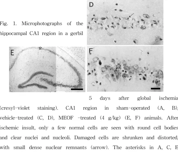
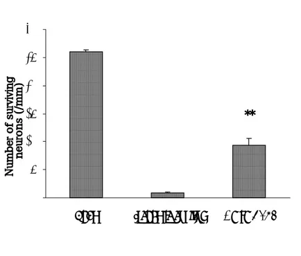
Histologial examination of the nervous system demonstrated marked cell damages in the hippocampal CA1 region of gerbils treated with the vehicle when compared with the sham-operated group. The CA1 pyramidal neurons showed pyknosis, eosinophilia, karyorrhexia, and chromosome condensation in the vehicle-treated group (Fig. 1). This neuronal cell damages was suppressed by high dose of MEOF (4 g/kg) administered orally (Figs. 1 and 2) which significantly reduced the neuronal damage induced by ischemia. The lower dose of MEOF had no significant reduction in hippocampal neuronal damage compared with vehicle (Fig. 2). The number of surviving neurons in the CA1 region of the sham- operated animals was 260±4 neurons/mm (Fig. 1A, B). In the ischemic control group, however, most of the CA1 neurons were damaged and only 9±2 neurons/mm survived (Fig. 1C, D). The mean numbers of surviving neurons following oral administration of MEOF (4 g/kg) was 94±13 neurons/mm (Fig. 1E, F).
Fig. 1. Microphotographs of the hippocampal CA1 region in a gerbil
5 days after global ischemia (cresyl-violet staining). CA1 region in sham-operated (A, B), vehicle-treated (C, D), MEOF -treated (4 g/kg) (E, F) animals. After ischemic insult, only a few normal cells are seen with round cell bodies and clear nuclei and nucleoli. Damaged cells are shrunken and distorted, with small dense nuclear remnants (arrow). The asterisks in A, C, E indicate the middle CA1 regions shown at higher magnification. Bar=500 ㎛ (A, C, E) and 25 ㎛ (B, D, F).
Fig. 2. Numbers of surviving neurons in the hippocampal CA1 region
after MEOF treatment. The neuroprotective effects with 4 g/kg
MEOF in gerbils were significant compared with the ischemic control
animals. The differences were evaluated with a one-way ANOVA
and
a
post-hoc
Student-Neuman-Keuls
procedure
for
multiple
comparisons (**; P<0.01 vs. ischemic control). Values are the mean
**
0
50
100
150
200
250
300
Sham
Ischemic control
4 g/kg MEOF
Number of surviving
2. Effects of methanol extracts of Opuntia Ficus-Indica on NMDA- and OGD-induced neurotoxicity
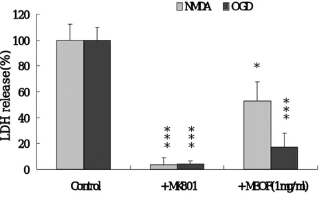
Exposure of mixed cortical cell cultures to 20 μM NMDA for 20∼24 h caused LDH release to increase by 70∼80%. When 1 mg/㎖ MEOF was present both before and during NMDA exposure, neuronal damage was reduced by 47% (p<0.05) (Fig. 3). MEOF also inhibited OGD-induced neurotoxicity by 83% (p<0.001) (Fig. 3). When cultures were subjected to OGD by switching from oxygenated MEM to BSS0 for 60 min, neuronal
swelling was observed soon after the switch. At 20∼24 h after OGD ceased, approximately 75∼85% of the neuronal cells were damaged. When 10 μM MK-801 was added to the cultures during the OGD period, neuronal injury completely inhibited (p<0.001) (Fig. 3). However, the lower concentrations of MEOF (0.01 and 0.1 mg/㎖) did not prevent the NMDA- and OGD-induced neurotoxicity.
Fig. 3. Methanol extracts of Opuntia Ficus-Indica (1 mg/㎖) attenuated NMDA-, and OGD-induced neurotoxicity. Bars represent LDH release (mean ± SEM, n=4) in sister cultures after 20-24 h of exposure to 20 μM NMDA or after 60 min of OGD (controls), or with the addition of 10 μM MK-801 or with the addition of 1 mg/㎖ MEOF. The differences were evaluated with a one-way ANOVA and the post-hoc
Student-Neuman-Keuls test for multiple comparisons (*; p<0.05, ***; 0 20 40 60 80 100 120 Control + MK801 + MEOF(1mg/ml) LDH release(%) NMDA OGD * * * * * * * * * *
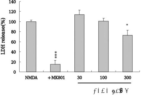
3. Effect of the butanol fraction of Opuntia Ficus-Indica (BFOF) on NMDA-induced neurotoxicity.
Mixed cortical cell cultures were exposed to 20 μM NMDA in the presence or absence of 30, 100 or 300 μg/㎖ butanol, H2O, haxane, or ethyl
acetate fractions of Opuntia Ficus-Indica. The maximun dose of BEOF (300 μg/㎖) only reduced the NMDA-induced neurotoxicity by 27% (p<0.05) (Fig. 4). However, pre- and co- treatment of cultured neurons with 30∼300 μg/㎖ H2O, haxane, or ethyl acetate fractions of Opuntia
Ficus-Indica did not have a neuroptrotective effect. MK-801 significantly
blocked the NMDA-induced neurotoxicity in same cultures (p<0.001) (Fig. 4).
Fig. 4. Butanol fractions of Opuntia Ficus-Indica (BFOF) (300 μg/㎖) attenuated NMDA-induced neurotoxicity. The Bars represent LDH release (mean ± SEM, n=4) in sister cultures after 20-24 h of exposure to 20 μM NMDA or with the addition of 10 μM MK-801 or with the addition of 30, 100, 300μg/㎖ BFOF. The differences were evaluated with a one-way ANOVA and the post-hoc Student-Neuman-Keuls test for multiple comparisons (*; p<0.05, ***; p<0.001 vs. controls). 0 20 40 60 80 100 120 140 NMDA + MK801 30 100 300 LDH release(%) *
BFOF(µg/ml)
* * *Ⅳ. Discussion
In this study, we found that a methanol extract of Opuntia Ficus-Indica (MEOF) exerted protective effects in both OGD- and global
ischemia-induced neuronal injury. As shown in other reports, it is well known that Opuntia Ficus-Indica contains beneficial bioflavonoids components that scavenge the harmful free radicals, which are believed to be involved in the mechanism of neurodegenerative diseases (Huk et al., 1998). Especially, the acceleration of arachidonic acid metabolism in cerebral ischemia is well known to trigger the massive production of free radicals, such as hydroxyl (OH-) or superoxide radicals (O2-) (Dajas et al., 1998; Wei et al., 1997). Previously, we observed that MEOF ameliorated the neurotoxicity induced by arachidonic acid, including its antioxidant action in cortical cell cultures (Wie, 2000). The results of the current study indicate that Opuntia Ficus-Indica extract has a neuroprotective effect against NMDA- and OGD-induced neurotoxicity in cortical culture system, which seems consistent with the in vivo global ischemia model. In this study, we used the Opuntia Ficus-Indica extract at dose of 0.1, 1 or 4g/kg, administrated orally immediately after ischemia. The lower doses of Opuntia
Ficus-Indica (0.1, 1g/kg) produced no significant reduction in hippocampal
neuronal damage compared with the control group. However, animals that received the 4 g/kg dose had significantly more surviving neurons in the hippocampal CA1 region. The dose required to achieve a protective effect against the hippocampal CA1 neuronal damage following global ischmia in gerbils appears to be rather high. It is known that NMDA receptor blocker,
such as by MK-801 (dizocilpine), exert their neuroprotective effect mainly via hypothermic action (Buchan et al., 1990; Corbett et al., 1990). Therefore, we sustained a normothermic state during ischemia and for 3∼4 h afterwords to eliminate the hypothermic effect. Interestingly, MEOF still had a neuroprotective effect. We feel that the neuroprotective effcet of
Opuntia Ficus-Indica extract is not due to the hypothermic effect. Our
results suggest that MEOF extracts its neuroprotection through the indirect modulation of NMDA neurotoxicity by another mechanism, not by NMDA receptors directly.
In conclusion, this study suggest that Opuntia Ficus-indica has a beneficial neuroprotective effect against the neuronal damage occurs following ischemic insult.
Ⅴ. References
Benveniste, H., Drejer, J., Schousboe, A. and Diemer, N. H. 1984. Elevation of the extracellular concentrations of glutamate and aspartate in rat hippocampus during transient cerebral ischemia monitored by intracerebral microdialysis. J. Neurochem. 43 : 1369∼1374.
Buchan, A. and Pulsinelli, W.A. 1990. Hypothermia but not the N-methyl-D-aspartate antagonist, MK-801 attenuates neuronal damages in gerbils subjected to transient global ischemia. J. Neurosci., 10 : 311∼316.
Choi D. W. 1990. Cerebral hypoxia : some new approaches and unanswered questions. J. Neurosci., 10 : 2493∼2501.
Corbett, D., Evans, S., Thomas, C., Wang, D. and Jonas, R.A. 1990. MK-801 reduces cerebral ischemic injury by inducing hypothermia. Brain
Res., 514 : 300∼304.
Dajas, B., Federico, A., Martignoni, B.A., Costa, G., Abina, J.A., Martignonib, E., Nappib, G. and Dajasa, F. 1998. Hydroxyl radical production in the substania nigra after 6-hydroxydapamine and hypoxia-reoxygenation. Brain Res., 813 : 18∼25.
Globus, M. Y., Busto, R., Martinez, E., Valdes, I., Dietrich, W.D. and Ginsberg, M.D. 1991. Comparative effect of transient globla ischemia on
extracellular levels of glutamate, glycine and gamma-aminobutyric acid in vulnerable and nonvulnerable brain regions in the rats. J.
Neurochem., 57 : 470∼478.
Huk, I., Brovkovych, V., Nanobash, V.J., Weigel, G., Neumayer, C., Partyka, L., Patton, S. and Malinski, T. 1998. Bioflavonoid quercetin scavenges superoxide and increases nitric oxide concentration in ischaemia-reperfusion injury: an experimental study. Br J Surg., 85 : 1080∼5
Kirino, T. 1982. Delayed neuronal death in the gerbil hippocampus following ischemia. Brain Res., 237 : 57∼69.
Kirino, T. and Sano, K. 1984. Selective vulnerablility in the gerbil hippocampus following transient ischemia. Acta Neruopathol., 62 : 201∼208.
Lee, H. J., Lee, Y. W. and Kim, J. H. 1998. A study on antiulcer effects of
Opuntia dillenii Haw. on stomach ulcer induced by water-immersion strees
in rats. J. Food Hyg. Safty., 13 : 53.
Moon, C.J., Kim, S.J., Ahn, M.J., Lee, S.J., Park, S.J., Jeong, K.S., Yoon, D.Y., Choe, Y.K. and Shin, T.K. 2000. Effects of Opuntia ficus-incus extract on immune cell activation. Kr. J of Life Science, 10 : 4.
Park, E. H., Kahng, J.H. and Paek, E.A. 1998. Studies on the pharmacological action of cactus : identification of its anti-inflammatory effect. Arch Pharm. Res., 21: 30.
Rothman, S. M. and Olney, J. W. 1986. Glutamate and the pathophysiology of hypoxic-ischemic brain damage. Ann. Neurol., 19 : 105∼111.
Shin, T., Kim, S. and Lee, S. 1998. Effect of Opuntia ficus-indica extract on the activation of immune cells with special reference to autoimmune disease models. Kor. J. vet. Pathol., 2 : 31.
Shin, T., Kim, S., Moon, C., Wie, M. and Hyun, B. 1999. Opuntia
ficus-indica ethanol extract ameliorates streptozoticin-induced hyperglycemia in rat. Kor. J. Gerontol., 9 : 78.
Siesjo, B.K. 1988. Calcium, ischemia, and death of brain cell. Ann. NY
Acad. Sci., 522 : 638∼661.
Wie, T., Huang, N.C. and Quast, M. J. 1997. Hydroxyl radical formation in hyperglycemic rats during middle cerebral artery artery occlusion/reperfusion. Free radical biol Med., 23 : 986.
Wie, M.B. 2000. Protective effects of Opuntia Ficus-Indica and Saururus
Chinensis on free radical-induced neuronal injury in mouse cortical cell
초 록
저빌 허혈시 세포 손상 및 피질세포배양에서
산소-포도당 결핍 유발 독성에 대한
손바닥선인장의 신경보호효과
(지도교수: 신 태 균)
김 정 훈
제주대학교 대학원 수의학과
손바닥선인장 (Opuntia Ficus-Indica)은 in vivo 와 in vitro 실험 시 다양 한 항산화 활성을 보고되고 있다. 본 연구는 손바닥선인장 추출물이 신경세포 손상에 대한 방어효과를 관찰하고자 in vitro 와 in vivo에서 실험하였다. 먼저 손바닥선인장 메탄올추출물(0.01, 0.1, 1 mg/㎖)이 생쥐 피질세포배양에서 N-methyl-D-aspartate(NMDA)와 oxygen-glucose deprivation (OGD)로 유도 된 신경세포 손상에 대해 방어효과가 있는지 실험하였다. 더불어, 손바닥선인
g/㎖에서 NMDA로 야기된 신경독성을 27% (P<0.05) 감소시켰다. 그러나 다 른 분획층의 어떠한 농도에서도 신경방어효과를 가지지 않았다. 우리는 또한 손바닥선인장 추출물이 저빌에서 뇌 허혈로 일어나는 해마 신 경세포 손상을 방어하는지 실험하였다. 저빌들은 하루에 한번, 3일 동안 0.1, 1 과 4g/kg씩 경구투여한 후 양측 경동맥을 5 분간 결찰하였다. 허혈 후 마지 막으로 각각의 그룹에 투여하였다. 해마 CA1 부위의 신경세포 손상은 허혈 후 5 일 뒤 정량적으로 평가하였다. 4 g/kg씩 투여한 그룹은 해마부위의 신경세 포 손상을 34% (P<0.01) 감소시켰으나, 그 이하의 적은 용량을 투여한 그룹은 CA1 신경세포 손상을 감소시키지 않았다. 이들 결과를 종합하여 볼 때, 손바닥선인장 추출물은 마우스 신경세포배양 을 이용한 산소-포도당 결핍으로 유발되는 신경독성 뿐만 아니라, 저빌에서 허 혈 후 손상되는 신경세포에 대해 우수한 보호효과가 있음을 알 수 있었다. 이 상의 소견으로 보아 손바닥선인장 추출물은 허혈성 신경세포의 손상을 예방하 거나 치료하는 데 기여할 것으로 여겨진다.