I
깨e뼈urnal이Medlcfneand Ule Science Vol. 10. No. 3{Febr배ry),2014I
Expression of Matrix Metalloproteinases
밍ld Tissue Inhibitor of
Metalloproteinase-l
in Carotid Stenosis
Young-Hee Maeng
’,
Jee Won Chang',
Jay Chol Choi',
Su Wan Kim' 10epartment01 Palhology. SChool01Medicine.Jeju Nalional Universily:Oepartment01Thoracic and Gardiovascularsu얘ery. School 01Medicine.Jeju Natior딩I Universily
’
Department이Neurology.Sch∞
1이Medicine,Jeju Nation히University(Received January 21. 2014: Revised Janu밍'y 28. 2014; A
∞
epted Febru81γ 4. 2014) AbstractIt is still controversial whether the specilic subtypes 01 matrix metalloproteinases are related to cli미cal characteristics in carolid stenosis palienls. Carolid atherosclerolic plaques were oblained Irom 34 patienls,21 males and 13 lemales with mean age 이71.1. Carotid endartereclomy was performed under the monitoring of EEG and car이id stump pressure. Expression 01 matrix metalloproteinase-2 ,8,9,and tissue inhibitor of metalloproteinase-1 in the atherosclerotic plaque was assessed by immunohistochemical slides stained wilh primary antibodies semi-아Jantitatively. Twenty patients (58.8%) were symptomatic
stenosis and 15 (44.1 %) had stable plaques withoul evidence 01 fibrous cap rupture. Strong stain (degree 3) 01 matrix
metal1oproteinase-2 and 9 was dominant in asymplomatic group (F느0.026 and p= 0.0007. respectively). Slrong slain 01 matrix metalloproteinase-2 was dominant in stable plaque group (P= 0.026). Specific subtypes 01 matrix metalloproteinases can be related to clini
∞
I characteristics such as symptoms or plaque morphology. (J Med 미e 501 2014;10(3);209-213)Key Words : Atherosclerosis Carotid Arteries Matrix metalloproteinases
Introduction
Carotid stenosis is part of 앙stemic atherosclerosis which causes various neurologic disabilities or deaths. Recent swdies have presented that various subtypes of the matrix metalloproteinases (MMPs) family showed increased or decreased expression in plasma or atherosclerotic plaquesl and they have suggested that MMPs mi양lt play divergent roles in atherosclerotic process such as remodeling of extracell비ar maσix or plaque rupture
‘
However. it still remains controversial whether the expression of specific subtypes of :MMPs family is related to clinical characteristics。
r plaque morphology in carotid stenosis patients πllS study is focused on analyzing the relationship between clinical characteristics and the degree of expression ofMMP-2. MMP-8. MMP-9 ,and tissue inhibitor of
me어1l0proteinase-1 (TIMP-1) in carotid stenosis
M씨3terials and Methods
Thïrty-four patients who underwent endarterectomy for 양rnptomalic or a앙mptomatic carotid stenosis from March 2010 to December 2012 were included. This study was approved by Institutional Review Board of Jeju Nationa1 University Hospital. The patients ’ characteristics are presented in Table 1
Table 1. Patients characteristics (n=34)
Age(Moon
앤",,)
71.1 80,
M 21 (61.7%) F 13 Hypertcnsionor d떠betes 28 (62,4") 어sli얘‘찌뼈끼뼈d_ 10 (29,4%) Atherosclerotic이양잉e' 9 (26.S%) α.ltTeJltsmoker 8 (23.S")‘
It means history of diagnosis or treatrnent for coronroγ atherosclerosis. atherosc1erotic anewγsm of aorta‘。
r 밍lY type of periphera1 arterial atherosclerotic diseaseCorrespondenceto : Jee Won Chang
Department이Thoracic and Gardiovasc비ar surgery. SChoolof Medicine,Jeju National University
1753-3. Ara-l Oon9,Jeju Si. Jeμ Republic이Korea,690-767 E-mall : jeewon71@naver
∞
mdifferences in expression of MMPs and Tη
,
jp-l between various c1inical characteristics: a p-value < 0.05 was considered statistically significant7\Nenty (58.8%) patients had symptoms of stroke or transient ischemic attack within 6 weeks of surgery Temporary shunt was used in nine (26.5%) patients during CEA 쩌tbou빼 two of them showed initi외 stump pressure
higher than 40mmHg‘temporary shunt was used unexpectedly because they showed abrupt slowing and asymmet2γ in EEG after c1arnpingof intem외 carotid artery
After temporary shunt was applied. asymmetric EEG disappeared immediately and they did not have any neurologic sequels postoperative1y.1ntracerebral hemorrhage was detected immediate1yafter the operation in one patient and no significant postoperative neurologic comp1ications were detected in otber patients
When we compare the asymptomatic group (n=14) ω
양mptomatic group '(n=20),야1e proportion of patients with hypertension or diabetes was significant1y higher in symptomatic양oup (P=0.021) and there were no sigr띠ìcant diITerences in other clinical characteristics. Vlhen we defme the ‘stable plaque' as p1aque with intact fibrous cap (without evidence of plaque미pture) and ‘'vulnerable plaque'
as p1aque with ruptured fibrous cap and exposed red thrombi or 1ipid core debris. there was no difference of p1aque morphology between a양mptomatic뻐d 양mptomatic
groups. When the degree of expression ‘3‘is considered to identify ‘strong stain‘and the degree of expression ‘'2,1,
and 0’are considered to identify ‘moderate to weak
st8ln‘.MMP-2 and MMP-9 showed significant difference in expression between asymptomatic and symptomatic group (P= 0.026 and P= O.αJ07. respectively) (Table 2) (Fig 1)
끼1ere were a 1arger number of ‘sσ
。
ng st8ln' of MMP-2 and MMP-9 in asymptomatic group. When we compare st8ble plaque group (n=15) to v1매nerable plaque group(n=19). there were no significant difference in age,or proportion of symptomatic patients. When we use the above-mentioned classification according to the degree of expression. MMP-2 showed si밍1ificant difference between stable and vulnerab1e p1aque group. There was a larger number of ‘sσ'ong s뻐
m
‘。
f MMP-2 in st8ble plaque (P= 0.026) <Teble3).Young-HceMaeng,Jee WonChtmg‘Jay CholChoi.Su WanKim
Syrnptomatic carotid stenosis patients who did not have contraindications for general anesthesia based on the medica1 history. careful physical examination,and routine laboratory sωdies were treated by carotid endarterectomy (CEA).A양mptomatic patien1s who showed rapid progression
。
f carotid stenosis on MRIor carotid dup1ex,and more than 70% of stenosis at initial evaluation also underwent CEA CEA was performed under general anesthesia and monitoring of EEG. Temporary carotid shunt (Pruitt-Inahara outlying c밍O디d shunι LeMaiσe V.앓cu]ar,Burlingtα,.USA)was se1ectively used based on the intraoperative EEG findings and carotid stump pressure. If carotid stump pressure was 1ess than 40mmHg or changes in EEG after clamping of carotid artery. the shunt was used Postoperative intensive care such as sb"1ctcontrol of blood pressure and systemic neurologic examination was perfonned and fol1ow-up diffusion MRI was used based on the mental status or neuro10gicsymptoms
lmmunohistochemical an머ysis for the e앵ression of the MMPs and πMP-l was peñonned as follows. Fonna1in-Hxed paraffm-embedded tissue b10cks of the endarterectomy specimens were cut at 4 mm slices. deparaffmized in xylene,
and rehydrated with graded ethanol. A standard immunohistochemical technique was performed using a Ventana Benchmark XT immunostainer (Ventana Medical
않stems Jnc.. AZ. USA). Heat epitope re미eval provided by
the immunostainer was perfonned for 60 minutes. 'I'he enzymatic reactivity was visua1ized with 3‘3‘
diaminobenzidine깨e primary antibodies used were
anti-MMP-2 (Diagnostic BioSystems. California. USA: clone mom 312: dilution 1:25),anti-MMP-8 (Sant8 Cruz Biotechnolo양
lnc,TX. USA: clone H-45: dilution '1:400),ar띠-MMP-9
(Abnova,Taipei. Taiwan: clone SBI5c,dilution 1:1αJO),and anti-πMP-1 (Abnova. Taipai. Taiw밍1: clone 2A5‘dilution
1:50). They were diluted
、
I.'itb PBS buffer-based dilution solution and incubated for 32minutes at 37t:. For the negative contro1s,the slides were stained by omitting the primary antibody from tbe protocol and substiωting it witb commercially available non-irrunune mouse immunoglobulin G serum (DAKO,CarPinteria,CA,USA)The immunohistochemical slides were evaluated and interpreted by one pathologist who was blinded to the patients’clinical findings. 'I'he protein expression was scored by staining intensity and positive와eas: 0 (no signal),
1 (focal we삶‘reaction). 2 (focal sσong or diffuse weak reaction). or 3 (diffuse strong reaction)
Expression of Matrix Metalloproteinases and 까ssue Inhibitor of Metalloproteinase-l in Carotid Stenosis
Table 2. Differences in clinica1 characteristics and :MMPs expression between the asymptomatic and symptomatic group
Table 3. Differences in clinicaJ characteristics and MMPs expression between stable plaque group and vulnerable plaque group
Asymplα떠lic group잉mplomalic grαIp
(0=14) (0=201
Slable plaq냐e
(n=lS)
Vulnerable plaQue (0=19)
A양(mean. yearn) 68.6 72.8 Age (m!!an앤뼈) 70.5 71.5 1': 0.7'J:1 80
,
M:P 7:7 M:P 14:6 sex M:F 9:6 M:F 12:7liypertension or이abe!as 10 (71.4") 18 (90%) P=O.021 Hypert!!nsionor diabctes 10 (68.7") 15 (78.9%) 1': 0.'J:I8 Current smoker 4 (28.6") 4 (20%) Curren
‘
smoker 3 5Stable plaque (n=lS) 6 (43%1 9 (45") """"_ ••••_ (n=14) 6 (40%1 8 (42.1") Ruptured미aque (n=1잉 8 (57"1 11 (55") Symptomaticpatients (n=ro) 9 (60'1>) 11 (57.9%)
MMP-2 MMP-2
Strong stain 8 4 Strong stain 12 8
Modemte to weak st8in 6 16 1': 0.lY26 Moderate to weslt5어m 3 11 1': 0.lY26
MMP-8 MMP-8
Slrong stain 4 8 Strong stain 5 7
Modemte ω wealu"밍n 10 12 P=O.493 Modemte to
‘
weak stain 10 12 P= 0.493k애P-9 MMP-9
Sσ
。
ng stain 11 4 Slrong stain 8 13M여rn-ateto weak stain 3 16 1':0.α111 Modemte ω weak slain 7 6 1': 0.371
TlMP-1 TlMP-l
Sσ
。
'ng stain 4 8 Slrong staÎn 8 4Moderate ωW않k """" 10 12 P=O.493 Modemte to weak stain 7 15 P= 0.050
*
lt me밍lS history of dia양losis or treatment for coronBlγ atherosclerosis ,atherosclerotic aneurysm of aoria ,or any type of peripheral arte 꺼외 atherosclerotic 이sease.‘
It means hiswry of d.ia밍losis or u-eatment for coronarγ atherosclerosis. atherosclerotie aneurysm of aorta ,or 와1y앙pe of periphera1 arteri 띠 atherosclerotic disease
.
h i(b)
Ifiκ
':J'!~.,: ← 끼 •r:1
깐
It•..
:‘
'~~~4‘"
‘;j'
j원진 ;
j?샤 샤‘
i.
ti-:- --사
x
‘ "
':;:
.
.τ
,
r〉 :’
r
‘@‘시
;-?tF
、
f→“
i?
;;,시‘
•
서[
• - ••' .Þ ••• ;". ‘•、 ,、 ; ← •‘ - … • ‘",i _“ “z ι* η‘'.;.1'
,:~:;::/t~.-...'.'~-
-!--~석,,、
ti
‘싸
~
--~,
.、‘-"
~
.'~‘.’.ιJ‘-, )
” •그 ;;싸
‘~,;~‘
기i:c
v ·t t •‘
ν;;:
~.-"I
".
‘.
‘.
’
‘
~~
.•..•
‘’r:~i;7' •셔영 .
‘
~
'.q-.: R훌κ
‘훌i
‘
γ
<.-‘
l!iI.)lIg" '. fI'..,
톨톨를얄
!/?it/
t(a)
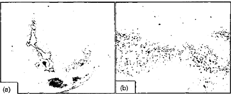
Figure 1.Photornicro 랑aphs of immunohistochemistly results: 8table plaque Cfibrocalcified plaque without rupture of fibrous cap) (a) shows sσong (degree 3) stain of MMP-2 (b)
Young-HeeMaeng,Jec WonChang,Jay CholChoi,Su WanKim
MMPs are a family of membrane-bound proteases which are structurally related and share a Zn2<-based catalytic mechanism' .. Because it has been suggested that
。
veracüvation of MMPs causes destruction of exσ'acellular matrix rather than remodelin냥,it is considered that MMPs play a role in rupture of fibrous cap leading to neurologic ischemic events. Therefore,it was initia1ly hypothesized that symptomatic carotid sìenosis patients may have a larger number of vulnerable plaques and show stronger expression。
fv 외i.ous subtypes of MMPs as well as we빼expression ofTIMP because uncontrolled expression MMPs and subsequent 야linning of fibrous cap by MMPS’degradation
of extracellular matrix w。띠d fmally lead to stroke. Sh빼el al reported that hurnan monocyte-derived macrophages induces collagen breakdown in fibrous cap of atherosclerotic plaque 밍ld such activities are suppressed by 1\αÆP inhibitors
in acute coron밍Y양nψomes.
“
1t has 떠so been shown thatm
링l1yi띠lamed atheromatous plaques presented increased glob외MMP activities and rupture-prone human plaques hadmcreas잉d MMP-8,11,14. and 16'.
However,there were no sigI피3
∞
nt differences in plaque morphology between asymptomatic and symptomatic patients. Shishkina et al reported that 77% of endarterectomy specimens removed from asymptomatic carotid stenosis patients showed ruptured plaques6 andJayasooriya et외suggested that silent cerebral infarcüon on brain CT and microemboli detected on transcranial doppler (TCD) are risk factors of future sσ
。
ke in asymptomatic carotid stenosis.’
Based on such studies. asymptomatic carotid sænosis patients can have silent ruptured plaques Asymptoma성c patients of this sbJdy were clinically silent atthe time of operation and may have unrecognized infarction
。
r microemboli. Although 1'CD was penonned as a p앙t ofroutine preoperative evaluation for the included patien잉,
microemboli were not det.ect.edin any asymptomatic patients A1thou방1 most of studies conceming the role of MMPs in atherosclerosis have suggested that they can promote macrophage invasion,plaque inflamrnation,뻐d밍19J.ogenesls
as well as thinning of fibrous cap,each subtype of MMPs may have different role in advances in atherosclerotic plaques. Sluiiter et 외 reported that levels of MMP-2 are
increased in fibrous rather than rupture-prone atheromatous human carotid plaques
’
밍ld this is in accord with the restùt。
f this sludy. 11was also suggesled lhal 2 and MMP-14 promote vasc비ar smooth muscle cell migration and께lis study is supported by the grant from Departrnent of
Indus뇨y-University Cooperation in Jeju National University to promoting plaque stabili냥 MMP m양 have du띠role in
plaque rupture and stability or vulnerability may be determined by expression of predominant subtype of MMP and their level of activitieslO.FurU1er study conceming the role of MMPs in carotid stenosis should focus on relationship between the selective inhibition or activation of speα.fic subtypes }o.αÆPsand cωlical features and it seems to be used as therapeutic 때rgeι In conclusion,MMP-2 and MMP-9 showed increased expression in asymptomatic carotid stenosis and MMP-2 showed increased expression in stable plaque rather than v버nerable plaque. FurU1er study
shouJd be penonned to investigate the role of each subtype of뻐ÆPs in carotid atheroscleroüc process
l)Nilsson L,Jonasson L,Nijm J,Hamslen A,Eriksson P Tncreased plasma concen'!ration of ma미X
meta11oproteinase-7 in patients with coron밍Y밍-tery
disease. Clin Chem 2006:52:1522-7
2) Newby AC. Dual role of maσi.x meta1loproteinases (matrix:ins)in inüma1 thickeniI)g and atherosclerotic plaque ruplure. Physiol Rev 2005:85:1-31
3) Newby AC. Metalloproleinases and 뻐nerable
atherosclerotic plaques Trends C밍ùiovasc Med
2007:17(8):253-8
4)Sh빼PK,F외k E,Badimon JJ et외 Human monocyte-derived macrophages induce collagen breakdown in fibrous caps of atherosclerotic plaques. Poæntial role of matrix-degrading metalloproteinases 와ld implications for
plaque ruplure. Circulation 1995:92:1565-9
5)Chou버1ary S,Higgins CL,Chen IY el al. Quantilation and localization of ma미x metalloprotienases and their
inhibitors in human carotid endart.erectomy tissues Arterioscler Thromb Vasc Biol 2006:26:2351-8 6) Shinshkina VS,Toklueva LR,Kashirina SV el 외
Comp밍1son of morphologica1 features of carotid 와teries
atherosclerotic plaques with clinica1-instrumenta1 data in syrnptomatic뻐d a양mp
∞
matic patients with severecarotid atherosclerosis. K와'C!iol。밍ia 2013:53:25-31 7)Jayasooriya G,η1apar A,Sh머houb J,Da'씨es AH. Silent
cerebral events in asymptomatic carotid stenosis. J Vasc
←← 4 Acknowledgemenl References 까 ---←---~-← ←←←?←←-←」←→←←←」
j
DiscussionExpression of Matrix Met.alloproteinases and 꺼ssue Inhibitor of Met.alloproteinase-l in Carotid StenosÍs
/
’
’
8) Sluijter JP,"Pulskens WP,Schoneveld AH et a1. Ma띠X
I
me떠lloproteinase 2 is associated -with stable and matrix me뎌llopro뼈inases 8 잉1d 9 with vlψ18rable e양。tid atheroselerotie lesions - a study in human"i! endartereetomy specimen pointing to a ro1e for different extraeellular matrix meta11oproteinases inducer
glycosylation forms. 8lroke 2006;37:235-9
9) Newby AC. Matrix metalloproteinases regulate migration ,
proliferation ,업ld death of vaseular smooth muscle eells
by degrading matrix and non-matrix substrates Cardiovasc Res 2006;69:614-24
lO)Hower CD,Dassow MS,Kajdacsy-Balla A,Seabrook GR, Jean-Claude J,Towne JB ,Cambria RA
Metalloproteinases levels are decreased in symptomatic carotid plaques. J 8urg Res 2000:88:155-9