Introduction
Temporomandibular disorder (TMD) is a broad term for chronic disorders with chief complaints and
symptoms of pain at the temporomandibular joint (TMJ) or masticatory muscles, joint clicking, trismus, and abnormal jaw movement1).
Also, TMD is multifactorial and its progress is am-J Korean Dent Sci. 2020;13(1):21-27 https://doi.org/10.5856/JKDS.2020.13.1.21 pISSN 2005-4742 ∙∙ eISSN 2713-7651
Corresponding Author: Sang-Sun Han, https://orcid.org/0000-0003-1775-7862
Department of Oral and Maxillofacial Radiology, Yonsei University College of Dentistry, 50-1 Yonsei-ro, Seodaemun-gu, Seoul 03722, Korea
TEL : +82-2-2228-8843, FAX : +82-2-363-5232, E-mail : sshan@yuhs.ac
Received for publication June 17, 2020; Returned after revision June 22, 2020; Accepted for publication June 25, 2020
Copyright © 2020 by Korean Academy of Dental Science
cc This is an open access article distributed under the terms of the Creative Commons Attribution Non-Commercial License (http://creativecommons.org/licenses/ by-nc/4.0) which permits unrestricted non-commercial use, distribution, and reproduction in any medium, provided the original work is properly cited.
The Analysis of Incidental Findings on
Temporomandibular Joint Magnetic Resonance
Imaging
Yoon Joo Choi , Chena Lee , Kug Jin Jeon , Sang-Sun Han
Department of Oral and Maxillofacial Radiology, Yonsei University College of Dentistry, Seoul, Korea
Purpose: The aim of this study was to investigate the types and frequency of the various incidental findings (IFs) on magnetic resonance images (MRI) taken from the patients with temporomandibular disorder (TMD) symptoms.
Materials and Methods: Temporomandibular joint (TMJ) MRI taken from 1,013 patients with TMD symptoms were evaluated retrospectively. IF was defined as imaging features that were accidentally or unexpectedly found, rather than degenerative bony changes of TMJ complex or disc derangement. They were classified into two groups as TMJ site-specific findings and unexpected findings at other regions. The frequency of the sub groups was analyzed.
Result: A total of 26 (2.57%) cases with IFs were classified into 13 cases with TMJ site-specific findings and 13 cases with unexpected findings at other region. TMJ site-specific findings included synovial chondromatosis in 6 cases, synovial cyst in 6 cases and osteochondroma in one case. Unexpected findings included salivary gland tumor in 3 cases, developmental cyst in 3 cases, vascular malformation in 2 cases, mastoiditis in 4 cases and sialadenitis on pa-rotid gland in one case.
Conclusion: When diagnosing TMD through TMJ MRI, clinicians should carefully read the image, considering the possibility of IFs because TMJ MRI can provide pathologic information in TMJ region and other oral and maxillofa-cial region.
biguous and difficult to predict, which requires accu-rate diagnosis and appropriate intervention from the outset1). Especially, it is mandatory to discriminate
the inflammatory or tumorous lesion of TMJ and its surrounding structures from disc-related dysfunc-tion or myofascial pain. According to the causes of TMD, its treatment plan and prognosis will change. Therefore, precise diagnosis is crucial for the selec-tion of appropriate treatment2-4). To assess
patho-logical findings in the TMJ area, several imaging modalities have been used including conventional radiography, cone-beam computed tomography (CBCT) or computed tomography (CT)5-9). CBCT or
CT are currently regarded as an essential modality for the accurate evaluation of the bony alteration on TMJ structures such as flattening, osteophyte forma-tion, subchondral sclerosis, subchondral cyst, ero-sion7-9). Magnetic resonance imaging (MRI) can be a
proper modality to detect the pathology of articular disc and the neighboring soft tissue such as muscles, ligaments. Particularly, it can be recommended as gold standard for assessing the disc displacement or joint effusion with the highly-proven contrast resolu-tion for soft tissue9,10).
Clinicians usually need MRI evaluation for precise diagnosis of suspected TMD patients and may en-counter unexpected radiologic findings in and out of the TMJ region11-15). In this study, we defined
‘in-cidental findings (IFs)’ as radiologic features that are accidentally and unexpectedly detected and distin-guished from disc displacement, joint effusion and pathology of the neighboring masticatory tissue16).
The purpose of this study is to suggest various IFs found in TMJ MRI of patients with suspected TMD. This is to inform the clinicians of the clinical signifi-cance of differential diagnosis for patients complain-ing of TMD-like symptoms.
Materials and Methods
1. Patients ReviewThis study was approved by Institutional Review Board of Yonsei Dental College Hospital (2-2019-0077), and the requirement for patient consent was waived because of the retrospective nature of the study.
A retrospective review of all patients who under-went MRI examination of TMJ at the Yonsei Uni-versity Dental Hospital from January 2019 to March 2020. Patients with a history of TMJ trauma or sys-temic disease were excluded. The total number of patients examined in this study was 1,013, with 238 male and 775 female patients and age range from 8 to 82 years.
2. Magnetic Resonance Imaging Protocol
The MRI examination of the TMJ was performed using a 3.0 T scanner (Pioneer; GE Healthcare, Waukesha, WI, USA). The imaging protocols were a 300×250 matrix, 210 mm field of view, and a pixel size of 0.449×0.449 mm. The axial slices were ob-tained with 4.0 mm thickness with T2-weighted images (repetition time [TR]/time to echo [TE]: 3,217/89). The oblique sagittal and coronal slices were obtained in proton density sequence (TR/ TE: 2,143/52) with 2.5 mm thickness. The oblique sagittal slices were also obtained in T2-weighted sequence (TR/TE: 3,120/84) with 2.5 mm thickness. The oblique sagittal images of all imaging sequences were obtained with open and closed mouth position.
3. Image Analysis and the Criteria of Incidental Findings
Experienced oral and maxillofacial radiologists evaluated all MRI images. IFs were defined as radio-logic features which the accidentally found without any relation to the clinical indication of imaging purpose in field of view of each scan. Because the purpose of diagnosis for suspected TMD patient is
to identify the pathologic findings of the articular disc or the surrounding masticatory tissue on TMJ area, the former pathologic findings were excluded from IFs. Mucosal thickening and small lymph node enlargement without any clinical significance were excluded as well. According to the location of the le-sion, IFs were classified into two groups; TMJ site-specific findings and unexpected findings at other regions.
1) Group of temporomandibular joint site-specific findings
TMJ site-specific findings included neoplastic le-sions that appear more frequently than other jaw bones due to the histological specificity of the TMJ site which consisted of synovium, cartilage, and articular disc tissue. Degenerative joint disease or morphological pathology of disc were excluded. Re-sultantly, the findings included such lesions as syno-vial chondromatosis, osteochondroma, synosyno-vial cyst, giant cell tumor of TMJ, and condylar hyperplasia.
2) Group of unexpected findings
Unexpected findings were defined as IFs that ap-pear at other sites than the TMJ in the field of TMJ MRI. This included neoplastic or inflammatory le-sions originated from the major or minor salivary glands and vascular malformations, and other con-genital lesions such as thyroglossal duct cyst.
4. Statistical Analysis
The prevalence analysis was performed for IFs. For each group, TMJ site-specific and unexpected find-ings, the prevalence of individual disease was pre-sented.
Result
Twenty-six cases (2.57%) of the 1,013 suspected TMD symptoms patients had IFs in TMJ region or outside the TMJ area. Thirteen cases were regarded
as the TMJ site-specific findings, and not pathologic findings on articular disc tissue. The other 13 cases were radiologic findings of clinical importance such as cyst and tumor detected in other areas than TMJ region. Each finding was described with its frequen-cy distribution in Table 1. The case report for each case is as follows.
1. TMJ Site-Specific Findings
1) Synovial chondromatosis
A 29-year-old male was referred to our dental hos-pital for the evaluation of osteoarthrosis of left TMJ from the local clinic. He was complaining pain and clicking sound on left TMJ when opening the mouth at the end of orthodontic treatment. MR images showed well-defined T2 hyperintensity on the left joint space. The lesion included multiple low signal foci suggesting calcifications. Imaging diagnosis was synovial chondromatosis and it was confirmed with histopathology after the mass excision (Fig. 1).
Table 1. The frequency by MR findings incidentally found during TMJ MRI screening
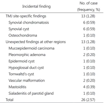
Incidental finding No. of case (frequency, %) TMJ site-specific findings 13 (1.28)
Synovial chondromatosis 6 (0.59)
Synovial cyst 6 (0.59)
Osteochondroma 1 (0.10)
Unexpected findings at other regions 13 (1.28) Mucoepidermoid carcinoma 1 (0.10)
Pleomorphic adenoma 2 (0.20)
Epidermoid cyst 1 (0.10)
Hypoglossal duct cyst 1 (0.10)
Tornwaltdʼs cyst 1 (0.10)
Vascular malformation 2 (0.20)
Mastoiditis 4 (0.39)
Sialadenitis of parotid gland 1 (0.10)
Total 26 (2.57)
2. Unexpected Findings at Other Regions
1) Mucoepidermoid carcinoma
A 61-year-old female was referred to our dental hospital complaining of pain and click sounds in both TMJ areas at the open mouth position. Addi-tional MR examination was performed for the differ-ential diagnosis of discal pathologic change and MRI described T2 hyperintense lesion with approximate-ly 1.3 cm size on the left parotid gland. The lesion was relatively well-defined while it showed irregular margin locally. Imaging diagnosis was made as
be-nign salivary gland tumor or low grade malignancy. The lesion was resected and histopathologically con-firmed as intermediate grade of mucoepidermoid carcinoma (Fig. 2).
2) Pleomorphic adenoma
A 20-year-old female visited our hospital com-plaining of consistent facial swelling and locking sensation on left preauricular region when mouth opening. The axial view of TMJ MR images showed 1.9 cm-sized lesion with heterogeneous T2 signal at the superficial lobe of left parotid gland. After then,
* *
A
B
Fig. 2. (A) The axial view of T2weighted image shows 1.3 cm sized T2 hyperintense lesion (asterisk) at the superficial lobe of left parotid gland. (B) The coronal view of proton density image presents mass with lobulating shape (asterisk). Imaging diagno sis is made as benign salivary gland tumor or low grade malig nancy and the final histopathology is confirmed as mucoepider moid carcinoma.
* *
A
B
Fig. 3. (A) The T2weighted axial image showed relatively well encapsulated round mass (asterisk) with heterogeneous high T2 signal. (B) The gadoliniumenhanced axial image reveals the lesion with increased enhancement (asterisk). Imaging diagnosis is made as benign salivary gland tumor, most likely pleomorphic adenoma and the histopathology is confirmed as pleomorphic adenoma.
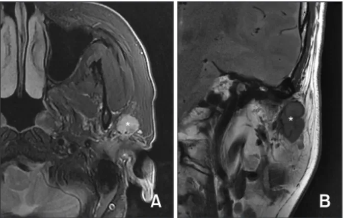
A
B
Fig. 4. (A) The axial T2wighed image shows T2 hyperintense lesion (arrows) at inferior border of left master muscle. The le sion includes multiple low signal foci. (B) The lesion (arrows) is appeared in the sagittal T2weighted image as well. Imaging diagnosis is made as venous malformation with phlebolith.
* * *
A
B
Fig. 1. (A, B) The T2weighted magnetic resonance image of sagittal view presents well defined T2 hyperintensity within the temporomandibular joint space (arrows). The lesion includes multiple low signal foci indicating calcification (asterisk). Imaging diagnosis is synovial chondromatosis.
additional gadolinium-enhanced MR images was obtained for differential diagnosis. The lesion was well-encapsulated and heterogeneously enhanced. The imaging diagnosis was made as benign salivary gland tumor, most likely pleomorphic adenoma. The mass resection was performed and final diagnosis was pleomorphic adenoma (Fig. 3).
3) Vascular malformation
A 61-year-old female visited our dental hospital with the chief complaint of the discomfort on the left preauricular region and masseter muscle area, espe-cially under stressful or insomnia condition. The TMJ MR images presented the T2 hyperintense lesion within inferior region of master muscle. It includes multiple low signal foci indicating calcifications. Im-aging diagnosis was made as venous malformation with multiple phlebolith (Fig. 4).
Discussion
TMD is caused mainly by structural or functional problems among the articular fossa, mandibular con-dyle, articular disc, and the adjacent muscles. TMD is a collective term encompassing a number of clini-cal problems involving both the masticatory muscles and TMJ. Typical complaints reported by TMD patients are pain in the masticatory muscles and/or the preauricular area particularly during mandibular movement, stiffness in the masticatory muscles, limi-tation in mandibular movement, and joint sounds1).
MRI has a pivotal role for the diagnosis of the TMD and the soft tissue pathology in head and neck im-aging9,10). From TMJ MRIs taken for the detection
of disc pathology, oral radiologists often encounter incidentally and unexpectedly detected radiologic features – ‘IFs’15). These findings vary in their clinical
importance, from common benign lesion to signifi-cant pathologic findings that may have an important impact on the health of the patient16).
IFs on TMJ MRI has been reported as a various
frequency from 0.072% to 26.85%11-14). Yanagi et al.11)
reported two tumor cases (0.072%) as IFs on MRI of consecutive TMD 2,776 patients in Japan, which was the first report of IFs of the TMJ MRI. Makdissi et al.12) explained total 53 cases (7.3%) including 11
cases of intracranial findings (1.5%) and 42 cases of extracranial findings (5.7%) in 730 symptomatic Brit-ish patients. Kamio et al.14) reported 461 cases (26.85%)
in a retrospective study of 1,717 MR images taken from suspected TMD patients in Japan. The higher frequency of IFs on previous study was attributed for the combination method using both typical TMJ coil and conventional coil for the head and neck. The superficial coil used in the former11) would have
a limit in obtaining deeper scans in oral and maxil-lofacial region than the conventional head coil used in the latter17,18). In our study, the frequency of IFs
detected on TMJ MRI was 2.57%, that was the result performed using the surface coil and 3.0 Tesla.
Apart from the difference of used coil, many vari-ables have made differences in the results among previous studies. The variables include the scan protocol, the slice thickness, the number of patients and the inclusion criteria of IFs19,20). Also the several
overestimated studies included lymph node enlarge-ment or mucosal thickening of sinus without any symptom, which may occur and regress spontane-ously12-14). In the present study, only IFs with clinical
significance to make a treatment or follow up were included21).
In conclusion, the prevalence of IFs was 2.57% in TMJ MRI scanning. In all cases of IFs, some were congenital lesions or anatomic variations, while other cases required scrutiny and urgent surgical intervention. When diagnosing TMD through TMJ MRI data, clinicians should carefully read the image, considering the possibility of IFs because TMJ MRI can provide the pathologic information in TMJ re-gion and other oral and maxillofacial rere-gions.
Conclusion
Advanced MRI could be regarded as a gold stan-dard for the evaluation of TMD and differential diagnosis of other mimicking lesions. The incidence of IFs of TMJ MRI were up to 2.57% among the sus-pected 1,013 TMD patients. This study is helpful to inform the clinicians of the need for precise differen-tial diagnosis using MRI during routine diagnosis of patients with TMD symptoms.
Conflict of Interest
No potential conflict of interest relevant to this ar-ticle was reported.
Acknowledgement
This work was supported by the National Research Foundation of Korea (NRF) grant funded by the Ko-rea government (MSIT) (No. 2019R1A2C1007508).
References
1. Okeson JP. Management of temporomandibular disorders and occlusion. 4th ed. St. Louis: Mosby; 1998.
2. Bavitz JB, Chewning LC. Malignant disease as tem-poromandibular joint dysfunction: review of the literature and report of case. J Am Dent Assoc. 1990; 120: 163-6.
3. Heo MS, An BM, Lee SS, Choi SC. Use of advanced imaging modalities for the differential diagnosis of pathoses mimicking temporomandibular disorders. Oral Surg Oral Med Oral Pathol Oral Radiol Endod. 2003; 96: 630-8.
4. Miyamoto H, Matsuura H, Wilson DF, Goss AN. Malignancy of the parotid gland with primary symptoms of a temporomandibular disorder. J Oro-fac Pain. 2000; 14: 140-6.
5. Epstein JB, Caldwell J, Black G. The utility of
pan-oramic imaging of the temporomandibular joint in patients with temporomandibular disorders. Oral Surg Oral Med Oral Pathol Oral Radiol Endod. 2001; 92: 236-9.
6. Mawani F, Lam EW, Heo G, McKee I, Raboud DW, Major PW. Condylar shape analysis using panoram-ic radiography units and conventional tomography. Oral Surg Oral Med Oral Pathol Oral Radiol Endod. 2005; 99: 341-8.
7. Hussain AM, Packota G, Major PW, Flores-Mir C. Role of different imaging modalities in assessment of temporomandibular joint erosions and osteo-phytes: a systematic review. Dentomaxillofac Ra-diol. 2008; 37: 63-71.
8. dos Anjos Pontual ML, Freire JS, Barbosa JM, Frazão MA, dos Anjos Pontual A. Evaluation of bone changes in the temporomandibular joint using cone beam CT. Dentomaxillofac Radiol. 2012; 41: 24-9. 9. Morales H, Cornelius R. Imaging approach to
tem-poromandibular joint disorders. Clin Neuroradiol. 2016; 26: 5-22.
10. Tasaki MM, Westesson PL. Temporomandibular joint: diagnostic accuracy with sagittal and coronal MR imaging. Radiology. 1993; 186: 723-9.
11. Yanagi Y, Asaumi J, Maki Y, Murakami J, Hisatomi M, Matsuzaki H, Konouchi H, Honda Y, Kishi K. Incidentally found and unexpected tumors discov-ered by MRI examination for temporomandibular joint arthrosis. Eur J Radiol. 2003; 47: 6-9.
12. Makdissi J, Pawar RR, Radon M, Holmes SB. Inci-dental findings on MRI of the temporomandibular joint. Dentomaxillofac Radiol. 2013; 42: 20130175. 13. Orhan K, Avsever H, Aksoy S, Seki U, Bozkurt P.
Temporomandibular joint MR images: incidental head and neck findings and pathologies. Cranio. 2019; 37: 121-28.
14. Kamio T, Yakushiji T, Takaki T, Shibahara T, Imoto K, Wakoh M. Incidental findings during head and neck MRI screening in 1717 patients with temporo-mandibular disorders. Oral Radiol. 2019; 35: 135-42. 15. Mirilas P, Skandalakis JE. Benign anatomical
mis-takes: incidentaloma. Am Surg. 2002; 68: 1026-8. 16. Price JB, Thaw KL, Tyndall DA, Ludlow JB, Padilla
RJ. Incidental findings from cone beam computed tomography of the maxillofacial region: a descrip-tive retrospecdescrip-tive study. Clin Oral Implants Res. 2012; 23: 1261-8.
17. Harms SE, Wilk RM, Wolford LM, Chiles DG, Mi-lam SB. The temporomandibular joint: magnetic resonance imaging using surface coils. Radiology. 1985; 157: 133-6.
18. Manoliu A, Spinner G, Wyss M, Filli L, Erni S, Ettlin DA, Ulbrich EJ, Kuhn FP, Gallo LM, Andreisek G. Comparison of a 32-channel head coil and a 2-chan-nel surface coil for MR imaging of the
temporoman-dibular joint at 3.0 T. Dentomaxillofac Radiol. 2016; 45: 20150420.
19. Held P, Moritz M, Fellner C, Behr M, Gmeinwieser J. Magnetic resonance of the disk of the temporoman-dibular joint. MR imaging protocol. Clin Imaging. 1996; 20: 204-11.
20. Honda E, Sasaki T, Simm FC, Maruyama K. An optimized fast protocol for magnetic resonance im-aging of the temporomandibular joint. Dentomaxil-lofac Radiol. 2001; 30: 126-30.
21. Illes J, Rosen AC, Huang L, Goldstein RA, Raffin TA, Swan G, Atlas SW. Ethical consideration of incidental findings on adult brain MRI in research. Neurology. 2004; 62: 888-90.