This is an open-access article distributed under the terms of the Creative Commons Attribution Non-Commercial License (http://creativecommons.org/ licenses/by-nc/4.0/), which permits unrestricted non-commercial use, distribution, and reproduction in any medium, provided the original work is properly cited. CC
Efficacy of local hyaluronidase administration in guided bone
regeneration surgery: a randomized controlled trial
Min-Jeong Kwoen1,*, Yong-Hoon Choi2,*, Keun-Suh Kim1, Na-Hee Chang3, Young-Kyun Kim4,5, Hyo-Jung Lee1
Departments of 1Oral and Periodontology, 2Conservative Dentistry, and 4Oral and Maxillofacial Surgery, Section of Dentistry, Seoul National University Bundang Hospital, 3Biomedical Research Institute, Seoul National University Bundang Hospital, 5Department of
Dentistry and Dental Research Institute, School of Dentistry, Seoul National University, Seoul, Korea
Abstract(J Korean Assoc Oral Maxillofac Surg 2021;47:91-98)
Objectives: Hyaluronoglucosaminidase (hyaluronidase) increases the local intercellular permeability of the peripheral lymphatic channel and capillar-ies, which may help reduce edema. In the present study, the effects of hyaluronidase on postoperative edema and pain reduction were evaluated.
Materials and Methods: The study included 38 patients who underwent guided bone regeneration (GBR) surgery before implantation. Patients were randomly assigned to either the control group (n=20) or the test group (n=18). Hyaluronidase was injected into the GBR site of subjects in the test group. Postoperative edema was evaluated by measuring the distance between specific facial landmarks immediately after surgery (T1) and 2-4 days after surgery (T2). The degree of pain at T2 and at 10-14 days after surgery (T3) was assessed.
Results: In the test group, the degree of swelling was lower than in the control group, however, only two measurements, from the tragus to the mouth corner and from the outer canthus to the mouth corner, showed statistically significant differences (P=0.012 and P=0.001, respectively). The anti-edema effect of hyaluronidase was more effective in the maxilla than in the mandible. In the maxilla, the percentage of facial swelling was significant for three measurements. However, in the mandible, the percentage of facial swelling was significant for only one measurement. Low levels of pain that were similar at T2 and T3 were reported in both groups.
Conclusion: The results indicate the degree of swelling was lower in the test group and hyaluronidase appeared to be more effective in the maxilla. The degree of pain reduction was similar between groups. Further in vivo and randomized controlled trials with larger sample sizes are warranted.
Key words: Hyaluronoglucosaminidase, Bone graft, Guided bone regeneration, Postoperative edema, Pain reduction
[paper submitted 2020. 11. 6 / revised 2021. 1. 5 / accepted 2021. 1. 11]
Copyright © 2021 The Korean Association of Oral and Maxillofacial Surgeons. All rights reserved.
I. Introduction
After tooth extraction, the alveolar bone undergoes dimen-sional and structural alterations. These changes are associ-ated with decreased blood supply from the periodontal liga-ment, resulting in a marked increase in osteoclastic activity1. Consequently, the bundle bone, which is a tooth-dependent
structure, gradually decreases, and the reduction is more pronounced in the buccal side than in the lingual side2. In addition, in the edentulous area, which does not have long-term functional power, the bone mass becomes insufficient when absorption of the alveolar bone continues due to non-use, resulting in atrophy. In this case, the quality of bone be-comes poorer and the amount of soft tissue is reduced3. These changes are often irreversible, thus impeding functional and aesthetic implantation.
Guided bone regeneration (GBR) surgery is one of the best established methods for augmenting the alveolar bone before or during implant placement. Flap dehiscence, membrane ex-posure, and poor quality and quantity of the regenerated bone are common complications. To prevent these complications, tension-free primary closure is a prerequisite for successful GBR surgery4. A periosteal releasing incision is a predict-able and easy way to advance the flap when the soft tissue is insufficient to achieve complete coverage5,6. However, as the Hyo-Jung Lee
Department of Oral and Periodontology, Section of Dentistry, Seoul National University Bundang Hospital, 82 Gumi-ro 173beon-gil, Bundang-gu, Seongnam 13620, Korea
TEL: +82-31-787-7547 E-mail: periolee@gmail.com
ORCID: https://orcid.org/0000-0002-0439-7389
extent of release increases, the degree of hematoma, swelling, and pain also increase which has a significant effect on the quality of life of patients after surgery and can lead to surgi-cal results that are not ideal6.
Edema is caused by the accumulation of serous fluid in the interstitial space in response to surgical trauma. The degree of edema depends on the patient, surgical method, degree of invasive surgery, and length of surgical intervention. Edema early during the healing process can cause severe pain to the patient and may lead to dehiscence of the predicate, result-ing in delayed healresult-ing. Various drugs and methods have been used to reduce postoperative edema, such as cold therapy, low-level laser therapy, and steroid and nonsteroidal anti-inflammatory agents.
Hyaluronic acid (HA) is a major component of the inter-stitial barrier and has strong hydrophilic and hydration ac-tivities. HA prevents the regression of extravascular fluid to the lymphatic system, thus contributing to the maintenance of edematous conditions7. Hyaluronidase is an enzyme that exerts an anti-inflammatory effect and is present in connec-tive tissue and extracellular matrix8. Hyaluronidase, which acts as a spreading factor, degrades HA to increase the local intracellular permeability of the peripheral lymphatic channel and reduce viscosity. Reportedly, these properties allow the spreading of fluid inside the interstitial space and help reduce edema9. In addition, the diffusion capacity of hyaluronidase increases the analgesic efficacy of local anesthetics and helps reduce postoperative pain10.
In the present study, topical hyaluronidase was applied when performing GBR to improve the hard tissue condi-tion prior to implant placement. Furthermore, we evaluated whether application of hyaluronidase is effective at reducing postoperative edema and pain.
II. Materials and Methods
This prospective, randomized, controlled clinical trial targeted patients with severe bone defects in the maxillary and mandibular posterior regions who required a bone graft before placement at Seoul National University Bundang Hos-pital between January 2018 and October 2018 (clinical trial registration No. 06-2017-198; registration date December 9, 2017). This study was conducted with the approval of the Bioethics Review Committee of Seoul National University Bundang Hospital (B-1708-415-005). The research was per-formed in adherence with the central tenets of the Declaration
was taken to avoid exposing the patient’s personal informa-tion and face. Informed consent was obtained from all par-ticipants and all research was performed in accordance with the relevant guidelines.
At the beginning of the trial, we aimed to evaluate 20 pa-tients in each group. The sample size was calculated based on health medicine statistics. We intended to achieve 80% power and a 5% significance level, assuming a dropout rate of 20%. However, during the trial period, two patients in the trial group were excluded because they were unable to follow the study protocol.
The inclusion criteria were as follows: bone graft required before maxillary and mandibular posterior implant surgery; 20 years of age or older with complete jaw bone growth; written informed consent to participate in this clinical trial; voluntary decision to participate in this clinical trial and writ-ten informed consent provided; well-controlled medical con-ditions (diabetes, hypertension, heart disease) managed with standard treatment; eligible to participate in clinical trials; and judged as suitable for participation in the clinical trial by the responsible researcher.
Exclusion criteria were as follows: autoimmune diseases adversely affecting bone metabolism; systemic diseases not medically controlled; suspected or demonstrated mental ill-ness; pregnancy or lactating; abusive alcohol consumption; and judged by the clinical researcher as not suitable for par-ticipation in the clinical trial.
Thirty-eight patients were randomly assigned to the control group (GBR sites untreated; n=20) or the test group. The test group received liquid hyaluronidase (Hirax; BMI Korea, Jeju, Korea) 1 mL; 0.5 mL was directly injected in the mesial area and 0.5 mL was directly injected in the distal area of the GBR sites (n=18) immediately after suturing was completed. The randomization results were kept confidential for all subjects.
Among the 20 controls, 10 patients underwent surgery of the maxilla and the other 10 underwent surgery of the mandi-ble. Eleven of the 18 patients in the test group underwent sur-gery of the maxilla and the remaining 7 underwent sursur-gery of the mandible. The 38 patients who agreed to participate in the study were between 31 and 79 years of age (mean age, 59.2 years). Among the 20 controls, 15 were male and 5 were fe-male. The mean age of the controls was 61.55 years. Among the 18 participants in the test group, 11 were male and 7 were female, with a mean age of 57.7 years.(Table 1)
The primary outcome was the degree of edema reduction, and the secondary outcome was the degree of pain
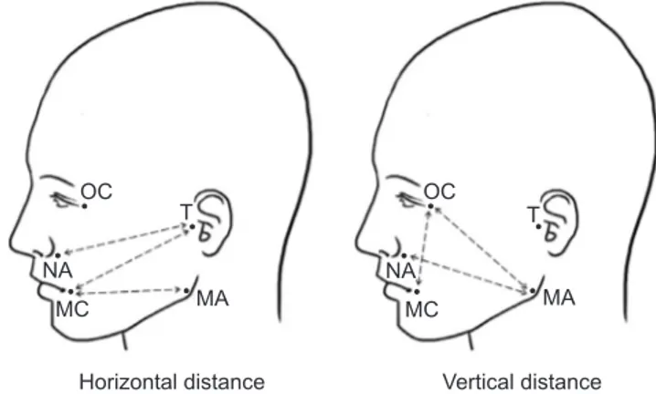
reduc-the length of reduc-the line connected to reduc-the specific landmark of the face immediately after surgery (T1) and 2-4 days after surgery (T2). After using an oil pen to draw a landmark on the patient’s face, a moisture-proof band was used to prevent the landmark from being erased. A flexible ruler was used to measure the distance between landmarks (recorded in millimeters). Measurement points included the tragus (T), mouth corner (MC), nasal alar (NA), mandible angle (MA), and outer canthus (OC). Six different measurements were recorded between the landmarks (Fig. 1): T-MC, T-NA, MA-MC (horizontal distances) and MA-NA, MA-OC, OC-MA-MC (vertical distances). The following equation was used to obtain the percentage of facial swelling: percentage of facial swelling=difference between the T2 and T1 values, divided by the T1 value, and multiplied by 100(T2 value–T1 value ×100T1 value ), as described by Cerqueira et al.11.
The degree of pain at T2 and the degree of pain at 10-14 days after surgery (T3) were assessed using the numerical rat-ing scale (NRS). The extent of pain reduction was evaluated by calculating the difference between T2 and T3.
All adverse events that occurred during the clinical trial were included in the safety assessment, recorded in the case report, and their abnormality evaluated. Treatment-emergent adverse events were recorded up to 14 weeks from the time of application of the clinical trial drug. Hyaluronidase is a product already licensed for use; therefore, no special side effects or safety risks were expected. However, if an abnor-mal reaction did occur, then the study was discontinued and treatment of the patient was considered top priority. An ex-planation was provided to every patient whenever there was a question, and the trial was stopped immediately if any patient decided to no longer participate.
1. Details regarding clinical schedules and methodology 1) Assessment 1 (baseline screening)
For patients who provided written consent, the demo-graphic information was collected and whether bone grafts were used was recorded. Registered subjects were numbered sequentially, starting from R01, and patients were assigned to the test group and control group. The randomization code was generated by a statistician using a block randomization method and a computer program; the contents were enclosed in an envelope and transmitted to the person in charge.
2) Assessment 2 (day of surgery)
Patients enrolled in the study underwent alveolar bone aug-mentation with or without hyaluronidase. The length of the line connecting a specific landmark on the face to the surgical site was measured using a flexible ruler.
3) Assessment 3 (2-4 days after surgery)
The occurrence of complications such as abnormal pain, wound healing, and edema at the surgical site, were investi-gated. The length of the line connecting a specific landmark on the face to the surgical site was measured using a flexible ruler. The degree of pain was assessed using the NRS.
4) Assessment 4 (10-14 days after surgery)
The presence or absence of side effects was evaluated. In addition, the degree of pain was assessed using the NRS. Table 1. Demographic characteristics
Variable Control group(n=20) Test group(n=18) Sex
Male 15 (75.0) 11 (61.1)
Female 5 (25.0) 7 (38.9)
Mean age (yr) 61.55 57.05
Site
Maxilla 10 (50.0) 11 (61.1)
Mandible 10 (50.0) 7 (38.9)
Values are presented as number (%) or mean.
Min-Jeong Kwoen et al: Efficacy of local hyaluronidase administration in guided bone regeneration surgery: a randomized controlled trial. J Korean Assoc Oral Maxillofac
Surg 2021 Horizontal distance Vertical distance
OC NA MC T MA OC NA MC MA T
Fig. 1. Measurement points: tragus (T), mouth corner (MC), nasal
alar (NA), mandible angle (MA), outer canthus (OC). Six different measurements were made between the landmarks: horizontal dis-tance (T-MC, T-NA, MA-MC), vertical disdis-tance (MA-NA, MA-OC, OC-MC).
Min-Jeong Kwoen et al: Efficacy of local hyaluronidase administration in guided bone regeneration surgery: a randomized controlled trial. J Korean Assoc Oral Maxillofac Surg 2021
2. Statistical analysis
Data associated with the test group or control group, con-sent acquisition date, name, initials, registration number, sex, date of birth, dates of the first, second, third, and fourth visits, edema, and pain were organized and saved using Microsoft Excel 2010 (Microsoft, Redmond, WA, USA). The Shapiro– Wilk test was used to evaluate the data distribution. Because the data followed a non-normal distribution, differences be-tween groups were assessed using the nonparametric Mann– Whitney U test. Statistical analyses were performed using IBM SPSS Statistics (ver. 20; IBM, Armonk, NY, USA). Descriptive statistics, including the median values and inter-quartile ranges (IQR), were determined for all variables of the control and test groups. The criterion for statistical sig-nificance was P<0.05.
III. Results
1. Postoperative swelling
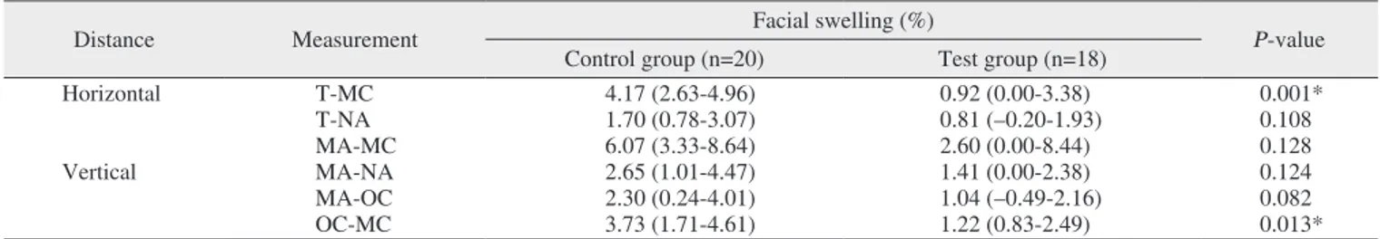
The degree of swelling in the test group was less than in the control group but was only significant for the T-MC (horizontal distance; median of the control group, 4.17% [IQR, 2.63%-4.96%]; median of the test group, 0.92% [IQR, 0.00%-3.38%]; P=0.001) and OC-MC (vertical distance; dian of the control group, 3.73% [IQR, 1.71%-4.61%]; me-dian of the test group, 1.22% [IQR, 0.83%-2.49%]; P=0.013) values.(Table 2, Fig. 2)
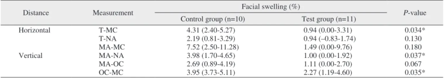
2. Postoperative swelling of the maxilla and mandible A topical hyaluronidase injection was more effective in the maxilla than in the mandible. In the maxilla, the percentage of facial swelling was significant for the T-MC (horizontal
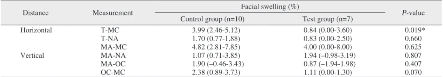
distance; median of the control group, 4.31% [IQR, 2.40%-5.27%]; median of the test group, 0.94% [IQR, 0.00%-3.31%]; P=0.034), MA-NA (vertical distance; median of the control group, 3.98% [IQR, 1.70%-4.65%]; median of the test group, 1.00% [IQR, 0.00%-1.92%]; P=0.037), and OC-MC (vertical distance; median of the control group, 3.95% [IQR, 3.73%-5.11%]; median of the test group, 2.27% [IQR, 1.19%-4.60%]; P=0.035) values.(Table 3, Fig. 3) In contrast, the percentage of facial swelling in the mandible was signifi-cant for the T-MC value (horizontal measurement; median of the control group, 3.99% [IQR, 2.46%-5.12%]; median of the test group, 0.84% [IQR, 0.00%-3.60%]; P=0.019).(Table 4, Fig. 4)
3. Pain evaluation and complications
Low levels of pain at T2 were reported in both the test and control groups (median of the control group, 3.00 [IQR,
Table 2. Postoperative swelling measurements
Distance Measurement Facial swelling (%) P-value
Control group (n=20) Test group (n=18)
Horizontal T-MC 4.17 (2.63-4.96) 0.92 (0.00-3.38) 0.001* T-NA 1.70 (0.78-3.07) 0.81 (–0.20-1.93) 0.108 MA-MC 6.07 (3.33-8.64) 2.60 (0.00-8.44) 0.128 Vertical MA-NA 2.65 (1.01-4.47) 1.41 (0.00-2.38) 0.124 MA-OC 2.30 (0.24-4.01) 1.04 (–0.49-2.16) 0.082 OC-MC 3.73 (1.71-4.61) 1.22 (0.83-2.49) 0.013*
(T-MC: tragus-mouth corner, T-NA: tragus-nasal alar, MA-MC: mandible angle-mouth corner, MA-NA: mandible angle-nasal alar, MA-OC: mandible angle-outer canthus, OC-MC: outer canthus-mouth corner)
*Statistical significance (P<0.05). T-MC 8 7 6 5 4 3 2 1 % 0
Postoperative swelling measurement
T-NA MA-MC MA-NA MA-OC OC-MC
Control (n=20) Test (n=18)
Fig. 2. Comparison of control and test for percentage of facial
swelling in six different measurements. Refer to Fig. 1 for the mea-surement points.
Min-Jeong Kwoen et al: Efficacy of local hyaluronidase administration in guided bone regeneration surgery: a randomized controlled trial. J Korean Assoc Oral Maxillofac Surg 2021
2.00-5.00]; median of the test group, 3.00 [IQR, 2.75-5.00]; P=0.941). However, only a few symptoms were reported at T3 (median of the control group, 0.00 [IQR, 0.00-0.00]; median of the test group, 0.00 [IQR, 0.00-1.00]; P=0.258). Although the mean extent of pain reduction was greater in the control group, statistically significant difference was not observed in the extent of the change.(Table 5) Medication-related side effects were not observed in the hyaluronidase group.
IV. Discussion
HA is a major carbohydrate component that constitutes the structural framework of the interstitial barrier of various tis-sues in the body. HA is an anionic, non-sulfated glycosami-noglycan. As a molecule constituting the extracellular matrix, HA has unique hygroscopic, rheological, and viscoelastic properties. Edema is a common complication after surgery.
The strong hydrophilic and hydration activities of HA prevent extravascular fluid from returning to the lymphatic system, thus contributing to the continued edematous condition7. Var-ious attempts have been made to reduce edema after swell-ing because edema early durswell-ing the healswell-ing phase can cause severe pain and dehiscence of the surgical site, thus delaying healing.
Hyaluronidase was first introduced by Duran-Reynals12; at that time, hyaluronidase was thought to reduce tissue im-permeability, therefore, it was called “spreading/permeability factor.” Later, the substance was discovered to selectively hydrolyze HA and loosen the extracellular matrix. Therefore, it was called “hyaluronidase”12,13. As an HA-metabolizing enzyme, hyaluronidase breaks the 1,4-glucosaminidic bond between the C1 and glucosamine moiety of HA, the ground substance of connective tissue, and C4 of glucuronic acid8. Consequently, the local intracellular permeability of peripher-al lymphatic channels and capillaries increases and viscosity decreases. This spreads the fluid inside the interstitial space and helps reduce edema. Due to this diffusion capacity, when administered adjunctively, hyaluronidase helps increase the analgesic efficacy of local anesthetics10.
The anti-inflammatory and anti-edema effects of topically applied hyaluronidase have been previously described. When topically administered, increased numbers of leukocytes, mononuclear cells, and neutrophils were observed, and its rolling adhesion capacity was increased. Reportedly, the lev-els of tumor necrosis factor-α, interleukin-8, and leukotriene C4 at the inflammation site were also reduced14. Because HA is a type of glycosaminoglycan with a half-life of approxi-mately 20 hours, the connective tissue is reportedly restored to its original structure within approximately 2 days after ad-ministration, therefore, there is no permanent site change15.
GBR surgery can be used to reconstruct hard tissue into an ideal form before implant placement. Tension-free primary Table 3. Swelling measurements of the maxilla
Distance Measurement Facial swelling (%) P-value
Control group (n=10) Test group (n=11)
Horizontal T-MC 4.31 (2.40-5.27) 0.94 (0.00-3.31) 0.034* T-NA 2.19 (0.81-3.29) 0.94 (–0.83-1.74) 0.130 MA-MC 7.52 (2.50-11.28) 1.49 (0.00-9.76) 0.180 Vertical MA-NA 3.98 (1.70-4.65) 1.00 (0.00-1.92) 0.037* MA-OC 2.69 (0.89-4.19) 1.11 (0.00-2.70) 0.067 OC-MC 3.95 (3.73-5.11) 2.27 (1.19-4.60) 0.035*
(T-MC: tragus-mouth corner, T-NA: tragus-nasal alar, MA-MC: mandible angle-mouth corner, MA-NA: mandible angle-nasal alar, MA-OC: mandible angle-outer canthus, OC-MC: outer canthus-mouth corner)
*Statistical significance (P<0.05).
Values are presented as median (25%-75% interquartile range).
Min-Jeong Kwoen et al: Efficacy of local hyaluronidase administration in guided bone regeneration surgery: a randomized controlled trial. J Korean Assoc Oral Maxillofac Surg 2021
T-MC 8 7 6 5 4 3 2 1 % 0
Swelling measurement of maxilla
T-NA MA-MC MA-NA MA-OC OC-MC
Control (n=10) Test (n=11)
Fig. 3. Comparison of control and test for percentage of facial
swelling of maxilla in six different measurements. Refer to Fig. 1 for the measurement points.
Min-Jeong Kwoen et al: Efficacy of local hyaluronidase administration in guided bone regeneration surgery: a randomized controlled trial. J Korean Assoc Oral Maxillofac Surg 2021
closure is a prerequisite for successful GBR surgery results. If a periosteal releasing incision is created to obtain primary wound closure, then postoperative edema due to the accumu-lation of serous fluid in the interstitial space in response to surgical trauma is inevitable. Postoperative edema is the most common complication and can lead to increased pain and dehiscence of the surgical site. Various methods to reduce edema have been introduced, however, the optimal approach to reduce edema has not yet been identified. In addition, postoperative inflammatory reactions reach a maximum on the second day after surgery and gradually disappear over the course of the following week. Eliminating other factors that may affect early wound healing is important because this may improve the quality of life during the first week after surgery and the surgical outcome16.
For several years, hyaluronidase has been used in various
et al.17 reported injecting hyaluronidase after hematoma and fibrosis due to facial trauma, which resulted in complete resolution of the hematoma, reduced fibrosis, and alleviation of pain. Because nasal bone fractures require surgery after edema is reduced, a considerable amount of time is required between the accident and surgery. Kim et al.18 applied hyal-uronidase to reduce edema before nasal bone fracture surgery during a case-control study of 181 patients and showed that when hyaluronidase was applied, a mean of 3 days from trauma to surgery was required and a mean of 8.6 days was required for the control group. Furthermore, a significant reduction in latency due to the edema-reducing effect of hy-aluronidase was reported18. The effect of hyaluronidase on postsurgical edema reduction has been well-demonstrated in animal studies. For example, Nekoroski et al.9 demonstrated the diffusion activity of accumulated postsurgical edematous fluid as a result of injecting recombinant human hyaluroni-dase sustained-release gel in mice with lymphedema.
Among the various methods used to reduce edema after surgery, corticosteroids inhibit phospholipase A2 and lower the activation of the arachidonic acid pathway. Corticoste-roids have an excellent anti-edema effect but also reduce leukocyte chemotaxis, fibroblast migration, and collagen Table 4. Swelling measurements of the mandible
Distance Measurement Facial swelling (%) P-value
Control group (n=10) Test group (n=7)
Horizontal T-MC 3.99 (2.46-5.12) 0.84 (0.00-3.60) 0.019* T-NA 1.70 (0.77-1.88) 0.83 (0.00-2.50) 0.660 MA-MC 4.82 (2.81-7.85) 4.00 (0.00-8.00) 0.625 Vertical MA-NA 1.07 (0.71-3.85) 1.94 (–0.98-3.19) 0.807 MA-OC 1.90 (–0.46-3.43) 0.87 (–1.94-1.98) 0.407 OC-MC 2.38 (0.89-3.73) 1.11 (0.00-1.30) 0.070
(T-MC: tragus-mouth corner, T-NA: tragus-nasal alar, MA-MC: mandible angle-mouth corner, MA-NA: mandible angle-nasal alar, MA-OC: mandible angle-outer canthus, OC-MC: outer canthus-mouth corner)
*Statistical significance (P<0.05).
Values are presented as median (25%-75% interquartile range).
Min-Jeong Kwoen et al: Efficacy of local hyaluronidase administration in guided bone regeneration surgery: a randomized controlled trial. J Korean Assoc Oral Maxillofac Surg 2021
T-MC 8 7 6 5 4 3 2 1 % 0
Swelling measurement of mandible
T-NA MA-MC MA-NA MA-OC OC-MC
Control (n=10) Test (n=7)
Fig. 4. Comparison of control and test for percentage of facial
swelling of mandible in six different measurements. Refer to Fig. 1 for the measurement points.
Min-Jeong Kwoen et al: Efficacy of local hyaluronidase administration in guided bone regeneration surgery: a randomized controlled trial. J Korean Assoc Oral Maxillofac Surg 2021
Table 5. Numerical rating scale
Time Control group (n=20) Test group (n=18) P-value
T2 3.00 (2.00-5.00) 3.00 (2.75-5.00) 0.941
T3 0.00 (0.00-0.00) 0.00 (0.00-1.00) 0.258
T2-T3 3.00 (2.00-5.00) 3.00 (1.00-4.00) 0.332
(T2: 2-4 days after surgery, T3: 10-14 days after surgery, T2-T3: difference between T2 and T3)
Values are presented as median (25%-75% interquartile range).
Min-Jeong Kwoen et al: Efficacy of local hyaluronidase administration in guided bone regeneration surgery: a randomized controlled trial. J Korean Assoc Oral Maxillofac Surg 2021
mune response. Consequently, corticosteroids are not the first choice of treatment after surgery. Koç and Er19 induced trau-matic edema in rats and compared the anti-edema effect of the topical administration of hyaluronidase and corticosteroid and reported that hyaluronidase can be used as a substitute for dexamethasone because it reduces edema more effectively.
In the field of dentistry, hyaluronidase has been used in only a few clinical studies to reduce postoperative edema. In the present study, whether hyaluronidase can be used as an adjunct in dental bone surgery to improve postoperative sat-isfaction with minimal side effects was evaluated; significant improvements in postoperative swelling after directly inject-ing hyaluronidase in the surgical site were observed.
The degree of edema in the oral and maxillofacial regions is generally assessed by two measurements. In the present study, four additional values were measured20,21 and com-parison of the two groups revealed that not all measurements were significantly different. However, the degree of edema in the test group tended to be lower than in the control group. Significant edema reduction was observed in the T-MC (hori-zontal; P=0.001) and OC-MC (vertical; P=0.013) values. Be-cause the mean age of the test group was 4.5 years younger than in the control group, a study with a larger sample size is needed to investigate the effect of age.
When comparing the maxilla and mandible of the test and control groups, three measurements of the maxilla and one measurement of the mandible showed significant edema reduction. Specifically, in the maxilla, the T-MC value (P=0.034), which is a horizontal measurement, and the MA-NA (P=0.037) and OC-MC (P=0.035) values, which are vertical measurements, showed statistically significant edema reduction. Only one T-MC value (horizontal measurement) of the mandible (P=0.019) was statistically significant. These findings indicate that edema reduction through the local in-jection of hyaluronidase is more effective in the maxilla than in the mandible.
Hyaluronidase was reported to have an analgesic effect in several studies10,8. However, in the present study, significant difference was not observed between the test and control groups. Patients were instructed to use painkillers before and after surgery. We suggest an additional method should be de-vised to control the masking effect of painkillers to accurately determine the degree of hyaluronidase-induced pain reduc-tion. In the present study, the NRS was used to evaluate the severity of pain. One limitation of the NRS is the degree of pain can be underestimated or overestimated. However, NRS
is a reliable method suitable for assessing self-reported pain because it is simple and straightforward22.
In this prospective study, the effect of the topical applica-tion of hyaluronidase during GBR surgery on postoperative edema and pain was evaluated. Although each step in the study was conducted according to the relevant guidelines, standardization of the location and size of the surgical site was not possible. Furthermore, the study was limited by its small sample size. The ability of a local injection of hyal-uronidase to reduce edema in the dental field has been as-sessed in only a few studies. Therefore, further in vivo studies with larger sample size and randomized controlled trials are needed to validate our results.
Despite the need for more research, when applied adjunc-tively, hyaluronidase has the tendency to reduce edema after surgery without any specific side effects. Therefore, the supplemental use of hyaluronidase in dentistry is considered highly beneficial.
V. Conclusion
Based on the present clinical trial, the differences in swell-ing were not statistically significant; however, the degree of swelling was lower in the group that received topical hyal-uronidase (test group). Hyalhyal-uronidase appeared more effec-tive in the maxilla; however, the degree of pain reduction was similar in the test and control groups.
ORCID
Min-Jeong Kwoen, https://orcid.org/0000-0002-0246-4090 Yong-Hoon Choi, https://orcid.org/0000-0001-8222-219X Keun-Suh Kim, https://orcid.org/0000-0002-5986-4810 Na-Hee Chang, https://orcid.org/0000-0001-8634-571X Young-Kyun Kim, https://orcid.org/0000-0002-7268-3870 Hyo-Jung Lee, https://orcid.org/0000-0002-0439-7389
Authors’ Contributions
H.J.L. and Y.K.K. conceived and designed the study. M.J.K., K.S.K., and N.H.C. collected and analyzed the data. M.J.K., Y.H.C., and K.S.K. interpreted the data. M.J.K. and Y.H.C. drafted the manuscript. M.J.K., Y.H.C., K.S.K., N.H.C., Y.K.K., and H.J.L. critically reviewed the manu-script. All authors read and approved the final version of the manuscript.
Acknowledgements
BMI Korea (Jeju, Korea) provided financial support for the research (clinical trial registration No. 06-2017-198; registra-tion date December 9, 2017).
We would like to thank Duck-won Kwon for assistance with the creation of Fig. 1.
Ethics Approval and Consent to Participate
This study was conducted with the approval of the Bioeth-ics Review Committee of Seoul National University Bundang Hospital (B-1708-415-005). Informed consent was obtained from all participants.
Conflict of Interest
BMI Korea (Jeju, Korea) supported this study. The funders had no influence in study design, data collection and analysis, decision to publish, or preparation of the manuscript.
No potential conflict of interest relevant to this article was reported.
References
1. Cardaropoli G, Araújo M, Lindhe J. Dynamics of bone tissue for-mation in tooth extraction sites. An experimental study in dogs. J Clin Periodontol 2003;30:809-18. https://doi.org/10.1034/j.1600-051x.2003.00366.x
2. Araújo MG, Lindhe J. Dimensional ridge alterations follow-ing tooth extraction. An experimental study in the dog. J Clin Periodontol 2005;32:212-8. https://doi.org/10.1111/j.1600-051X.2005.00642.x
3. Reich KM, Huber CD, Lippnig WR, Ulm C, Watzek G, Tangl S. Atrophy of the residual alveolar ridge following tooth loss in an historical population. Oral Dis 2011;17:33-44. https://doi. org/10.1111/j.1601-0825.2010.01699.x
4. Lim G, Lin GH, Monje A, Chan HL, Wang HL. Wound healing complications following guided bone regeneration for ridge aug-mentation: a systematic review and meta-analysis. Int J Oral Max-illofac Implants 2018;33:41-50. https://doi.org/10.11607/jomi.5581 5. Romanos GE. Periosteal releasing incision for successful coverage
of augmented sites. A technical note. J Oral Implantol 2010;36:25-30. https://doi.org/10.1563/AAID-JOI-D-09-00068
6. Moslemi N, Khorsand A, Torabi S, Shahnaz A, Soleimani Shayes-teh Y, Fekrazad R. Periosteal releasing incision with diode laser in guided bone regeneration procedure: a case series. J Lasers Med Sci 2016;7:259-64. https://doi.org/10.15171/jlms.2016.46
7. Stern S, Lindenhayn K, Schultz O, Perka C. Cultivation of porcine cells from the nucleus pulposus in a fibrin/hyaluronic acid matrix. Acta Orthop Scand 2000;71:496-502. https://doi. org/10.1080/000164700317381207
8. Khan N, Niazi ZR, Rehman FU, Akhtar A, Khan MM, Khan S, et al. Hyaluronidases: a therapeutic enzyme. Protein Pept Lett
2018;25:663-76. https://doi.org/10.2174/092986652566618062912 1823
9. Nekoroski T, Paladini RD, Sauder DN, Frost GI, Keller GA. A recombinant human hyaluronidase sustained release gel for the treatment of post-surgical edema. Int J Dermatol 2014;53:777-85. https://doi.org/10.1111/ijd.12304
10. Buhren BA, Schrumpf H, Hoff NP, Bölke E, Hilton S, Gerber PA. Hyaluronidase: from clinical applications to molecular and cellular mechanisms. Eur J Med Res 2016;21:5. https://doi.org/10.1186/ s40001-016-0201-5
11. Cerqueira PR, Vasconcelos BC, Bessa-Nogueira RV. Compara-tive study of the effect of a tube drain in impacted lower third molar surgery. J Oral Maxillofac Surg 2004;62:57-61. https://doi. org/10.1016/s0278-2391(03)00675-x
12. Duran-Reynals F. Studies on a certain spreading factor existing in bacteria and its significance for bacterial invasiveness. J Exp Med 1933;58:161-81. https://doi.org/10.1084/jem.58.2.161
13. Hobby GL, Dawson MH, Meyer K, Chaffee E. The relation-ship between spreading factor and hyaluronidase. J Exp Med 1941;73:109-23. https://doi.org/10.1084/jem.73.1.109
14. Fronza M, Muhr C, da Silveira DS, Sorgi CA, Rodrigues SF, Far-sky SH, et al. Hyaluronidase decreases neutrophils infiltration to the inflammatory site. Inflamm Res 2016;65:533-42. https://doi. org/10.1007/s00011-016-0935-0
15. Frost GI. Recombinant human hyaluronidase (rHuPH20): an enabling platform for subcutaneous drug and fluid adminis-tration. Expert Opin Drug Deliv 2007;4:427-40. https://doi. org/10.1517/17425247.4.4.427
16. van Wijk A, Kieffer JM, Lindeboom JH. Effect of third molar surgery on oral health-related quality of life in the first postopera-tive week using Dutch version of Oral Health Impact Profile-14. J Oral Maxillofac Surg 2009;67:1026-31. https://doi.org/10.1016/ j.joms.2008.12.041
17. Han JH, Kim J, Yoon KC, Shin HW. Treatment of post-traumatic hematoma and fibrosis using hyaluronidase injection. Arch Cranio-fac Surg 2018;19:218-21. https://doi.org/10.7181/acfs.2017.01396 18. Kim JH, Yang H, Oh SH, Song SH, Kyung H. The efficacy of
hyal-uronidase in early surgery of nasal bone fracture. J Craniofac Surg 2019;30:e617-9. https://doi.org/10.1097/SCS.0000000000005646 19. Koç O, Er N. Can hyaluronidase be an alternative postoperative
anti-edema agent to dexamethasone? Preliminary results of an animal study. J Oral Maxillofac Surg 2018;76:1653-9. https://doi. org/10.1016/j.joms.2018.03.010
20. Amin MM, Laskin DM. Prophylactic use of indomethacin for pre-vention of postsurgical complications after removal of impacted third molars. Oral Surg Oral Med Oral Pathol 1983;55:448-51. https://doi.org/10.1016/0030-4220(83)90227-x
21. Chukwuneke FN, Oji C, Saheeb DB. A comparative study of the effect of using a rubber drain on postoperative discomfort fol-lowing lower third molar surgery. Int J Oral Maxillofac Surg 2008;37:341-4. https://doi.org/10.1016/j.ijom.2007.11.016 22. Ferreira-Valente MA, Pais-Ribeiro JL, Jensen MP. Validity of four
pain intensity rating scales. Pain 2011;152:2399-404. https://doi. org/10.1016/j.pain.2011.07.005
How to cite this article: Kwoen MJ, Choi YH, Kim KS, Chang
NH, Kim YK, Lee HJ. Efficacy of local hyaluronidase administra-tion in guided bone regeneraadministra-tion surgery: a randomized controlled trial. J Korean Assoc Oral Maxillofac Surg 2021;47:91-98. https:// doi.org/10.5125/jkaoms.2021.47.2.91