서 론
저등급 골육종에는 방골성 골육종과 저등급 중심부 골육종이 있 으며 전체 골육종의 4-5%를 차지한다.1) 저등급 중심부 골육종은 방골성 골육종보다 더 드물며 전체 골육종의 1-2% 정도를 차지한 다.2) 방골성 골육종은 방사선학적으로 비교적 진단이 용이하며, 특징적으로 원위 대퇴 후방이나 근위 상완골, 경골, 비골 순으로 호발한다. 조직학적으로는 화골성 근염, 기괴 방골성 골연골성 증 식증(bizarre parosteal osteochondromatous proliferation), 유골 골종 등과 감별하여야 한다. 반면 저등급 중심부 골육종은 진단이 용이 하지 않다. 영상학적으로도 양성 종양의 양상과 비슷하여, 약간의 공격적인 성향을 띠는 양성 종양으로 오인되는 경우가 있다.3-6) 70 예를 분석한 한 대규모 연구에서는 저등급 중심부 골육종의 영상 학적 특징으로 다양한 정도의 두껍고 거친 소주골을 동반한 골융저등급 중심부 골육종의 진단, 치료 및 예후
Diagnosis, Treatment and Prognosis of Low Grade Central Osteosarcoma
송원석 • 조완형 • 이광열 • 공창배 • 고재수* • 전대근 • 이수용
원자력병원 정형외과학교실, *병리학교실 목적: 저등급 중심부 골육종 환자의 진단, 치료 및 예후에 대하여 알아보고자 하였다. 대상 및 방법: 1994년부터 2011년까지 저등급 중심부 골육종으로 진단받고 본원에서 치료받은 16명의 환자를 대상으로 하였다. 결과: 환자 분포는 남자가 4명 여자가 12명이었으며 평균 연령은 26세였다. 초기 진단은 11명의 환자가 중심부 저등급 골육종으로 맞게 진단 되었으나 나머지 5명의 환자는 각각 유골 골종, 비골화성 섬유종, 골모세포종, 동맥류성 골낭종, 결합조직형성 섬유종 등으로 오진되었다. 15 명의 환자가 최종적으로 광범위 절제술을 시행하였으며 그 중 한 명은 수술 전 항암치료를 시행하였다. 그 중 14명의 환자가 치료 후 재발 없 이 추시중이며, 한 명은 기존에 앓던 신세포암의 악화로 수술 후 21개월 후 사망하였다. 나머지 한 명은 다발성 종양 환자로, 부분적으로만 광 범위 절제술을 시행하였으며 잔존 종양에 방사선 치료만을 시행한 후 7년째 생존 중이다. 9명(56%)의 환자가 종양이 피질골 밖까지 파급되어 있는 소견을 보였으며 그 중 한 명은 구획 외로까지의 파급을 보였다. 결론: 저등급 중심부 골육종은 진단이 어려우나 임상적 의심과 함께 조직병리학적, 영상학적인 특징을 고려하여 주의 깊은 감별이 요구된다. 치료에 있어서는 광범위 종양 절제술이 권장되며, 양성 종양으로 오진하여 병소내 절제술만 시행한 경우라도 국소 재발이나 고등급으로의 악 성 전환 가능성이 있으므로 광범위 재절제술을 시행할 것을 권고하는 바이다. 색인단어: 저등급 중심부 골육종, 진단, 치료 해 소견(31%), 소주골이 얇고 불명확하며 골융해 소견이 뚜렷한 경우(30%), 조밀한 경화성 소견(24%), 골융해 소견과 골경화 소견 이 혼합된 경우(14%) 등을 보고하였다.7) 조직학적으로도 섬유성 골 이형성증, 비골화성 섬유종, 결합조직형성 섬유종(desmoplastic fibroma) 등 다른 양성 종양과 유사한 소견을 보여 이들과의 감별 이 중요하다.8,9) 저등급 중심부 골육종의 영상학적 소견이 양성 골종양과 비슷 하기 때문에 절개 생검술 없이 골소파술만을 시행하는 경우가 종 종 발생하며, 이 경우 종양의 파급으로 이어질 수 있다.9-11) 또한 절개 생검술을 적절히 시행했다 하더라도 병리학자에 의해 제대 로 진단되지 못하는 경우도 있다.10-12) 본 연구에서는 저등급 중심부 골육종 환자 16예를 경험하여 진 단, 치료 및 예후에 관해 분석해보고자 한다.대상 및 방법
1994년부터 2011년까지 저등급 중심부 골육종으로 진단받은 18명 의 환자를 선별하였으며, 두 명의 숙련된 병리학자가 조직 슬라이 드를 재검하여 확인하였다. 조직학적 진단의 기준으로는 피질골 과 하버시안계로의 종양 침범 및 병변 주변 소주골의 포착 여부로 접수일 2014년 9월 16일 심사수정일 2014년 11월 26일 게재확정일 2014년 11월 28일 교신저자 전대근 서울시 노원구 공릉동 215-4, 원자력병원 정형외과 TEL 02-970-1242, FAX 02-970-2427 E-mail dgjeon@kcch.re.krCopyrights © 2014 by The Korean Bone and Joint Tumor Society
“This is an Open Access article distributed under the terms of the Creative Commons Attribution Non-Commercial License (http://creativecommons.org/licenses/by-nc/3.0/) which permits unrestricted non-commercial use, distribution, and reproduction in any medium, provided the original work is properly cited.”
서 결정하였다.2,8) 병리학자의 재검 후 두 예가 제외되었는데, 각각 일부만이 저등급 소견을 보인 고등급 골육종과 고등급 섬유모세 포성 골육종이 저등급 골육종으로 오진되었던 경우였다. 재검 후 확진된 16예는 나이, 성별, 종양의 위치, 단순 방사선 소견, 종양의 크기, 골 외로의 파급여부, 초기 진단 및 초기 치료, 최종 진단 및 최종 치료, 절제연, 종양학적 예후 등을 조사하였다.
결 과
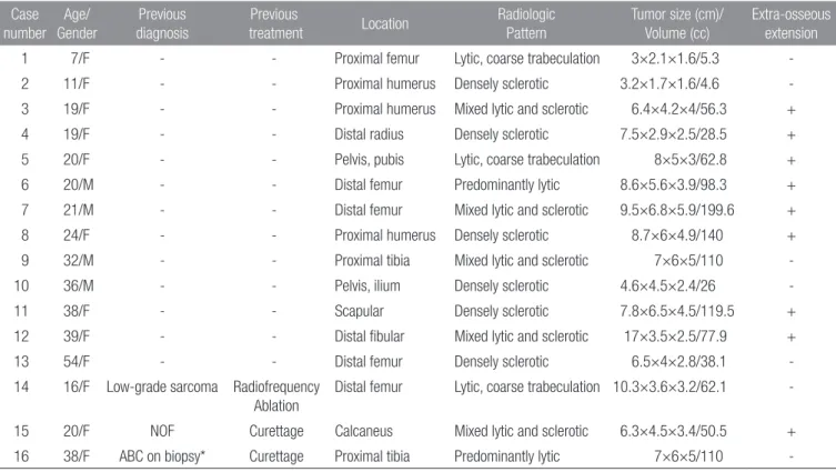
12명은 여자, 4명은 남자였으며 평균 나이는 26세(7-54)였다. 13명 의 환자가 치료 전 본원으로 직접 의뢰되었으나 나머지 3명은 타 병원에서 진단 및 치료 후 본원으로 전원되었다. 추시 기간은 평 균 81개월(21-171)이었다. 종양의 위치는 대퇴부가 5명, 근위 상완 골이 3명, 근위 경골이 2명, 골반골이 2명, 원위 비골, 종골, 견갑골, 원위 요골이 각각 1명이었다. 초기 진단은 16명 중 14명의 환자가 중심부 저등급 골육종 혹은 저등급 육종으로 진단되었으나 나머 지 2명의 환자는 각각 비골화성 섬유종, 동맥류성 골낭종 등으로 오진되었다(Table 1). 첫 진단 시 전이를 보였던 환자는 없었으며 추시 중 제1 흉추로의 전이를 보였던 한 환자는 본래 앓던 신세포 암에서 유래한 것으로 판명되어, 결과적으로 모든 환자에서 전이 는 없었다. 영상학적으로는 상기한 Andresen의 기준에 따라 다음의 네 그 룹으로 분류하여 조사하였다;조밀한 골경화성 소견(6예), 골융해 소견과 골경화 소견이 혼합된 경우(5예), 다양한 정도의 두껍고 거 친 소주골을 동반한 골융해 소견(3예), 소주골이 얇고 불명확하 며 골융해 소견이 뚜렷한 경우(2예). 종양의 크기는 자기 공명 영 상 검사를 이용하여 측정하였는데, 종양의 길이는 평균 7.6 cm (3-17), 종양의 부피는 평균 74 cc (5.3-200)이었다. 종양의 Enneck-ing’s criteria에 따른 병기는 7예가 stage IA, 9예가 stage IB였다. 한 환자는 수술 전 항암 화학 요법을 2회 시행하였는데, 이는 이 환자 의 영상학적 패턴이 공격적이고, 종양의 크기가 커서(200 cc) 고등 급의 악성이 의심되었고, 절개 생검술을 시행한 부위가 이 종양의 고등급 악성 부위를 놓쳤을 가능성을 고려했기 때문이었다(Fig. 1). 그러나 종양 절제술 후 시행한 전체 종양의 조직 검사상 저등 급 중심부 골육종으로 확진되어, 이에 수술 후 항암 화학 요법은 시행하지 않았다. 이 환자(증례 14)를 제외한 모든 환자가 본원에 서 광범위 절제술을 시행받았다. 저자들은 저등급의 악성도를 감 안하여, 절제연을 2 cm로 하되 연부 조직의 절제를 최소화 하였 고, 종양이 관절과 가까웠던 두 환자(증례 1, 2)에서는 절제연을 1 cm 이하로 하여 수술을 시행하였다. 15명 중 세 명의 환자는 종양 절제술만을 시행 받았으며, (내골반골 절제술 2예, 견갑골 절제술 1예) 나머지 12명의 환자는 재건술을 시행하였다(종양대치물 5예, 중첩 동종골 3예, 동종골 혹은 재사용 자가골과 종양 대치물 복합 체 2예, 자가 비골 이식 1예, 골 시멘트 보형물 1예). 절제술을 시Table 1. Patient Characteristics and Radiologic Feature Case number Age/ Gender Previous diagnosis Previous treatment Location Radiologic Pattern Tumor size (cm)/ Volume (cc) Extra-osseous extension
1 7/F - - Proximal femur Lytic, coarse trabeculation 3×2.1×1.6/5.3
-2 11/F - - Proximal humerus Densely sclerotic 3.2×1.7×1.6/4.6
-3 19/F - - Proximal humerus Mixed lytic and sclerotic 6.4×4.2×4/56.3 +
4 19/F - - Distal radius Densely sclerotic 7.5×2.9×2.5/28.5 +
5 20/F - - Pelvis, pubis Lytic, coarse trabeculation 8×5×3/62.8 +
6 20/M - - Distal femur Predominantly lytic 8.6×5.6×3.9/98.3 +
7 21/M - - Distal femur Mixed lytic and sclerotic 9.5×6.8×5.9/199.6 +
8 24/F - - Proximal humerus Densely sclerotic 8.7×6×4.9/140 +
9 32/M - - Proximal tibia Mixed lytic and sclerotic 7×6×5/110
-10 36/M - - Pelvis, ilium Densely sclerotic 4.6×4.5×2.4/26
-11 38/F - - Scapular Densely sclerotic 7.8×6.5×4.5/119.5 +
12 39/F - - Distal fibular Mixed lytic and sclerotic 17×3.5×2.5/77.9 +
13 54/F - - Distal femur Densely sclerotic 6.5×4×2.8/38.1
-14 16/F Low-grade sarcoma Radiofrequency Ablation
Distal femur Lytic, coarse trabeculation 10.3×3.6×3.2/62.1
-15 20/F NOF Curettage Calcaneus Mixed lytic and sclerotic 6.3×4.5×3.4/50.5 +
16 38/F ABC on biopsy* Curettage Proximal tibia Predominantly lytic 7×6×5/110
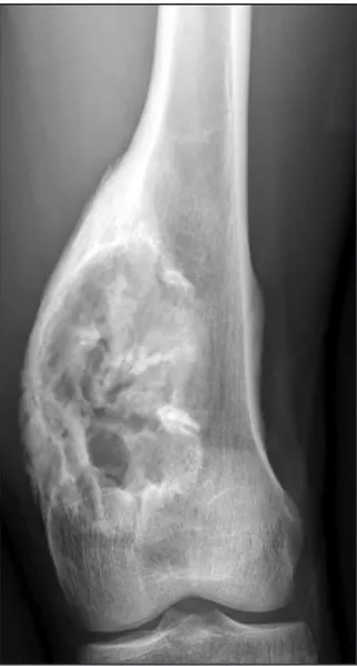
행하지 않은 나머지 한 명(증례 3)의 환자는 다발성 저등급 중심 부 골육종 환자로, 타병원에 처음 방문하여 경골 근위부의 병변만 을 발견하였으며, 이 부위에서 두 차례 조직검사 시행 후 저등급 중심부 골육종으로 진단되었다. 이에 이 부위의 병변을 절제하고 비골 이식 및 골이식을 시행하였으며 이후 재발은 없었다. 그러나 추시 중 동측 대퇴골두 일부, 경부와 간부에서도 병변이 발견되었 다. 다만 첫 방문 당시 이 부위에 대한 검사를 시행하지 않아 이 병 변이 언제부터 존재했었는지는 알 수가 없었다. 이에 대퇴 골두와 경부 및 간부에 골소파술을 시행한 후 전기 고주파 소작술을 시 행하였으며, 대퇴 간부는 이후 전기 소작으로 인한 병적 골절이 발생하여 내고정술 및 골이식을 시행하였다. 그러나 이후 시행한 PET-CT 검사에서 동측 경골 하부 관절면 부위와 동측 비구 부위 에도 전이 소견이 발견되어 본원으로 의뢰되었다. 본원에서는 전 이 부위에 대한 근치적 절제술은 예후상의 이득이 없을 것으로 판 단하여 방사선 치료만을 시행하였다. 16명 중 14명의 환자가 치료 후 재발이나 전이 없이 현재까지 추시 중이다. 나머지 두 명중 한 명은 기존에 앓던 신세포암의 제 1 흉추로의 전이 및 이후 종양의 전신 파급으로 사망하였다. 절제 술을 시행하지 않고 방사선 치료만을 시행한 환자(증례 14)는 7년 이 지난 현재까지 종양의 파급 없이 생존 중이다(Table 2). 종양의 영상학적 파급 범위는 9명(56%)의 환자가 피질골 밖까지 파급된 종양 소견을 보였다. 그 중 한 명은 구획 외로의 파급을 보였는데, 본원에서 시행한 절개 생검술의 조직 검사상 결합조직형성 섬유 종(desmoplastic fibroma)으로 진단되었다. 그러나 영상 소견의 공 격성을 감안한 임상적 판단 하에 악성 종양에 준하는 광범위 절제 Figure 1. Plain radiograph of a 21-year-old man showing expansile
mixed osteolytic and sclerotic lesion in the distal meta-diaphysis of femur. Radiologic differential diagnosis included osteosarcoma, fibrosarcoma, adamantinoma, and giant osteoblastoma.
Table 2. Surgical Procedure, Immune-Histochemical Analysis, and Clinical Course
Case number Chemotherapy Definitive treatment Surgical margin Local recurrence Metastasis Followup (months) Final status
1 - Intercalary resection, overlapping allograft Wide - - 56 CDF 2 - Intercalary resection, overlapping allograft Wide - - 57 CDF
3 - Allograft prosthesis composite Wide - - 35 CDF
4 - Excision, fibula graft Wide - - 171 CDF
5 - Internal pelvectomy Marginal - - 141 CDF
6 - Tumor prosthesis Wide - - 74 CDF
7 + Tumor prosthesis Wide - - 44 CDF
8 - Tumor prosthesis Wide - - 45 CDF
9 - Tumor prosthesis Wide - - 107 CDF
10 - Internal pelvectomy Marginal - - 21 DOC
11 - Scapulectomy Wide - - 122 CDF
12 - Resection, overlapping allograft Wide - - 46 CDF
13 - Tumor prosthesis Wide - - 36 CDF
14 - Radiotherapy - + - 142 AWD
15 - Cement spacer Marginal - - 46 CDF
16 - Pasteurized bone-prosthesis
composite
Wide - - 162 CDF
술을 시행하였고, 최종 조직병리 또한 저등급 중심부 골육종으로 진단되었다.
고 찰
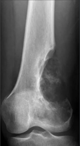
중심부 저등급 골육종은 피질골과의 상대적 위치에 따라 방골성 골육종과 저등급 중심부 골육종으로 구분된다. 두 종류의 종양 모 두 30대에 호발하고, 무증상 기간이 길며, 거친 소주골을 동반하 는 섬유성 간질(fibrotic stroma)에 비정형 방추세포의 증식 소견이 관찰되고, 염색체 12번이 증폭(amplified)되는 유전적 특질을 보이 며, 재발률, 전이율, 탈분화(dedifferentiation), 무병 생존률이 비슷 하다는 유사점을 갖는다.1,13-15) 반면, 저등급 중심부 골육종은 방 골성 골육종보다 빈도가 더 드물고, 영상학적으로도 양성 종양의 양상을 띠거나, 간혹 더 공격적인 악성 종양의 모습을 보이기도 한다(Fig. 2-4). 때문에 저등급 중심부 골육종은 과소진단(under-diagnosis) 혹은 과잉진단(over-과소진단(under-diagnosis)될 가능성이 더 높다. 이전의 연구들은 이러한 드문 중심부 저등급 골육종의 진단에 대한 난점을 보고하였다(Table 3). 약 30%의 환자가 처음 방문한 병원에서 유골 골종, 간엽종(mesenchymoma), 골낭종, 결합조직형 성 섬유종(desmoplastic fibroma), 섬유성 이형성증, 섬유골성 병소 (fibro-osseous lesion), 거대세포종, 악성 섬유성 조직구증 등으로 오진된다.10-12) 게다가 골연부종양 전문 기관에서조차도 조직 생 검 후 옳은 병리학적 진단을 내는 경우가 50-54% 밖에 되지 않는 다.3,11) 이러한 오진으로 병소내 절제술이나 소파술의 빈도가 33-56%까지 높게 나타난다. 이 경우 국소 재발이 잘 일어날 뿐만 아 니라, 재발한 종양은 고등급 골육종으로 전환될 가능성이 있기 때 문에 그 중 39-56% 가량은 수술 전 항암 치료와 함께 광범위 재절 제술을 시행하게 된다.11,12) 적절한 최종 절제술을 시행한 후에는 재발율이 약 0-14%로 감소한다고 한다. 폐전이의 비율은 이전 연 구 중 Yoshida의 연구(43%)를 제외한 세 연구에서 0-20%로 비교 적 낮게 보고되었는데, 절제연이나 항암 치료 여부가 서로 차이 가 있었음에도 유사한 결과를 보였다. 또한 Choong 등은 수술만 을 시행한 20명의 환자에서 4명(20%)의 환자만이 전이를 보였다 고 하였다.3) 즉, 고등급의 전형적 골육종에서는 항암 치료가 폐 전 이를 유의하게 낮추어 줄 수 있지만, 저등급 중심부 골육종에 있 어서는 항암 치료의 효과가 아직 불확실한 실정이다. 저자들도 이 전의 연구에서, 재발 후 공격적인 성향을 보이는 방골성 골육종Figure 3. Plain radiograph of a 20-year-old man showing predominantly lytic lesion in the distal femur. Radiologic differential diagnosis included osteosarcoma, fibrosarcoma, chondrosarcoma, and giant cell tumor with aneurismal bone cystic change.
Figure 4. (A) Plain radiograph of a 54-year-old woman showing sclerotic lesion in the distal femur. (B) Mature bone trabeculae is admixed with permeating spindle cell stroma (×100).
B A
Figure 2. (A) Plain radiograph of a 19-year-old girl showing mixed osteolytic and sclerotic lesion in the proximal humerus. (B) The typical histologic appearance of low-grade osteosarcoma, permeation of osseous trabeculae by spindle cell stroma (hematoxylin-eosin, ×100).
환자에게 항암 치료를 추가한 경우를 분석하였고, 그 결과 예후의 유의한 이득이 없었음을 확인한 바 있다.16) Yoshida의 연구에서도 56%의 저등급 골육종 환자가 항암 치료를 받았지만 43%의 환자 가 전이를 보였다.12) 저자들은 이러한 결과들을 종합하여 볼 때 저 등급에서 고등급으로 악성도의 변이를 보인 골육종에서는 항암 치료가 예후에 별다른 도움이 되지 않을 것이라고 생각하였다. 하 지만 정확한 관련성은 더 많은 연구가 필요하리라 사료된다. 중심부 저등급 골육종에 대한 방사선 치료는 이전의 연구에서 는 잘 언급되어 있지 않지만, 본 연구에서는 근치적 광범위 절제 술을 시행하기 어려웠던 다발성 저등급 중심부 골육종 환자에게, 종양의 안정화를 위하여 시행하였다. 이 증례의 경우 7년 추시 기 간 동안 더 이상의 파급 없이 생존해 있다. 그러나 이렇게 방사선 치료가 불가피한 경우를 제외하고는 중심부 저등급 골육종의 치 료에 있어서 원칙적으로 광범위 절제술이 권고된다.
결 론
저등급 중심부 골육종은 진단이 어려우나 임상적 의심과 함께 조 직병리학적, 영상학적인 특징을 고려하여 주의 깊은 감별이 요구 된다. 치료에 있어서는 광범위 종양 절제술이 권장되며, 양성 종 양으로 오진하여 병소내 절제술만 시행한 경우라도 국소 재발이 나 고등급으로의 악성 전환 가능성이 있으므로 광범위 재절제술 을 시행할 것을 권고하는 바이다.참고문헌
1. Schwab JH, Antonescu CR, Athanasian EA, Boland PJ, Healey JH, Morris CD. A comparison of intramedullary and juxta-cortical low-grade osteogenic sarcoma. Clin Orthop Relat Res. 2008;466:1318-22.
2. Kurt AM, Unni KK, McLeod RA, Pritchard DJ. Low-grade intraosseous osteosarcoma. Cancer. 1990;65:1418-28.
3. Choong PF, Pritchard DJ, Rock MG, Sim FH, McLeod RA, Unni KK. Low grade central osteogenic sarcoma. A long-term followup of 20 patients. Clin Orthop Relat Res. 1996;(322): 198-206.
4. Franceschina MJ, Hankin RC, Irwin RB. Low-grade central osteosarcoma resembling fibrous dysplasia. A report of two cases. Am J Orthop (Belle Mead NJ). 1997;26:432-40.
5. Kenan S, Ginat DT, Steiner GC. Dedifferentiated high-grade osteosarcoma originating from low-grade central osteosar-coma of the fibula. Skeletal Radiol. 2007;36:347-51.
6. Muramatsu K, Hashimoto T, Seto S, Gondo T, Ihara K, Ta-guchi T. Low-grade central osteosarcoma mimicking fibrous dysplasia: a report of two cases. Arch Orthop Trauma Surg. 2008;128:11-5.
7. Andresen KJ, Sundaram M, Unni KK, Sim FH. Imaging fea-tures of low-grade central osteosarcoma of the long bones and pelvis. Skeletal Radiol. 2004;33:373-9.
8. Bertoni F, Bacchini P, Fabbri N, et al. Osteosarcoma. Low-grade intraosseous-type osteosarcoma, histologically resem-bling parosteal osteosarcoma, fibrous dysplasia, and desmo-plastic fibroma. Cancer. 1993;71:338-45.
9. Kumar A, Varshney MK, Khan SA, Rastogi S, Safaya R. Low grade central osteosarcoma--a diagnostic dilemma. Joint Bone Spine. 2008;75:613-5.
10. Hayashi K, Tsuchiya H, Yamamoto N, et al. Diagnosis and treatment of low-grade osteosarcoma: experience with nine cases. Int J Clin Oncol. 2014;19:731-8.
11. Malhas AM, Sumathi VP, James SL, et al. Low-grade central osteosarcoma: a difficult condition to diagnose. Sarcoma. 2012;2012:764796.
12. Yoshida A, Ushiku T, Motoi T, et al. Immunohistochemical analysis of MDM2 and CDK4 distinguishes low-grade osteo-Table 3. Comparison with Previous Studies
Author Number of patients Definitive treatment Intralesional operation* (%) Chemotherpy LR
† Metastasis Final status
Current study 16 13/3 3/16 (19%) 1/16 (6%) 0% 0% NED (15), DOC (1), AWD (1)
Choong et al8) 20 12/8 12/20 (60%) - 14% 20% NED (16), DOD (4)
Malhas et al12) 18 11/7 10/18 (56%) 7/18 (39%) 11% 11% NED (16), AWD (1), DOD (1)
Yoshida et al14) 9 3/6 3/9 (33%) 5/9 (56%) 14% 43% NED (4), AWD (2), DOD (1)
Dujardin et al18)
6 NA NA NA NA NA NA
Hayashi et al13) 5 NA 2/5 (40%) NA 0% 0% NED (5)
*Either at primary or referral center; †LR, local recurrence after definitive treatment; NED, no evidence of disease; DOC, died of other cause; AWD,
sarcoma from benign mimics. Mod Pathol. 2010;23:1279-88. 13. Antonescu CR, Huvos AG. Low-grade osteogenic sarcoma
arising in medullary and surface osseous locations. Am J Clin Pathol. 2000;114 Suppl:S90-103.
14. Gisselsson D, Pålsson E, Höglund M, et al. Differentially am-plified chromosome 12 sequences in low- and high-grade osteosarcoma. Genes Chromosomes Cancer. 2002;33:133-40.
15. Tarkkanen M, Böhling T, Gamberi G, et al. Comparative ge-nomic hybridization of low-grade central osteosarcoma. Mod Pathol. 1998;11:421-6.
16. Song WS, Jeon DG, Kong CB, Cho WH, Lee SY. Outcome of re-excision for intralesionally treated parosteal osteosarcoma. J Surg Oncol. 2011;103:264-8.
Diagnosis, Treatment and Prognosis of
Low Grade Central Osteosarcoma
Won Seok Song, Wan Hyeong Cho, Kwang-Youl Lee, Chang-Bae Kong,
Jae-Soo Koh*, Dae-Geun Jeon, and Soo-Yong Lee
Departments of Orthopedic Surgery and *Pathology, Korea Cancer Center Hospital, Seoul, Korea
Purpose: We analyzed the diagnosis and the treatment outcomes of patients with central low grade osteosarcoma.
Materials and Methods: We retrospectively reviewed 16 patients with central low grade osteosarcoma were treated at out institu-tion between 1994 and 2011.
Results: There were 4 men and 12 women with mean age of 26 years. Eleven patients were correctly diagnosed but 5 patients were misdiagnosed as osteoid osteoma, non ossifying fibroma, aneurysmal bone cyst, desmoplastic fibroma. 15 patients finally received wide margin en bloc excision and one of them treated under neoadjuvant chemotherapy. Final survival status was continuous disease free in 14 and 1 patient died of renal cell cancer. Remaining 1 with multifocal lesions is alive with disease for 7 years only treated ra-diation therapy on residual tumors. Nine (56%) of 16 tumors showed extra-osseous extension of tumor (56%) and 1 of them showed extra-compartmental tumors.
Conclusion: The diagnosis of central low grade osteosarcoma is challenging, however, considering of the clinical suspicion, the typi-cal findings of radiologic and pathologic features, proper diagnosis is needed. This tumor should be treated with wide excision, even after an intralesional excision, to avoid local recurrence or transformation to higher histologic grade.
Key words: central low grade osteosarcoma, diagnosis, treatment
Received September 16, 2014 Revised November 26, 2014 Accepted November 28, 2014 Correspondence to: Dae-Geun Jeon
Department of Orthopedic Surgery, Korea Cancer Center Hospital, 215-4, Gongneung-dong, Nowon-gu, Seoul 139-706, Korea TEL: +82-2-970-1242 FAX: +82-2-970-2427 E-mail: dgjeon@kcch.re.kr
Copyrights © 2014 by The Korean Bone and Joint Tumor Society
“This is an Open Access article distributed under the terms of the Creative Commons Attribution Non-Commercial License (http://creativecommons.org/licenses/by-nc/3.0/) which permits unrestricted non-commercial use, distribution, and reproduction in any medium, provided the original work is properly cited.”