Diffuse splenic FDG uptake is
predictive of clinical outcomes in
patients with rectal cancer
Sang Yoon Kim
1, Chang Mo Moon
1,2, Hai-Jeon Yoon
3, Bom Sahn Kim
3, Ji Young Lim
1,
Tae Oh Kim
1, A. Reum Choe
1, Chung Hyun tae
1, Seong-Eun Kim
1, Hye-Kyung Jung
1,
Ki-Nam shim
1& Sung-Ae Jung
1This study aimed to investigate the correlations between diffuse splenic Fluorine-18-fluorodeoxyglucose (18F-FDG) uptake on positron emission tomography/computed tomography (PET/CT) and inflammatory
markers and to evaluate the prognostic significance of splenic FDG uptake in rectal cancer patients who underwent curative surgery. We retrospectively analyzed the data from 161 patients who underwent splenic FDG PET/CT staging and subsequent curative surgical resection of rectal cancer between July 2006 and September 2014. The spleen-to-liver uptake ratio (S/L ratio) was calculated by dividing the spleen SUVmean by liver SUVmean. We found significant positive correlations between the S/L ratio and
neutrophil-to-lymphocyte ratio (P = 0.013) and platelet-to-lymphocyte ratio (P = 0.007). In a Kaplan– Meier analysis, patients with S/L ratio ≤0.815 had a significantly higher recurrence-free survival rate than those with S/L ratio >0.815 (P = 0.028). Also, patients with S/L ratio ≤0.731 had a significantly higher overall survival rate than those with S/L ratio >0.731 (P = 0.036). In multivariate analysis, higher S/L ratio, as well as male, poor differentiation, higher TNM stage, perineural invasion, and larger tumor size, was independently predictive of cancer recurrence (>0.815 vs ≤0.815, hazard ratio [HR]: 2.04,
P = 0.046). With regard to OS, S/L ratio was also an independent prognostic factor for death during follow-up (>0.731 vs ≤0.731, HR: 3.81, P = 0.017). Our results show significant correlations between S/L ratio on PET/CT and systemic inflammatory markers. Further, S/L ratio was an independent prognostic factor for predicting recurrence and death in patient with rectal cancer after curative surgery.
Fluorine-18-fluorodeoxyglucose (18F-FDG) positron emission tomography/computed tomography (PET/CT) has
been widely used for staging1,2, evaluating treatment response3, and predicting prognosis in patients with rectal
cancer4,5. In the clinical setting, diffusely increased splenic FDG uptake is sometimes observed by chance while
interpreting PET/CT. In healthy individuals, FDG uptake in the spleen is commonly decreased as compared with in the liver and dose not change with age6–8 or sex9. Due to the fact that hepatic FDG is useful as an internal
reference in clinical diagnostic settings, increased splenic FDG uptake greater than hepatic uptake is considered an unusual finding9,10. Focal splenic uptake mainly represents pathologic findings including primary splenic
neo-plasm, metastasis, or inflammation/infection such as human immunodeficiency virus infection or infectious mononucleosis6,7,11. In contrast, diffusely increased splenic FDG uptake is usually an incidental findings and its
clinical significance remains unclear.
The spleen, the body’s largest secondary lymphoid organ, is responsible for mounting the innate and adap-tive immune responses to antigens12. Inflammation has been known to play a pivotal role in the pathogenesis and
progression of rectal cancer13,14. Several studies have reported that systemic inflammatory markers including the
neutrophil-to-lymphocyte ratio (NLR), the platelet-to-lymphocyte ratio (PLR), C-reactive protein (CRP), and albu-min are significantly correlated with poor overall survival (OS) in patients with rectal cancer15–17. Additionally,
sec-ondary (splenic) erythropoiesis in the red pulp of the spleen can be activated in chronic anemia18. Some studies have
reported that anemia was associated with diffusely increased splenic FDG uptake in cancer patients on PET scan19–23.
1Department of internal Medicine, college of Medicine, ewha Womans University, Seoul, Republic of Korea. 2tissue
injury Defense Research center, ewha Womans University, Seoul, Republic of Korea. 3Department of nuclear
Medicine, college of Medicine, ewha Womans University, Seoul, Republic of Korea. Sang Yoon Kim, chang Mo Moon, Hai-Jeon Yoon and Bom Sahn Kim contributed equally. Correspondence and requests for materials should be addressed to c.M.M. (email: mooncm27@ewha.ac.kr) or B.S.K. (email: kbomsahn@ewha.ac.kr)
Received: 5 July 2018 Accepted: 5 November 2018 Published: xx xx xxxx
We hypothesized that the underlying mechanism of diffuse splenic FDG uptake is associated with systemic inflam-mation. Thus, we aimed to investigate the correlations between diffuse splenic FDG uptake on PET/CT and systemic inflammatory markers and to evaluate prognostic efficacy of diffuse splenic FDG uptake in patients with rectal cancer.
Materials and Methods
Patients.
The electronic medical records of 182 patients with rectal cancer who underwent FDG PET at Ewha University Mokdong Hospital in Seoul, the Republic of Korea were retrospectively reviewed from July 2006 to September 2014. Of these patients, we finally analyzed 161 who underwent PET staging prior to neoad-juvant chemoradiotherapy or curative surgical resection of rectal cancer. None of the patients had concomitant malignancies, such as lymphoma, or acute or chronic inflammatory disease. Patients with synchronous hepatic metastasis were included in the present study if they underwent curative hepatic resection at the same time. Patients were excluded from the current study if they (1) received any neoadjuvant treatment or surgery before PET staging; (2) showed multiple or huge hepatic metastasis, which hamper measurement of the FDG uptake of normal liver parenchyme; (3) underwent palliative surgery; or (4) demonstrated unmeasurable PET data (Fig. 1). The presence of hypertension (HTN) and diabetes mellitus (DM) was determined using the medical records. This study was approved by the Institutional Review Board of Ewha University Mokdong Hospital. Written informed consent was not obtained from participants because this study had a retrospective design. However, patients’ records were anonymized and deidentified prior to analysis to protect their privacy.Laboratory studies and cancer staging.
Laboratory data, including white blood cell (WBC) (/mm3,normal range 4,000–10,000), lymphocyte (%, normal range 20.5–51.1) platelet (/mm3, normal range 150,000–
450,000), hemoglobin (g/dL, normal range 12.0–16.0), albumin (g/dL, normal range 3.5–5.1), carcinoembryonic antigen (CEA) (U/ml, normal range –5.0), and carbohydrate antigen 19-9 (CA19-9) (U/ml, normal range –27.0) were collected from laboratory studies performed within 10 days before or after 18F-FDG PET/CT study. The
median period from FDG PET/CT to surgery was 36 days (range 1–237 days). Tumor–node–metastasis (TNM) stages of the study subjects were assessed according to the American Joint Committee on Cancer (AJCC) staging guidelines24.
18
F-FDG PET and image analysis.
18F-FDG PET images were obtained using an Allegro PETscan-ner (Philips-ADAC Medical Systems, Cleveland, OH, USA) or a Biograph mCT (Siemens Medical Solutions, Erlangen, Germany). Patients were instructed to fast for at least 6 hours before visiting our institutional PET center. About 5.18 MBq/kg of FDG was intravenously injected after confirming the level of fasting blood glucose was less than 140 mg/dL in all patients. Patients were asked to rest for 60 minutes prior to image acquisition. The covering range of PET or PET/CT was from the skull base to the thigh with the patient in the supine posi-tion. For PET, a transmission scan using the point source of 137Cs was performed for attenuation correction,
which was then followed by an emission scan. For PET/CT, a low-dose CT scan was obtained without contrast enhancement for attenuation correction and, subsequently, an emission scan of 2 minutes per bed positions using a three-dimensional mode was acquired and reconstructed using a three-dimensional OSEM iterative algorithm. The PET images of all patients were interpreted by two experienced nuclear medicine physicians who were una-ware of the clinical outcomes. Regions of interest (ROIs) confined to the spleen were drawn, and mean stand-ardized uptake values (SUV) were measured and defined as spleen SUVmean. The liver SUVmean was measured in
ROIs placed on the right lobe of the liver. To prevent respiratory motion-induced artifacts and uptake by adjacent organs (e.g., the bowel), the ROI was placed in the middle part of the organ in the cranio-caudal direction21.
In patients with a single hepatic metastasis, the liver SUVmean was measured in a ROI that did not encompass Figure 1. A flow diagram of patient selection in this study. 18FDG, Fluorine-18-fluorodeoxyglucose; PET,
metastatic lesions. The spleen-to-liver uptake ratio (S/L ratio) was calculated by dividing the spleen SUVmean
by liver SUVmean for each patient. For the precise measurement of metabolic activity, the placement of ROI was
carefully determined and adjusted to avoid possible spillover uptake from neighboring tissue (e.g., the bowel).
Statistical analysis.
Spearman correlation coefficients were calculated for the S/L ratio with regard to NLR, PLR, WBC, hemoglobin, and albumin levels. Differences in variables between patient groups (recur-rence vs no recur(recur-rence) were analyzed by using the Student’s t-test, chi-squared test or Mann-Whitney U test. Recurrence-free survival (RFS) and OS rates were analyzed using the Kaplan-Meier method and compared via a log-rank test. Survival time was defined as the time between the day of surgical resection and the day of cancer recurrence or death. Patients without recurrence or death were censored at the last follow-up visit. Receiver operating characteristic (ROC) curve analysis of recurrence was performed to assess the optimal cutoff values of continuous variables such as tumor size, NLR, PLR, S/L ratio, hemoglobin, and albumin. Specific cutoff values of all continuous variables in the survival analysis were determined by ROC curve analysis. Aforementioned prognostic factors with P values of <0.05 in univariate analysis were included in the multivariate analysis using a Cox proportional hazards regression model which evaluated independent prognostic factors of recurrence or death. The statistical analyses were carried out using MedCalc for Windows version 8.1 and SPSS version 21.0 for Windows software (SPSS Inc, Chicago, IL, USA). Statistical significance was defined as a P values of <0.05 under the two-tailed test. Data are available from the corresponding author upon request.Results
Patient characteristics.
The characteristics of the 161 patients with rectal cancer in this study are shown in Table 1. Of these individuals, 104 (64.6%) patients were men and 57 (35.4%) patients were women. The mean age of the study participants was 63.6 years ± 11.1 years (range 28–87) in this study. The median follow-up period was 54 months (range 0.4–130.3 months). Significant differences were observed in the histopathologic differen-tiation (poor vs well & moderate, P = 0.026), TNM stage (P < 0.001), tumor size (P = 0.030), lymphatic invasion (P < 0.001), perineural invasion (P < 0.001), and CEA level (P < 0.001) between the patient group with and the group without recurrence. However, the other clinical characteristics including DM, HTN, concomitant therapy and S/L ratio were not different between the two groups.Correlations between diffuse splenic FDG uptake and inflammatory markers.
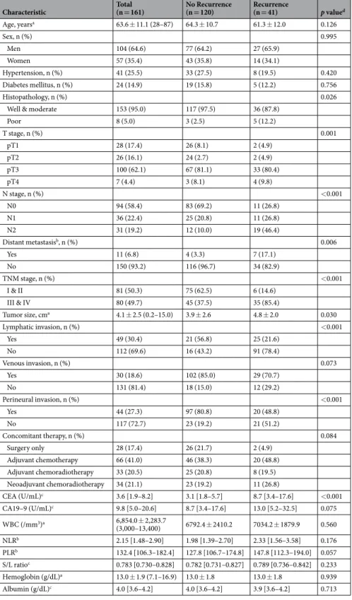
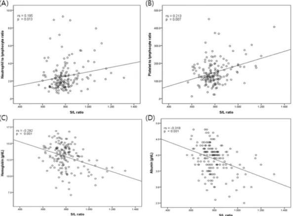
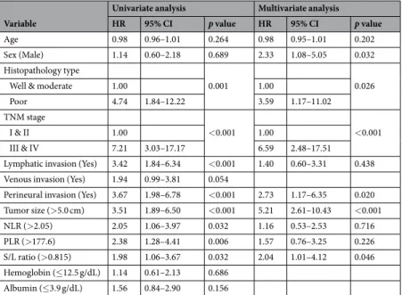
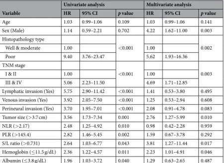
To evaluate the association between diffuse splenic FDG uptake and systemic inflammatory markers, we assessed S/L ratio, WBC, NLR, and PLR. In addition, we also assessed albumin and hemoglobin (Table 2). We found that S/L ratio was pos-itively correlated with NLR (rs = 0.195, P = 0.013, Fig. 2A) and PLR (rs = 0.213, P = 0.007, Fig. 2B). In addition,a negative correlation was observed between the S/L ratio and hemoglobin (rs = −0.292, P < 0.001, Fig. 2C) and
albumin (rs = −0.318, P < 0.001, Fig. 2D).
Clinical factors predicting recurrence during follow-up.
To evaluate the association of clinical fea-tures, systemic inflammatory markers and diffuse splenic FDG uptake with recurrence, Cox proportional hazards regression analyses were performed (Table 3). The optimal cutoff values determined by the ROC curve analysis were 5.0 cm for tumor size, 2.05 for NLR, 177.6 for PLR, 0.815 for S/L ratio, 12.5 g/dL for hemoglobin, and 3.9 g/ dL for albumin for the RFS.In the univariate analysis, histopathologic differentiation (poor vs well & moderate, P = 0.001), TNM stage (III & IV vs I & II, P < 0 0.001), lymphatic invasion (P < 0.001), perineural invasion (P < 0.001), tumor size (P < 0.001), NLR (P = 0.032), PLR (P = 0.006), and S/L ratio (P = 0.032) were significantly associated with the prognosis of recurrence during follow-up. In particular, on the basis of the Kaplan–Meier survival analysis, patients with S/L ratio ≤0.815 had significantly higher 2-year RFS rates (84.3% vs 77.7%) than did patients with S/L ratio >0.815 (P = 0.028, Fig. 3A). Multivariate analysis was subsequently carried out including variables with statistical significance in univariate analysis. Male gender (hazard ratio [HR]: 2.33, 95% confidence interval [CI]: 1.08–5.05, P = 0.032), histopathologic differentiation (poor vs well & moderate, HR: 3.59, 95% CI: 1.17–11.02,
P = 0.026), TNM stage (III & IV vs I & II, HR: 6.59, 95% CI: 2.48–17.51, P < 0.001), perineural invasion (HR:
2.73, 95% CI: 1.17–6.35, P = 0.020), tumor size (>5.0 cm, HR: 5.21, 95% CI: 2.61–10.43, P < 0.001), and S/L ratio (>0.815, HR: 2.04, 95% CI: 1.01–4.12, P = 0.046) were independent prognostic factors for recurrence (Table 3).
Clinical factors predicting mortality during follow-up.
The optimal cutoff values determined by the ROC curve analysis were 3.7 cm for tumor size, 2.17 for NLR, 145.4 for PLR, 0.731 for S/L ratio, 11.5 g/dL for hemoglobin, and 3.8 g/dL for albumin for the OS.In the univariate analysis, histopathologic differentiation (P < 0.001), TNM stage (P < 0.001), lymphatic invasion (P < 0.001), venous invasion (P < 0.001), perineural invasion (P < 0.001), tumor size (P = 0.001), NLR (P = 0.010), PLR (P = 0.002), S/L ratio (P = 0.043), hemoglobin (P = 0.011), and albumin (P = 0.040) were sig-nificantly associated with the prognosis of death during follow-up. Patients with S/L ratio ≤0.731 had signifi-cantly higher 2-year OS rates (94.8% vs 92.0%) than did patients with S/L ratio >0.731 (P = 0.036; Fig. 3B). The multivariate analysis including significant variables revealed that male gender (HR: 4.22, 95% CI: 1.62–11.00,
P = 0.003), histopathology (poor vs well & moderate, HR: 5.62, 95% CI: 1.93–16.36, P = 0.002), TNM stage (III
& IV vs I & II, HR: 4.69, 95% CI: 1.71–12.85, P = 0.003), tumor size (>3.7 cm, HR: 2.76, 95% CI: 1.27–5.99,
P = 0.010), S/L ratio (>0.731, HR: 3.81, 95% CI: 1.27–11.44, P = 0.017), and hemoglobin (≤11.5, HR: 2.23, 95%
Characteristic Total(n = 161) No Recurrence(n = 120) Recurrence(n = 41) p valued Age, yearsa 63.6 ± 11.1 (28–87) 64.3 ± 10.7 61.3 ± 12.0 0.126 Sex, n (%) 0.995 Men 104 (64.6) 77 (64.2) 27 (65.9) Women 57 (35.4) 43 (35.8) 14 (34.1) Hypertension, n (%) 41 (25.5) 33 (27.5) 8 (19.5) 0.420 Diabetes mellitus, n (%) 24 (14.9) 19 (15.8) 5 (12.2) 0.756 Histopathology, n (%) 0.026
Well & moderate 153 (95.0) 117 (97.5) 36 (87.8)
Poor 8 (5.0) 3 (2.5) 5 (12.2) T stage, n (%) 0.001 pT1 28 (17.4) 26 (8.1) 2 (4.9) pT2 26 (16.1) 24 (2.7) 2 (4.9) pT3 100 (62.1) 67 (81.1) 33 (80.4) pT4 7 (4.4) 3 (8.1) 4 (9.8) N stage, n (%) <0.001 N0 94 (58.4) 83 (69.2) 11 (26.8) N1 36 (22.4) 25 (20.8) 11 (26.8) N2 31 (19.2) 12 (10.0) 19 (46.4) Distant metastasisb, n (%) 0.006 Yes 11 (6.8) 4 (3.3) 7 (17.1) No 150 (93.2) 116 (96.7) 34 (82.9) TNM stage, n (%) <0.001 I & II 81 (50.3) 75 (62.5) 6 (14.6) III & IV 80 (49.7) 45 (37.5) 35 (85.4) Tumor size, cma 4.1 ± 2.5 (0.2–15.0) 3.9 ± 2.6 4.8 ± 2.0 0.030 Lymphatic invasion, n (%) <0.001 Yes 49 (30.4) 21 (56.8) 25 (21.6) No 112 (69.6) 16 (43.2) 91 (78.4) Venous invasion, n (%) 0.073 Yes 30 (18.6) 102 (85.0) 29 (70.7) No 131 (81.4) 18 (15.0) 12 (29.2) Perineural invasion, n (%) <0.001 Yes 44 (27.3) 97 (80.8) 20 (48.8) No 117 (72.7) 23 (19.2) 21 (51.2) Concomitant therapy, n (%) 0.084 Surgery only 28 (17.4) 26 (21.7) 2 (4.9) Adjuvant chemotherapy 66 (41.0) 46 (38.3) 20 (48.8) Adjuvant chemoradiotherapy 33 (20.5) 25 (20.8) 8 (19.5) Neoadjuvant chemoradiotherapy 34 (21.1) 23 (19.2) 11 (26.8) CEA (U/mL)c 3.6 [1.9–8.2] 3.1 [1.8–5.7] 8.7 [3.4–17.6] <0.001 CA19–9 (U/mL)c 9.8 [5.0–20.6] 8.7 [3.4–17.6] 13.0 [5.2–32.5] 0.075 WBC (/mm3)a 6,854.0 ± 2,283.7 (3,000–13,400) 6792.4 ± 2410.2 7034.2 ± 1879.9 0.560 NLRb 2.15 [1.48–2.90] 1.98 [1.39–2.70] 2.33 [1.56–3.58] 0.176 PLRb 132.4 [106.3–182.4] 127.8 [106.7–174.8] 147.8 [112.3–194.0] 0.057 S/L ratioc 0.783 [0.730–0.828] 0.782 [0.731–0.827] 0.789 [0.736–0.842] 0.233 Hemoglobin (g/dL)a 13.0 ± 1.9 (7.1–16.9) 13.0 ± 1.8 13.0 ± 1.8 0.939 Albumin (g/dL)c 4.0 [3.6–4.2] 4.0 [3.6–4.2] 3.9 [3.6–4.2] 0.713
Table 1. Clinical characteristics of the study subjects. Abbreviations: CEA, carcinoembryonic antigen; CA19-9,
carbohydrate antigen 19-9; WBC, White Blood Cells; NLR, neutrophil-to-lymphocyte ratio; PLR, platelet-to-lymphocyte ratio; S/L ratio, spleen-to-liver mean standardized uptake ratio; TNM, tumor–node–metastasis
aNormally distributed variables are expressed as mean ± standard deviation (range). bAmong the 11 patients
with distant metastasis, the single liver metastasis was noted in 5 patients, single or multiple lung metastases in 4 patients, and ovary metastasis in 2 patients. cNon-normally distributed variables are expressed as median
Discussion
In the present study, we found a notable association between S/L ratio and systemic inflammatory markers existed. S/L ratio, NLR, and PLR were significantly associated with recurrence and death in the univariate anal-ysis, while only S/L ratio was an independent prognostic factor for recurrence and death in patients with rectal cancer after curative surgical resection. Several studies have reported that the degree of splenic FDG uptake is related to inflammation and hematological parameters19,21–23,25. Meanwhile, we considered that the spleen SUV
itself would not be appropriate parameter because it can be influenced by various biological and technical fac-tors26. Instead, we calculated the S/L ratio by dividing the spleen SUV
mean by the liver SUVmean, which is widely
used as an internal reference. The hepatic FDG uptake is known as an useful internal reference in clinical diag-nostic settings due to its homogeneous values8,9,27.
In our study, S/L ratio was significantly correlated with NLR, PLR, and hemoglobin as well as albumin. Previous studies have revealed that a significant positive associations of S/L ratio with systemic inflammatory markers such as WBC, neutrophil and CRP were present in patients diagnosed with lung cancer and cholangio-carcinoma21,28,29. According prior investigations, presumably, diffusely increased splenic uptake FDG is
consid-ered to be a systemic inflammatory response-related phenomenon28–31. The spleen consists of red pulp and white
pulp, which are separated by the marginal zone containing macrophages and marginal zone B cells32,33. The white
pulp is involved in adaptive immunity and the marginal zone is involved in both innate and adaptive immunity12.
An increased splenic FDG uptake can indicate the activation of immune systems in the spleen and can reflect the presence of active systemic inflammation34–37. Inflammation has powerful effect on cancer promoters, facilitating
genomic instability, and stimulating angiogenesis14,38,39. Among inflammatory cells, neutrophils can contribute
to cancer promoters and cancer-activated platelets can drive cancer progression and invasion in rectal cancer, whereas lymphocytes can act on cancer-killing responses40–42. Albumin is also related to the cancer cell-induced
inflammatory reaction43. The level of albumin, high NLR, and high PLR have already been reported as systemic
NLR PLR WBC Hemoglobin Albumin
S/L ratio rs = 0.195
p = 0.013 rp = 0.007s = 0.213 p = 0.253rs = 0.091 rp < 0.001s = −0.292 rp < 0.001s = −0.318
Table 2. Correlation between Fluorodeoxyglucose uptake of spleen-to-liver and systemic inflammatory and
hematologic markers. Abbreviations: rs, Spearman correlation coefficients; NLR, neutrophil-to-lymphocyte
ratio; PLR, platelet-to-lymphocyte ratio; WBC, White Blood Cells; S/L ratio, spleen-to-liver mean standardized uptake ratio.
Figure 2. A scatter plot showing the correlations between S/L ratio and hematological and inflammatory
markers. S/L ratio, spleen-to-liver mean standardized uptake value; rs, Spearman correlation coefficient (A)
inflammatory markers39,44,45. Similarly to 18F- FDG uptake of the spleen, 18F-FDG uptake of bone marrow (BM)
in patients with cancer has been reported to reflect the degree of systemic inflammation19. In that study, BM SUV
was positively associated with systemic inflammatory markers including NLR, PLR, and CRP19,31,35. Further, BM
was found to be an independent predictor for recurrence in non-small cell lung cancer, lymphoma, and colorectal cancer10,23,31,46.
In this study, we also identified the prognostic value of S/L ratio for predicting recurrence and death in rectal cancer. Regarding this issue, 18F-FDG uptake of the spleen has been already found to be significantly associated
with death in patients with unresectable cholangiocarcinoma29. Furthermore, we reported previously that
dif-fuse splenic 18F-FDG uptake is significantly related to the RFS and OS of patients with gastric cancer25. In the
current study, univariate analyses demonstrated that S/L ratio, NLR, and PLR were significantly associated with recurrence and death. Among them, only S/L ratio was independently predictive of recurrence and death in multivariate analysis. This suggests that S/L ratio can be more helpful in the prediction of clinical outcome as compared with other systemic inflammatory markers. Patients with a high S/L ratio showed worse RFS and OS than did those with a low S/L ratio. Cancer recurrence occurred in 22.3% of patients with a high S/L ratio within 2-years after surgery, whereas it developed in 15.7% of those with a low S/L ratio. Similarly, 8.0% of patients with a high S/L ratio died within 2-years of surgical resection, whereas 5.2% of patients with a low S/L ratio died within that period. We thus hypothesize that among patients with rectal cancer, a higher S/L ratio is predictive of poor
Variable
Univariate analysis Multivariate analysis
HR 95% CI p value HR 95% CI p value
Age 0.98 0.96–1.01 0.264 0.98 0.95–1.01 0.202
Sex (Male) 1.14 0.60–2.18 0.689 2.33 1.08–5.05 0.032
Histopathology type
0.001 0.026
Well & moderate 1.00 1.00
Poor 4.74 1.84–12.22 3.59 1.17–11.02
TNM stage
<0.001 <0.001
I & II 1.00 1.00
III & IV 7.21 3.03–17.17 6.59 2.48–17.51 Lymphatic invasion (Yes) 3.42 1.84–6.34 <0.001 1.40 0.60–3.31 0.438 Venous invasion (Yes) 1.94 0.99–3.81 0.054
Perineural invasion (Yes) 3.67 1.98–6.78 <0.001 2.73 1.17–6.35 0.020 Tumor size (>5.0 cm) 3.51 1.89–6.50 <0.001 5.21 2.61–10.43 <0.001 NLR (>2.05) 2.05 1.06–3.97 0.032 1.16 0.53–2.53 0.716 PLR (>177.6) 2.38 1.28–4.41 0.006 1.57 0.76–3.25 0.226 S/L ratio (>0.815) 1.98 1.06–3.67 0.032 2.04 1.01–4.12 0.046 Hemoglobin (≤12.5 g/dL) 1.14 0.61–2.13 0.686 Albumin (≤3.9 g/dL) 1.56 0.84–2.90 0.156
Table 3. Clinical factors associated with the recurrence during follow-up. Abbreviations: HR, hazard ratio; CI,
confidence interval; TNM, tumor–node–metastasis; NLR, neutrophil-to-lymphocyte ratio; PLR, platelet-to-lymphocyte ratio; S/L ratio, spleen-to-liver mean standardized uptake ratio.
Figure 3. Kaplan-Meier analysis (A) Recurrence free survival of patients with rectal cancer in S/L ratio and (B)
prognosis after curative surgical resection. Followed by previous study for gastric cancer25, this suggests that
dif-fuse splenic FDG uptake, using hepatic FDG uptake as internal reference, has prognostic utility in patients with cancer of the gastrointestinal tract.
In addition, we found a significant negative correlation between the hemoglobin level and the S/L ratio. Therefore, anemia is predictive of death in patients with rectal cancer. Few studies have reported that anemia is associated with diffuse splenic FDG uptake on PET/CT in cancer patients21,22,28. We assume that concurrent
anemia during PET/CT study may be one of the causes of diffuse splenic FDG uptake. This assumption can be explained by the fact that spleen is a secondary hematopoietic organ12,32,33. Thus, when diffuse splenic FDG
uptake is observed on clinical PET/CT study, the degree of anemia in patients with rectal cancer who underwent curative surgical resection should be checked for correct interpretation.
The present study has several limitations. First, the number of the study subjects was relatively small, thus weakening the strength of our results. Further larger scaled studies are needed to clarify the implications of dif-fuse splenic FDG uptake. Second, to homogenize the patient population, we included 161 rectal cancer patients who underwent FDG PET/CT scan for staging and who had hematological data within 10 days before or after PET/CT scan. We excluded patients with colon cancer in order to avoid heterogeneity in the sample population, which might have led to selection bias. Third, diffuse splenic FDG uptake may be influenced by other factors, particularly hemoglobin level. Because anemia stimulates hematopoiesis, hemoglobin level can be associated with FDG uptake of the spleen12,32. Fourth, we did not consider pre- or post-operative chemotherapy or radiotherapy,
which may affect the prognosis of patients with rectal cancer. Fifth, hepatic micrometastasis, which cannot be detected by PET or PET/CT, might bias the liver SUVmean data, despite exclusion of patients with multiple or huge
hepatic metastases. In patients with a single hepatic metastasis, the liver SUVmean was measured in ROIs that did
not encompass metastatic lesions. Sixth, the use of two different PET system, a stand-alone PET and PET/CT, is a major limitation. Though the SUV is the most common method in PET studies, using the parameter is not appro-priate in our study because technologic factors from different scanners are known to affect SUV measurement. Thus, we used the SUV ratio (S/L ratio) rather than the SUV itself. Last, information on serum CRP results is needed to confirm the correlations between diffuse splenic FDG uptake and the systemic inflammatory markers. The level of CRP, an important acute-phase protein, provides a more accurate estimate of the inflammatory status than other systemic markers of inflammation16,19,21,35,45. In our hospital the CRP level is not evaluated during the
preoperative work-up of patients with rectal cancer not suspected to have infection or inflammation. The patients in this study did not have infection or inflammation and, thus, did not undergo measurement of the CRP level.
In conclusion, S/L ratio on FDG PET/CT imaging was significantly correlated with systemic inflammatory markers. Moreover, S/L ratio was an independent prognostic factor for the prediction of recurrence or death in rectal cancer during follow-up after curative surgery. Accordingly, we suggest that physicians need to pay par-ticular attention to patients with a high S/L ratio among those with rectal cancer who undergo curative surgical resection.
References
1. Briggs, R., Chowdhury, F., Lodge, J. & Scarsbrook, A. Clinical impact of FDG PET-CT in patients with potentially operable metastatic colorectal cancer. Clinical radiology 66, 1167–1174 (2011).
2. Ye, Y. et al. Pre-operative TNM staging of primary colorectal cancer by 18F-FDG PET-CT or PET: a meta-analysis including 2283 patients. International journal of clinical and experimental medicine 8, 21773–21785 (2015).
Variable
Univariate analysis Multivariate analysis
HR 95% CI p value HR 95% CI p value
Age 1.03 0.99–1.06 0.109 1.03 0.99–1.06 0.141
Sex (Male) 1.14 0.59–2.21 0.702 4.22 1.62–11.00 0.003
Histopathology type
<0.001 0.002
Well & moderate 1.00 1.00
Poor 9.40 3.76–23.47 5.62 1.93–16.36
TNM stage
<0.001 0.003
I & II 1.00 1.00
III & IV 5.06 2.23–11.50 4.69 1.71–12.85
Lymphatic invasion (Yes) 5.75 2.90–11.42 <0.001 1.41 0.53–3.80 0.495 Venous invasion (Yes) 3.92 2.05–7.50 <0.001 1.25 0.53–2.94 0.608 Perineural invasion (Yes) 3.70 1.95–7.01 <0.001 2.08 0.91–4.78 0.083 Tumor size (>3.7 cm) 3.56 1.73–7.34 0.001 2.76 1.27–5.99 0.010 NLR (>2.17) 2.48 1.25–4.92 0.010 0.98 0.42–2.28 0.959 PLR (>145.4) 2.82 1.46–5.45 0.002 1.59 0.67–3.78 0.292 S/L ratio (>0.731) 2.64 1.03–6.77 0.043 3.81 1.27–11.44 0.017 Hemoglobin (≤11.5 g/dL) 2.36 1.22–4.57 0.011 2.23 1.01–4.91 0.046 Albumin (≤3.8 g/dL) 1.96 1.03–3.72 0.040 1.29 0.63–2.65 0.487
Table 4. Clinical factors associated with the death during follow-up. Abbreviations: HR, hazard ratio; CI,
confidence interval; TNM, tumor–node–metastasis; NLR, neutrophil-to-lymphocyte ratio; PLR, platelet-to-lymphocyte ratio; S/L ratio, spleen-to-liver mean standardized uptake ratio.
3. de Geus-Oei, L.-F., Vriens, D., van Laarhoven, H. W., van der Graaf, W. T. & Oyen, W. J. Monitoring and predicting response to therapy with 18F-FDG PET in colorectal cancer: a systematic review. Journal of Nuclear Medicine 50, 43S–54S (2009).
4. Byun, B. H. et al. Prognostic value of 18 F-FDG uptake by regional lymph nodes on pretreatment PET/CT in patients with resectable colorectal cancer. European journal of nuclear medicine and molecular imaging 41, 2203–2211 (2014).
5. Bang, J.-I. et al. Comparison of quantitative methods on FDG PET/CT for treatment response evaluation of metastatic colorectal cancer. Nuclear medicine and molecular imaging 51, 147–153 (2017).
6. Tomas, M., Tronco, G., Karayalcin, G. & Palestro, C. 22. FDG uptake in infectious mononucleosis. Clinical Positron Imaging 3, 176 (2000).
7. Scharko, A. M. et al. Whole-body positron emission tomography in patients with HIV-1 infection. The Lancet 362, 959–961 (2003). 8. Meier, J. M. et al. Assessment of age-related changes in abdominal organ structure and function with computed tomography and
positron emission tomography. Seminars in Nuclear Medicine 37, 154–172 (2007).
9. Zasadny, K. R. & Wahl, R. L. Standardized uptake values of normal tissues at PET with 2-[fluorine-18]-fluoro-2-deoxy-D-glucose: variations with body weight and a method for correction. Radiology 189, 847–850 (1993).
10. Rini, J. N. et al. F-18 FDG versus Ga-67 for detecting splenic involvement in Hodgkin’s disease. Clinical nuclear medicine 27, 572–577 (2002).
11. Metser, U. et al. Solid splenic masses: evaluation with 18F-FDG PET/CT. Journal of Nuclear Medicine 46, 52–59 (2005). 12. Mebius, R. E. & Kraal, G. Structure and function of the spleen. Nature Reviews Immunology 5, 606–616 (2005).
13. Elinav, E. et al. Inflammation-induced cancer: crosstalk between tumours, immune cells and microorganisms. Nature Reviews
Cancer 13, 759–771 (2013).
14. Coussens, L. M. & Werb, Z. Inflammation and cancer. Nature 420, 860–867 (2002).
15. Seong, M.-K. Prognostic inflammation score in surgical patients with colorectal cancer. Journal of Korean medical science 30, 1793–1799 (2015).
16. Køstner, A. H. et al. The prognostic role of systemic inflammation in patients undergoing resection of colorectal liver metastases: C reactive protein (CRP) is a strong negative prognostic biomarker. Journal of surgical oncology 114, 895–899 (2016).
17. Kwon, H.-C. et al. Clinical significance of preoperative neutrophil-lymphocyte versus platelet-lymphocyte ratio in patients with operable colorectal cancer. Biomarkers 17, 216–222 (2012).
18. Cesta, M. F. Normal structure, function, and histology of mucosa-associated lymphoid tissue. Toxicologic pathology 34, 599–608 (2006).
19. Nunez, R. et al. Correlation of hematologic parameters with bone marrow and spleen uptake in FDG PET. Revista espanola de
medicina nuclear 24, 107–112 (2005).
20. Dicato, M., Plawny, L. & Diederich, M. Anemia in cancer. Annals of Oncology 21, vii167–vii172 (2010).
21. Nam, H.-Y. et al. The clinical implication and prediction of diffuse splenic FDG uptake during cancer surveillance. Clinical nuclear
medicine 35, 759–763 (2010).
22. Pak, K. et al. Impact of cytokines on diffuse splenic 18F-fluorodeoxyglucose uptake during positron emission tomography/computed tomography. Nuclear medicine communications 34, 64–70 (2013).
23. Salaun, P. Y. et al. Analysis of 18 F-FDG PET diffuse bone marrow uptake and splenic uptake in staging of Hodgkin’s lymphoma: a reflection of disease infiltration or just inflammation? European journal of nuclear medicine and molecular imaging 36, 1813–1821 (2009).
24. Edge, S. B. & Compton, C. C. The American Joint Committee on Cancer: the 7th edition of the AJCC cancer staging manual and the future of TNM. Annals of surgical oncology 17, 1471–1474 (2010).
25. Yoon, H.-J. et al. Prognostic value of diffuse splenic FDG uptake on PET/CT in patients with gastric cancer. PloS one 13, e0196110 (2018).
26. Thie, J. A. Understanding the standardized uptake value, its methods, and implications for usage. Journal of Nuclear Medicine 45, 1431–1434 (2004).
27. Paquet, N., Albert, A., Foidart, J. & Hustinx, R. Within-patient variability of 18F-FDG: standardized uptake values in normal tissues.
Journal of Nuclear Medicine 45, 784–788 (2004).
28. Kim, K. et al. Factors associated with diffusely increased splenic F-18 FDG uptake in patients with cholangiocarcinoma. Nuclear
medicine and molecular imaging 48, 137–143 (2014).
29. Pak, K. et al. Splenic FDG uptake predicts poor prognosis in patients with unresectable cholangiocarcinoma. Nuklearmedizin 53, 26–31 (2014).
30. Ryuk, J. P. et al. Predictive factors and the prognosis of recurrence of colorectal cancer within 2 years after curative resection. Annals
of surgical treatment and research 86, 143–151 (2014).
31. Lee, J. W., Na, J. O., Kang, D.-Y., Lee, S. Y. & Lee, S. M. Prognostic Significance of FDG Uptake of Bone Marrow on PET/CT in Patients With Non–Small-Cell Lung Cancer After Curative Surgical Resection. Clinical lung cancer 18, 198–206 (2017).
32. Macneal, W. J. The circulation of blood through the spleen pulp. American Medical Association 7, 215–227 (1929). 33. Bronte, V. & Pittet, M. J. The spleen in local and systemic regulation of immunity. Immunity 39, 806–818 (2013).
34. Love, C., Tomas, M. B., Tronco, G. G. & Palestro, C. J. FDG PET of infection and inflammation. Radiographics 25, 1357–1368 (2005). 35. Murata, Y. et al. Correlations between 18F-FDG uptake by bone marrow and hematological parameters: measurements by PET/CT.
Nuclear medicine and biology 33, 999–1004 (2006).
36. Aktas, G. E., Sarikaya, A. & Demir, S. S. Diffusely Increased Splenic Fluorodeoxyglucose Uptake in Lung Cancer Patients. Turk
Toraks Dergisi 18, 6–10 (2017).
37. Liu, Y. Clinical significance of diffusely increased splenic uptake on FDG-PET. Nuclear medicine communications 30, 763–769 (2009).
38. Hanahan, D. & Weinberg, R. A. Hallmarks of cancer: the next generation. cell 144, 646–674 (2011).
39. Xu, Z. et al. The prognostic role of the platelet-lymphocytes ratio in gastric cancer: a meta-analysis. PloS one 11, e0163719 (2016). 40. Grivennikov, S. I., Greten, F. R. & Karin, M. Immunity, inflammation, and cancer. Cell 140, 883–899 (2010).
41. Piccard, H., Muschel, R. & Opdenakker, G. On the dual roles and polarized phenotypes of neutrophils in tumor development and progression. Critical reviews in oncology/hematology 82, 296–309 (2012).
42. Yan, M. & Jurasz, P. The role of platelets in the tumor microenvironment: from solid tumors to leukemia. Biochimica et Biophysica
Acta (BBA)-Molecular Cell Research 1863, 392–400 (2016).
43. Roxburgh, C. S. & McMillan, D. C. Role of systemic inflammatory response in predicting survival in patients with primary operable cancer. Future oncology 6, 149–163 (2010).
44. Hu, Z.-D. et al. Prognostic value of neutrophil to lymphocyte ratio for gastric cancer. Annals of translational medicine 3, 50 (2015). 45. Peng, B., Wang, Y.-H., Liu, Y.-M. & Ma, L.-X. Prognostic significance of the neutrophil to lymphocyte ratio in patients with
non-small cell lung cancer: a systemic review and meta-analysis. International journal of clinical and experimental medicine 8, 3098–3106 (2015).
46. Lee, J. W., Baek, M.-J., Ahn, T. S. & Lee, S. M. Fluorine-18-fluorodeoxyglucose uptake of bone marrow on PET/CT can predict prognosis in patients with colorectal cancer after curative surgical resection. European journal of gastroenterology & hepatology 30, 187–194 (2018).
Acknowledgements
This work was supported by the National Research Foundation of Korea (NRF) grant funded by the Korea government (MSIT) (2010-0027945, Chang Mo Moon) and was supported by Basic Science Research Program through the National Research Foundation of Korea (NRF), funded by the Ministry of Education (2014R1A1A2058325 & 2017R1D1A1B03035311, Chang Mo Moon; 2018R1D1A1B07045321, Bom Sahn Kim; 2018R1D1A1B07049400, Hai-Jeon Yoon).
Author Contributions
C.M. Moon and B.S. Kim supervised this study. S.Y. Kim, H.J. Yoon, B.S. Kim, J.Y. Lim, T.O. Kim, A.R. Choe, C.H. Tae, S.E. Kim, H.K. Jung, K.N. Shim, and S.-A. Jung collected the data. S.Y. Kim, H.J. Yoon, C.M. Moon, and B.S. Kim conducted the analysis and interpreted the data. S.Y. Kim drafted the manuscript. C.M. Moon and B.S. Kim critically revised the manuscript. All of the authors contributed to the manuscript revision.
Additional Information
Competing Interests: The authors declare no competing interests.
Publisher’s note: Springer Nature remains neutral with regard to jurisdictional claims in published maps and
institutional affiliations.
Open Access This article is licensed under a Creative Commons Attribution 4.0 International
License, which permits use, sharing, adaptation, distribution and reproduction in any medium or format, as long as you give appropriate credit to the original author(s) and the source, provide a link to the Cre-ative Commons license, and indicate if changes were made. The images or other third party material in this article are included in the article’s Creative Commons license, unless indicated otherwise in a credit line to the material. If material is not included in the article’s Creative Commons license and your intended use is not per-mitted by statutory regulation or exceeds the perper-mitted use, you will need to obtain permission directly from the copyright holder. To view a copy of this license, visit http://creativecommons.org/licenses/by/4.0/.