CASE REPORT
Acinar cell carcinoma with fatty change arising from the pancreas
1W-S CHUNG,
MD
,
1M-S PARK,
MD,
2D W KIM,
MDand
1K W KIM,
MD1Department of Diagnostic Radiology, Institute of Gastroenterology, Research Institute of Radiological Science, and 2Department of Pathology, Severance Hospital, Yonsei University Healthcare System, Seoul, South Korea
ABSTRACT. Acinar cell carcinoma of the pancreas is a rare malignant tumour
developing from acinar cells, accounting for approximately 1% of pancreatic exocrine tumours. We experienced a case of an acinar cell carcinoma with fatty change. To the best of our knowledge, this is the first case report of an acinar cell carcinoma with fatty change in the clinical literature.
Received 3 January 2011 Revised 21 January 2011 Accepted 31 January 2011 DOI: 10.1259/bjr/15914752
’2011 The British Institute of Radiology
Acinar cell carcinoma (ACC) of the pancreas is a malignant tumour developing from acinar cells. It is a rare tumour accounting for approximately 1% of pan-creatic exocrine tumours. ACC with fatty change is extre-mely rare in the literature. This is a report on a 72-year-old female who was found to have ACC with fatty change.
Case report
A 72-year-old female underwent a partial gastrectomy with Billroth I for early gastric cancer in September 2009. When a pancreatic mass was found during the 6-month follow-up CT, the patient was admitted for further investigation.
Test results showed elevated levels of alpha-fetoprotein (AFP): 1945 IU ml21 (normal range: 0–7 IU ml21) and normal levels of cancer antigen 19-9: 8.5 U ml21(normal range: 0–37 U ml21) and carcinoembryonic antigen (CEA): 2.31 ng ml21(normal range: 0–5 ng ml21).
An abdominal ultrasound showed a 2.5-cm hypoechoic mass in the neck of the pancreas. The mass was ill-delimitated and heterogeneous. Contrast-enhanced CT showed a 2.5-cm mass in the neck of the pancreas with distal pancreatic duct dilatation and parenchymal atro-phy. The mass was adjacent to the portal vein and showed less intense enhancement than the pancreas. No dilatation of the bile ducts, focal hepatic lesions or abdominal lymphadenopathies was found. The initial differential diagnoses were pancreatic cancer and metastasis.
Positron emission tomography (PET) showed no significant increase in fluorodeoxyglucose (FDG) uptake. MRI showed a 2.5-cm hypovascular well-defined mass at the neck of the pancreas with distal pancreatic duct dilatation and parenchymal atrophy. This mass had low
signal intensity at T2 weighted imaging with fat sup-pression, and had a fat component at in-phase and out-of-phase images (Figure 1). The portal vein was com-pressed and displaced by the mass, which remained permeable without invasion.
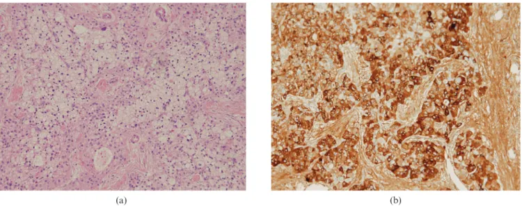
A Whipple procedure was performed on the patient. The specimen revealed an ill-defined lobulating mass of 1.961.7 cm, which was confined to the pancreas. Histo-logically, the mass showed an acinar pattern of cells with nucleoli (Figure 2). The tumour cells showed extensive and various degrees of clear cell change and microvesi-cular and macrovesimicrovesi-cular fatty change. The tumour cells showed strong and diffuse immunoreactivity for AFP and were negative for CD56, chromogranin A and synapto-physin. Pathological diagnosis was ACC with fatty change.
Discussion
ACC is an uncommon solid epithelial exocrine tumour. There are very few reports on this type of tumour. ACC typically occurs during the fifth to seventh decades of life, with a male predilection [1]. It can occur in any part of the pancreas, but the most common site is the head of the pancreas. A diagnosis of ACC can be made on the characteristic positive periodic acid–Schiff staining and immunohistochemical results.
The most common histological patterns seen in ACC are acinar and solid [2]. In a study by Klimstra et al [3], ACCs were positive for at least one immunohistochemical marker (i.e. trypsin, lipase, amylase, chymotrypsin, a1-antitrypsin, keratin, epithelial membrane antigen, carci-noembryonic antigen and AFP). ACCs, unlike endocrine cell tumours, are believed to originate from transformed acinar cells [2]. ACC with fatty change showed no evidence of an endocrine differentiation and diffuse immunoreactivity for AFP.
The imaging diagnosis of ACC varies owing to the rarity of the disease: a well-marginated, large, solid mass with a varied degree of cystic components; thin enhancing
Address correspondence to: Dr Mi-Suk Park, Department of Diagnostic Radiology, Institute of Gastroenterology, Research Institute of Radiological Science, Severance Hospital, Seodaemun-ku, Shinchon-dong 134, Seoul 120-752, Korea. E-mail: radpms@ yuhs.ac
The British Journal of Radiology, 84 (2011), e226–e228
(a) (b)
(c) (d)
Figure 1. A 72-year-old female. (a) Contrast-enhanced CT showing a mass in the neck of the pancreas, which had less intense enhancement than the pancreas. (b) Axial T2weighted MRI with fat suppression showed low signal intensity in this mass. (c)
In-phase and (d) out-of-In-phase MRI showing a fat component in this mass.
(a) (b)
Figure 2. Microscopic findings. (a) Pancreatic mass showing an acinar pattern of cells with nucleoli. The tumour cells with extensive and various degrees of clear cell change and fatty change (haematoxylin and eosin stain) (6100). (b) The tumour cells showed strong and diffuse immunoreactivity for alpha-fetoprotein (6200).
Case report: ACC with fatty change
capsule; occasional central calcification; intralesional haemorrhage and less intense enhancement than a normal pancreas on both CT and MRI [4, 5]. ACC is unique among pancreatic tumours as it is characterised by increased AFP levels. Our case also showed increased AFP levels and less intense enhancement than the pancreas on both CT and MRI.
Although ACC shows increased blood AFP levels and positive AFP stain, AFP can also be expressed by hepatoid carcinomas. The pathogenesis of hepatoid carcinomas of the pancreas is not fully understood. Paner et al [6] thought that the potentiality of hepatic differentiation may arise from any of the three main pancreatic cells (i.e. acinar, ductal and islet cells). AFP can also be expressed by pancreatic ductal carcinoma, acinar cell carcinoma, islet cell tumour and poorly differentiated pancreatic adenocarci-noma [7]. Therefore, the diagnosis of hepatoid carciadenocarci-noma is mainly provided by the cancer’s histological appearances on haematoxylin and eosin-stained material [8].
In our case, fat-suppressed MRI confirmed that the pancreatic mass contained a fat component. The pancreas is a very rare location for fat-containing tumours [9]. Differential diagnosis of fat-containing pancreatic tu-mours with direct invasion of pancreatic tissue should be mentioned (i.e. malignant fibrous histiocytoma, leiomyo-sarcoma, desmoids tumour, cystic teratoma, fibrolipoma, liposarcoma and lipoblastoma) [10–12]. However, as in our case, fat-containing tumours originating from the pancreas may be ACC with fatty change.
To our knowledge, this is the first reported case of ACC with fatty change. MRI is very important in detecting the fat component of tumours. ACC with fatty change must be considered in differential diagnosis of a fat-containing pancreatic tumour with elevated levels of AFP.
References
1. Holen KD, Klimstra DS, Hummer A, Gonen M, Conlon K, Brennan M, et al. Clinical characteristics and outcomes from an institutional series of acinar cell carcinoma of the pancreas and related tumors. J Clin Oncol 2002;20:4673–8. 2. Jena M, Shariff S, Jeyachandran P. Cystic variant of acinar
cell carcinoma of the pancreas presenting as pseudo-pancreatic cyst. Indian J Pathol Microbiol 2010;53:190–2. 3. Klimstra DS, Heffess CS, Oertel JE, Rosai J. Acinar cell
carcinoma of the pancreas. A clinicopathologic study of 28 cases. Am J Surg Pathol 1992;16:815–37.
4. Hsu MY, Pan KT, Chu SY, Hung CF, Wu RC, Tseng JH. CT and MRI features of acinar cell carcinoma of the pancreas with pathological correlations. Clin Radiol 2010;65:223–9. 5. Imamura M, Kimura Y, Ito H, Nobuoka T, Koito K, Sasaki A,
et al. Acinar cell carcinoma of the pancreas with intraductal growth: report of a case. Surg Today 2009;39:1006–9. 6. Paner GP, Thompson KS, Reyes CV. Hepatoid carcinoma of
the pancreas. Cancer 2000;88:1582–9.
7. Kawamoto S, Hiraoka T, Kanemitsu K, Kimura M, Miyauchi Y, Takeya M. Alpha-fetoprotein producing pancreatic cancer—a case report and review of 28 cases. Hepatogastro-enterology 1992;39:282–6.
8. Liu CZ, Hu SY, Wang L, Zhi XT, Jin B, Zhu M, et al. Hepatoid carcinoma of the pancreas: a case report. Chin Med J (Engl) 2007;120:1850–2.
9. Barutcu O, Cihangiroglu M, Yildirim T, Kayaselcuk F, Noyan T. Fat containing unusual tumor of the pancreas. Eur Radiol 2002;12:770–3.
10. Itai Y, Saida Y, Kurosaki Y, Kurosaki A, Fujimoto T. Focal fatty masses of the pancreas. Acta Radiol 1995;36:178–81. 11. Ferrozzi F, Zuccoli G, Bova D, Calculli L. Mesenchymal
tumors of the pancreas: CT findings. J Comput Assist Tomogr 2000;24:622–7.
12. Elliott TE, Albertazzi VJ, Danto LA. Pancreatic liposarcoma: case report with review of retroperitoneal liposarcomas. Cancer 1980;45:1720–3.
W-S Chung, M-S Park, D W Kim and K W Kim