Original Article
Copyright © 2020 The Korean Society of Plastic and Reconstructive Surgeons
This is an Open Access article distributed under the terms of the Creative Commons Attribution Non-Commercial License (https://creativecommons.org/
licenses/by-nc/4.0/) which permits unrestricted non-commercial use, distribution, and reproduction in any medium, provided the original work is properly cited. www.e-aps.org
Comparison of breast volume change between
oncoplastic breast-conserving surgery with
radiation therapy and a simultaneous contralateral
balancing procedure through the inverted-T scar
technique
Min Wook Kim
1, Won Seok Oh
1, Jae Woo Lee
1, Hyun Yul Kim
2, Youn Joo Jung
2, Ki Seok Choo
3,
Kyung Jin Nam
3, Seong Hwan Bae
1, Choongrak Kim
4, Su Bong Nam
1, Ji Hyeon Joo
51Department of Plastic and Reconstructive Surgery, Pusan National University School of Medicine, Yangsan; 2Department of Surgery, Pusan
National University Yangsan Hospital, Yangsan; 3Department of Radiology, Pusan National University School of Medicine, Yangsan; 4Department of Statistics, Pusan National University, Busan; 5Department of Radiation Oncology, Research Institute for Convergence of
Biomedical Science and Technology, Pusan National University Yangsan Hospital, Yangsan, Korea
Background Reduction mammoplasty or mastopexy is performed as an additional balancing procedure in patients with large or ptotic breasts who undergo breast-conserving surgery (BCS). Radiation therapy on breasts that have undergone surgery may result in changes in the volume. This study presents a comparative analysis of patients who received post-BCS bal-ancing procedures to determine whether volume changes were larger in breasts that received radiation therapy than on the contralateral side.
Methods Thirty-six participants were selected among patients who received BCS using the inverted-T scar technique between September 2012 and July 2017, were followed up for 2 or more years, and had pre-radiation therapy computed tomography images and post-radiation therapy images taken between 12 and 18 months after completion. The average age of the participants was 53.5 years, their average body mass index was 26.62 kg/m2.
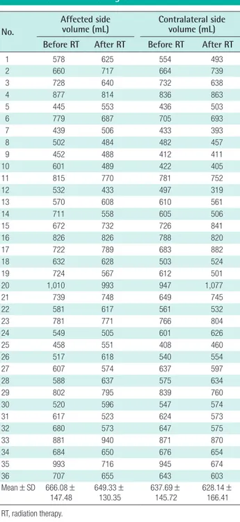
Results The pre- and post-radiation therapy volumes of the breasts receiving BCS were 666.08± 147.48 mL and 649.33± 130.35 mL, respectively. In the contralateral breasts, the vol-ume before radiation therapy was 637.69± 145.72 mL, which decreased to 628.14± 166.41 mL after therapy. The volume ratio of the affected to the contralateral breasts was 1.05± 0.10 before radiation therapy and 1.06± 0.12 after radiation therapy.
Conclusions The ratio of the volume between the two breasts immediately after surgery and at roughly 18 months postoperatively was not significantly different (P=0.98). For these reasons, we recommend a simultaneous single-stage balancing procedure as a reasonable option for pa-tients who require radiation therapy after BCS without concerns regarding volume change.
Keywords Mammaplasty / Breast reconstruction / Radiotherapy
Correspondence: Su Bong Nam Department of Plastic and Reconstructive Surgery, Pusan National University Yangsan Hospital, Pusan National University School of Medicine, 20 Geumo-ro, Mulgeum-eup, Yangsan 50612, Korea Tel: +82-55-360-1439 Fax: +82-55-360-1154 E-mail: subong71@hanmail.net Ji Hyeon Joo
Department of Radiation Oncology, Pusan National University Yangsan Hospital, 20 Geumo-ro, Mulgeum-eup, Yangsan 50612, Korea
Tel: +82-55-360-3454 Fax: +82-55-360-3449 E-mail: hi_juji@daum.net
This work was supported by a 2-year Research Grant of Pusan National University.
Received: June 15, 2020 • Revised: September 23, 2020 • Accepted: October 13, 2020
INTRODUCTION
The number of patients receiving breast-conserving surgery (BCS) is on the rise, as BCS plays a crucial role in aesthetic breast reconstruction and the early detection of breast cancer is becoming more common [1-3]. The inverted-T scar technique, a type of BCS, is used for ptotic or large breasts in procedures involving reconstruction of the abnormal breast and simultane-ous mastopexy or reduction mammoplasty on the contralateral side as a balancing procedure to obtain aesthetically symmetric results.
The contralateral balancing procedure can be either done si-multaneously with the reconstruction procedure or as a second-ary procedure after reconstruction [1,2,4]. Carrying out the bal-ancing procedure simultaneously with the reconstruction pro-cedure may generally increase the risk of asymmetric breasts compared to performing it as a delayed secondary procedure. However, previous research, including a study conducted by Smith et al. [1], reported good aesthetic results without addi-tional procedures when balancing was performed immediately.
BCS requires adjuvant radiation therapy, which can cause changes in breast volume. In order to achieve symmetry be-tween two breasts with an immediate balancing procedure, the normal breast should be reconstructed to be slightly smaller considering the potential for volume contraction after radiation therapy [5]. Very few studies have reported volume changes us-ing objective measures [5]. Against this background, this study aimed to provide a reference for determining the extent of exci-sion on the normal-side breast by comparing volume changes in breasts that received radiation therapy with those in the contra-lateral breasts among patients who received an immediate bal-ancing procedure using the inverted-T scar technique.
METHODS
Participants
In total, 48 patients received BCS using the inverted-T scar tech-nique for the abnormal breast accompanied by a simultaneous balancing procedure using the same method between Septem-ber 2012 and July 2017. Among them, 36 participants were se-lected, with the exclusion of patients who received an extended latissimus dorsi flap, implant, or acellular dermal matrix or pa-tients who did not undergo a follow-up computed tomography (CT) scan (Siemens, Munich, Germany) within 2 years postop-eratively. The average age of the patients was 53.5 years, and their average body mass index was 26.62 kg/m2. The purpose of the balancing procedure was reduction mammoplasty in 35 cas-es and mastopexy in the remaining case. The excised weight for
reduction was between 35 g and 675 g, except for the masto-pexy case, and the excision volume between the two sides dif-fered by 0 to 200 g. In 12 patients, the difference was 50 g or higher due to asymmetry between both sides. All patients re-ceived adjuvant radiation therapy and 30 patients underwent chemotherapy prior to radiation therapy. Only patients who had CT scans taken both before radiation therapy and 12 to 18 months after completing radiation therapy were selected as par-ticipants. Since breast volume is susceptible to changes after the completion of radiation therapy in response to changes in eating habits, exercise, and hormone therapy, the effects of radiation therapy were confirmed by comparing the volume of both breasts before and after radiation therapy. The reconstruction procedures were performed by one surgeon (SBN) and the CT measurements of breast were confirmed by single radiologist (KSC).
Radiation therapy
Radiation therapy was performed after surgery and the comple-tion of systemic cancer treatment. Radiacomple-tion therapy was ad-ministered to whole breast with or without regional lymphatics, using a 4–15 MV X-ray and/or 6–16 MeV electron beam from a linear accelerator (Varian Medical Systems, Palo Alto, CA, USA). Total radiation doses of 4,500–5,080 cGy in fractions of 180–250 cGy were typically administered, and boost doses of 540–2,540 cGy were directed to the tumor bed.
Surgical method
Prior to surgery, the design was made with the patient in a sit-ting position using the inverted-T scar technique, and the final excision area was marked. Surgery was performed under general anesthesia. The breast surgeon performed partial mastectomy after excision along part of the design, and then performed a re-construction using the remaining breast tissue and a deepitheli-alized cutaneous flap to minimize uneven results. The non-le-sion side was restored with a focus on maintaining symmetry using the inverted-T scar technique and excising the same area as the other side, with a similar excision volume. For breasts that were already asymmetric before surgery, the initial correction was made at the preoperative design stage, followed by a sec-ondary correction during surgery with the patient in the sitting position by controlling the excision volume so that both sides were as close in size as possible. The weight of the removed tis-sue was measured with a digital scale.
Breast volume measurements
Breast volume was measured using CT images (Fig. 1). Patients were instructed to take a supine position with arms abducted
and a lead wire was attached on the body surface to double-check the boundaries of the breasts by appearance. No contrast medium was used, and images were taken at a 5-mm slice thick-ness including the entire chest area. Special software for radia-tion image analysis (MIM Maestro ver. 6.6; MIM Software Inc., Cleveland, OH, USA) was used for image registration to com-pare the images taken before and after surgery. All measure-ments were performed by a single radiation oncologist to mini-mize individual differences in measurements. On each CT set, breasts were delineated according to ESTRO (European Soci-ety of Therapeutic Radiology and Oncology) guideline and Danish guideline, with modification of cranial and caudal defi-nition to maintain objectivity. Following anatomical borders were used: (1) cranial, 5 cm above nipple; (2) caudal, 5 cm be-low nipple; (3) ventral, skin surface; (4) dorsal, anterior surface of major pectoral muscle or costae and intercostal muscles where no muscle exist; (5) medial, lateral to the edge of ster-num; or (6) lateral, anterior to the lateral thoracic artery.
Statistical analysis
The ratio between the volume of the breast on which BCS was performed and that of the contralateral breast was calculated from the pre-radiation therapy CT scan, and the same ratio was calculated from the CT scan taken between 12 and 18 months post-radiation therapy. The Shapiro-Wilk test was conducted to test whether these values had a normal distribution, and the pre-radiation therapy values (P= 0.07) and the post-therapy values
(P= 0.69) yielded P-values larger than 0.05. Therefore, both parametric (paired t-test) and non-parametric (Wilcoxon signed-rank test and the sign test) methods were used. SPSS version 21.0 (IBM Corp., Armonk, NY, USA) was used.
RESULTS
The p and post-radiation therapy volumes of the breasts re-ceiving BCS were 666.08± 147.48 mL and 649.33± 130.35 mL, respectively. In the contralateral breasts, the volume before radi-ation therapy was 637.69 ± 145.72 mL, which decreased to 628.14± 166.41 mL after therapy. When the breast affected by cancer was compared to the contralateral breast pre- and post-radiation therapy, it was observed that the volumes of both sides decreased on CT imaging after radiation therapy, showing sta-tistically significant changes (Pipsi=0.004, Pcont=0.002) (Table 1). However, no statistically significant change was found in the volume ratio of the affected to the contralateral breasts, which was 1.05± 0.10 before radiation therapy and 1.06± 0.12 after ra-diation therapy (Table 2). To verify these results statistically, the paired t-test (35 degrees of freedom, P=0.97), Wilcoxon signed-rank test (P=0.81), and sign test (P=0.61) were performed, and all the results indicated equivalence. The pre- to post-radiation therapy ratio was 1.03±0.14 on the affected side, and 1.03±0.15 on the contralateral side.
In the clinical results from patients with at least 2 years of fol-low-up, both patients and surgeons were satisfied with the breast
Fig. 1. Breast volume measurements using CT images
Example of breast volume measurements on simulation computed tomography (CT) of patient who underwent right breast-conserving surgery. A transverse slice is shown (A), with corresponding projection view (B). Blue, ipsilateral breast; cyan, contralateral breast; magenta, contralateral nipple.
shape in 33 cases (Figs. 2, 3). A slight asymmetry in breast shape resulting from a deformity following radiation therapy was ob-served in one case. Three patients with a contraction deformity presented with a thin residual skin flap immediately after mas-tectomy, but these three patients were not dissatisfied with the shape of the breast and did not request reoperation. In addition, one patient wanted correction of a dog ear, one patient request-ed scar correction, and one patient wantrequest-ed correction of nipple-areolar complex deviation. The post-radiation therapy volume was larger than that of the contralateral side in one case.
DISCUSSION
An important goal of breast reconstruction surgery is to achieve symmetry. However, since each individual has a unique breast shape, when performing reconstruction surgery, it is challenging to reconstruct a symmetric shape resulting in patient satisfac-tion. As it is thought to be a safe way of achieving symmetry, many surgeons still prefer to perform a balancing procedure on the contralateral side as a second-stage operation [2,4,6]. How-ever, secondary revision breast surgery imposes an economic
No.
Affected and contralateral
sides volume ratio Pre- to post-RT volume ratio Before RT After RT Affected side Contralateral side
1 1.04 1.27 0.92 1.12 2 0.99 0.97 0.92 0.90 3 0.99 1.00 1.14 1.15 4 1.05 0.94 1.08 0.97 5 1.02 1.10 0.80 0.87 6 1.10 0.99 1.13 1.02 7 1.01 1.29 0.87 1.10 8 1.04 1.06 1.04 1.05 9 1.10 1.19 0.93 1.00 10 1.42 1.21 1.23 1.04 11 1.04 1.02 1.06 1.04 12 1.07 1.36 1.23 1.56 13 0.93 1.08 0.94 1.09 14 1.18 1.10 1.27 1.20 15 0.93 0.87 0.92 0.86 16 1.05 1.01 1.00 0.96 17 1.06 0.89 0.92 0.77 18 1.26 1.20 1.01 0.96 19 1.18 1.13 1.28 1.22 20 1.07 0.92 1.02 0.88 21 1.14 1.00 0.99 0.87 22 1.04 1.16 0.94 1.05 23 1.02 0.96 1.01 0.95 24 0.91 0.81 1.09 0.96 25 1.12 1.20 0.83 0.89 26 0.96 1.12 0.84 0.97 27 0.95 0.96 1.06 1.07 28 1.02 1.00 0.92 0.91 29 0.96 1.05 1.01 1.10 30 0.95 1.04 0.87 0.95 31 0.99 0.91 1.18 1.09 32 1.05 1.00 1.19 1.13 33 1.01 1.08 0.94 1.00 34 1.01 0.99 1.05 1.03 35 1.05 1.06 1.39 1.40 36 1.10 1.09 1.08 1.07 Mean ± SD 1.05 ± 0.10 1.06 ± 0.12 1.03 ± 0.14 1.03 ± 0.15 RT, radiation therapy.
Table 2. Change in the volume ratio No.
Affected side
volume (mL) Contralateral side volume (mL)
Before RT After RT Before RT After RT
1 578 625 554 493 2 660 717 664 739 3 728 640 732 638 4 877 814 836 863 5 445 553 436 503 6 779 687 705 693 7 439 506 433 393 8 502 484 482 457 9 452 488 412 411 10 601 489 422 405 11 815 770 781 752 12 532 433 497 319 13 570 608 610 561 14 711 558 605 506 15 672 732 726 841 16 826 826 788 820 17 722 789 683 882 18 632 628 503 524 19 724 567 612 501 20 1,010 993 947 1,077 21 739 748 649 745 22 581 617 561 532 23 781 771 766 804 24 549 505 601 626 25 458 551 408 460 26 517 618 540 554 27 607 574 637 597 28 588 637 575 634 29 802 795 839 760 30 520 596 547 574 31 617 523 624 573 32 680 573 647 575 33 881 940 871 870 34 684 650 676 654 35 993 716 945 674 36 707 655 643 603 Mean ± SD 666.08 ± 147.48 649.33 ±130.35 637.69 ±145.72 628.14 ±166.41 RT, radiation therapy.
and psychological burden on the patient. In addition, it has been reported that the outcomes did not differ between balancing procedures performed simultaneously with the first breast re-construction and those performed as a staged operation in terms of minor surgical complications or major complications such as transfusion, deep vein thrombosis, and readmission.
The author performed oncoplastic surgery with the inverted-T scar technique for large, ptotic breasts eligible for BCS. inverted-The inverted-T scar technique has the advantage of a wide operative field, and the residual breast tissue or deepithelialized dermal tissue can be reconstructed by transposition in the desired di-rection with a relatively reasonable margin. Therefore, when the
balancing procedure is performed simultaneously, it allows the surgeon to accurately inspect the affected area on the breast un-dergoing oncoplastic surgery, and to utilize every flap available to create a breast with the most natural shape possible. At the same time, in the intraoperative field, a similar amount and range of excision are determined by comparing the size and shape of the original breast and the excision amount during sur-gery. Intraoperatively, the sizes of both breasts and the projec-tion and the degree of ptosis are adjusted and completed with a symmetric scar in the sitting position. However, radiation thera-py can trigger dermatitis, fibrosis, scar formation, and seroma buildup; thus, it has been reported that radiation therapy is
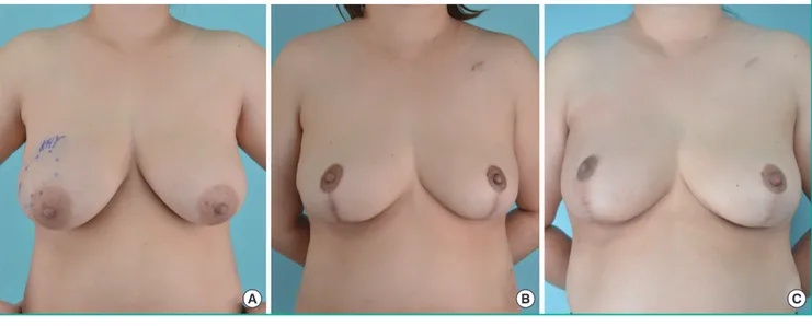
Fig. 2. Follow-up photographs after reconstruction
The patient was 34 years old and had right breast cancer (T2N0M0). Her body mass index was 27.7 kg/m2. The excised volume was 242 g on right
side and 248 g on left side. Photographs were taken at (A) pre-surgery, (B) pre-radiotherapy, and (C) 3-year post-radiotherapy.
A B C
Fig. 3. Follow-up photographs after reconstruction
The patient was 51 years old and had left breast cancer (T2N0M0). Her body mass index was 20.4 kg/m2. The excised volume was 119 g on right
side and 172 g on left side. Photographs were taken at (A) pre-surgery, (B) pre-radiotherapy, and (C) 3-year post-radiotherapy.
linked to unpredictable volume changes within 1 year after completion of radiation therapy, which affect the cosmetic out-comes of breast reconstruction [5,7,8]. Therefore, in order to create optimal symmetry, it is essential to analyze the predicted volume change, and to control the appropriate volume of the contralateral breast accordingly.
In patients who have undergone breast reconstruction using an extended latissimus dorsi muscle flap decreased latissimus dorsi muscle mass after radiation therapy is frequently observed due to factors including muscle atrophy and fibrosis [9,10]. However, in BCS, as muscle tissue is not used for breast recon-struction, it is predicted that the amount of volume decrease would be relatively small. In general, when performing balanc-ing reduction (or mastopexy) of the contralateral breast after BCS, the affected breast is made to be slightly larger than the contralateral breast. In the procedures performed by the author, the majority of the patients showed reduced volume in both breasts more than a year after radiation therapy completion [9]. However, there were also many cases with different results. In some cases, the size of the radiation-treated breast did not be-come smaller as expected, and often remained slightly larger than the contralateral breast. Therefore, this study was conduct-ed to examine whether breast volume would change significant-ly if both breasts are reconstructed to have similar intraoperative volumes by comparing volume changes between breasts that re-ceived BCS and the contralateral breasts that did not receive ra-diation therapy.
Patients were encouraged to start exercising when the sensa-tion of heat and redness had disappeared after the radiasensa-tion therapy. In general, upper body muscular workouts were carried out gradually starting 6 months after radiation therapy, and whole-body workouts such as swimming were encouraged starting 1 year after treatment. The degrees of swelling in both breasts was different, which frequently created a temporary asymmetry, but most of the swelling disappeared before radia-tion therapy.
No significant postoperative change was found in volume symmetry between both breasts after single-breast radiation therapy (P= 0.97). That is, although the sizes of both breasts changed with other factors, such as weight loss, few cases showed asymmetry due to volume reduction after radiation therapy in comparison with the contralateral breast. Needless to say, uneven contracture deformities remain after radiation ther-apy in some situations, such as in patients with a very thin resid-ual skin flap during surgery, those who undergo intensive exci-sion in the inferior or internal area of the breast, or in patients for whom the excision range is too large in relation to the breast size. In these cases, the shape of both breasts appears to be
simi-lar immediately after surgery, but that the deformity progresses gradually for at least 1 year after radiation therapy. Except for those specific circumstances, it is thought that radiation therapy is unlikely to cause asymmetry, even if the breast is reconstruct-ed to a similar size as that of the contralateral breast, and it is not necessary to reconstruct the affected breast to be larger based on the predicted effects of radiation therapy.
After surgery, it was observed that the patients’ weight fre-quently changed, resulting from postoperative hormonal thera-py, patient’s individual activity, and occupational factors. As in breast cosmetic surgery, when body weight changes, the breasts may appear to be have relatively different sizes. Therefore, dif-ferent size changes in both breasts may be observed after radia-tion therapy. In this study, it was observed that the breast treated with radiation became smaller in some patients, and that the contralateral breast that did not receive radiation therapy some-times became larger. In general, however, the radiation-treated breasts did not become smaller than expected. Moreover, the results changed minimally even during a follow-up period of more than 2 years. Therefore, since there was no statistically sig-nificant difference in breast volume on both sides, and the size changes for both breasts showed a similar relationship with weight modifications, it can be argued that it is not necessary to reconstruct one breast to be larger than the contralateral side during BCS.
In cases of fat necrosis, the patient can feel the lump on the re-constructed breast, and may complain of discomfort or present with fear of breast cancer recurrence. This occurs particularly often with thin residual skin flaps. Furthermore, severe partial uneven deformation and deviation of the nipple areolar com-plex may appear after radiation treatment. In addition, since fol-low-up CT, ultrasonography, or magnetic resonance imaging can differentiate it from breast cancer recurrence, the possibility of small areas of fat necrosis must be clearly explained to the pa-tient. Unlike patients who undergo breast cosmetic plastic sur-gery, patients with breast cancer who undergo breast recon-struction often have passive attitudes towards fat necrosis re-moval. Furthermore, unless they experience pain or discomfort, they usually do not want secondary surgery to modify the over-all shape of the breast.
Despite the measures taken to reduce bias, such as limiting the cases analyzed to those with a follow-up period exceeding 2 years, a single operator, and identical surgical methods, radiation therapy, and CT, this study has the limitation of analyzing only 36 cases, which may not be sufficient for statistical significance. In addition, there were various amounts and ranges of excision in patients with asymmetric breasts, and their surgical results may have been affected by the surgeon’s experience. However,
most of the surgical procedures in this study were performed with a minimal breast excision range and amount compared to the overall breast size. Therefore, this study is expected to be helpful for reconstruction surgery in patients who are con-cerned about the size of both breasts.
In a comparison of volume changes in both breasts using CT scans taken preoperatively and between 12 months and 18 months after radiation therapy, no statistically significant differ-ence in volume was found between both breasts after BCS with an immediate balancing procedure with the inverted-T scar technique and single-breast radiation therapy. This suggests that it is not necessary to reconstruct the radiation-treated breast to be larger than the contralateral breast. In addition, in patients re-quiring such surgical treatment, it can be considered that asym-metry is unlikely to occur, even when immediate balancing is performed.
NOTES
Conflict of interest
No potential conflict of interest relevant to this article was re-ported.
Ethical approval
The study was approved by the Institutional Review Board of Pusan National University Yangsan Hospital (IRB No. 05-2020-123) and performed in accordance with the principles of the Declaration of Helsinki. Written informed consents were ob-tained.
Patient consent
The patients provided written informed consent for the publica-tion and the use of their images.
Author contribution
Conceptualization: MW Kim, SB Nam, JH Joo. Data curation: MW Kim, WS Oh, SB Nam. Formal analysis: KS Choo, SH Bae, C Kim, SB Nam. Methodology: WS Oh, KS Choo, KJ Nam. Project administration: MW Kim. Visualization: MW Kim, YJ Jung, KS Choo, KJ Nam, SH Bae. Writing-original draft: JW Lee, JH Joo. Writing-review & editing: WS Oh, JW Lee, HY Kim, YJ Jung, KJ Nam, C Kim.
ORCID
Min Wook Kim https://orcid.org/0000-0001-8024-3608
Won Seok Oh https://orcid.org/0000-0002-1741-7369
Jae Woo Lee https://orcid.org/0000-0002-0945-6966
Hyun Yul Kim https://orcid.org/0000-0001-9008-1278 Youn Joo Jung https://orcid.org/0000-0002-1311-4950 Ki Seok Choo https://orcid.org/0000-0001-5072-4259 Kyung Jin Nam https://orcid.org/0000-0001-5118-1903 Seong Hwan Bae https://orcid.org/0000-0002-7203-8978 Choongrak Kim https://orcid.org/0000-0002-3096-7875
Su Bong Nam https://orcid.org/0000-0002-9661-0879
Ji Hyeon Joo https://orcid.org/0000-0001-9275-3197
REFERENCES
1. Smith ML, Clarke-Pearson EM, Vornovitsky M, et al. The efficacy of simultaneous breast reconstruction and contra-lateral balancing procedures in reducing the need for second stage operations. Arch Plast Surg 2014;41:535-41.
2. Nahabedian MY. Managing the opposite breast: contralater-al symmetry procedures. Cancer J 2008;14:258-63. 3. Song WJ, Kang SG, Kim EK, et al. Current status of and
trends in post-mastectomy breast reconstruction in Korea. Arch Plast Surg 2020;47:118-25.
4. Nahabedian MY. Symmetrical breast reconstruction: analy-sis of secondary procedures after reconstruction with im-plants and autologous tissue. Plast Reconstr Surg 2005;115: 257-60.
5. Mukesh M, Harris E, Jena R, et al. Relationship between ir-radiated breast volume and late normal tissue complica-tions: a systematic review. Radiother Oncol 2012;104:1-10. 6. Panettiere P, Marchetti L, Accorsi D, et al. Aesthetic breast
reconstruction. Aesthetic Plast Surg 2002;26:429-35. 7. Spear SL, Ducic I, Low M, et al. The effect of radiation on
pedicled TRAM flap breast reconstruction: outcomes and implications. Plast Reconstr Surg 2005;115:84-95.
8. Clough KB, Cuminet J, Fitoussi A, et al. Cosmetic sequelae af-ter conservative treatment for breast cancer: classification and results of surgical correction. Ann Plast Surg 1998;41:471-81. 9. Park TS, Seo JY, Razzokov AS, et al. Volumetric change of the
latissimus dorsi muscle after postoperative radiotherapy in immediate breast reconstruction with an extended latissimus dorsi musculocutaneous flap. Arch Plast Surg 2020;47:135-9. 10. Du Z, Zhou Y, Chen J, et al. Retrospective observational
study of breast reconstruction with extended latissimus dor-si flap following skin-sparing mastectomy. Medicine (Balti-more) 2018;97:e10936.