저작자표시-비영리-변경금지 2.0 대한민국 이용자는 아래의 조건을 따르는 경우에 한하여 자유롭게 l 이 저작물을 복제, 배포, 전송, 전시, 공연 및 방송할 수 있습니다. 다음과 같은 조건을 따라야 합니다: l 귀하는, 이 저작물의 재이용이나 배포의 경우, 이 저작물에 적용된 이용허락조건 을 명확하게 나타내어야 합니다. l 저작권자로부터 별도의 허가를 받으면 이러한 조건들은 적용되지 않습니다. 저작권법에 따른 이용자의 권리는 위의 내용에 의하여 영향을 받지 않습니다. 이것은 이용허락규약(Legal Code)을 이해하기 쉽게 요약한 것입니다. Disclaimer 저작자표시. 귀하는 원저작자를 표시하여야 합니다. 비영리. 귀하는 이 저작물을 영리 목적으로 이용할 수 없습니다. 변경금지. 귀하는 이 저작물을 개작, 변형 또는 가공할 수 없습니다.
A DISSERTATION
FOR THE DEGREE OF DOCTOR OF PHILOSOPHY
Development of defined culture condition
for pig pluripotent stem cells
and their potential application
돼지 만능성줄기세포 배양 조건 개발과
그 응용 가능성에 대한 연구
August, 2017
By
KWANG-HWAN CHOI
Department of Agricultural Biotechnology
Graduate School
농학박사학위논문
Development of defined culture condition
for pig pluripotent stem cells
and their potential application
돼지 만능성줄기세포 배양 조건 개발과
그 응용 가능성에 대한 연구
2017년 8월
서울대학교 대학원
농생명공학부
최 광 환
i
ABSTRACT
Development of defined culture condition
for pig pluripotent stem cells
and their potential application
KWANG-HWAN CHOI
Department of Agricultural Biotechnology
Graduate School
Seoul National University
Derivation of pluripotent cells can be accomplished by in vitro-culture of early embryos. Pluripotent stem cells (PSCs) have been considered as a candidate for regenerative medicine and cell therapy. PSC lines derived from domestic animals such as pigs and cattle are useful tools in the production of transgenic animals. Especially, because of the physiological and immunological similarities between pigs and humans, porcine PSCs have been identified as a useful candidate for a certain human disease. So, in this study, pig PSCs were derived from various origins including embryos and somatic cells to find their application. Firstly, I tried to
ii
analyze stem cells derived from embryos and fetus for unveiling mechanism of pluripotency in pig. So, pig embryonic stem cells (ESCs) were derived from in vitro-produced embryos by supplementing FGF2. And reprogramming of PGCs and maintenance of EGCs were achieved by FGF2 signaling. The results showed that FGF2 signaling has a pivotal roles in establishing and maintaining pluripotency in pig both PSCs. Next, pig somatic cells were reprogrammed into pluripotent state using Yamanaka’s factors. During reprogramming, FGF2 treatment strongly up-regulated specific pluripotent genes such as SOX2, KLF4, REX1, and epithelial-specific markers when compared to LIF treatment, and blocking FGF2 signaling down-regulated KLF4 and NANOG. Then, optimization of culture media for pig ESCs were conducted by using various metabolic components and signaling molecules. As a result, pig ESCs were successfully established by chemically defined media supplemented with FGF2, ACTIVIN A and WNT activator. These cells expressed pluripotent genes such as OCT4, SOX2 and NANOG, and could be maintained for extended periods. Next, transgenic pluripotent cell lines were generated by lentiviral vector harboring enhanced green fluorescence protein (EGFP). Transgenes were successfully introduced into ESCs and transfection was the most efficient under multiplicities of infection (MOI) of 75. It was apparent that the
iii
expression of inserted lentiviral transgenes was controlled by DNA methylation. Neuronal progenitor cells were derived from pig embryonic germ cells. Similar with other species, neuronal progenitor cells were successfully induced by treatment of retinoic acid and these cells expressed neuronal markers such as PAX6, NESTIN and
SOX1. Taken together, I found that, as a non-permissive species, pig PSCs are maintained by mainly FGF signaling, and additional signaling molecules such as ACTIVIN and WNT are required for supporting pluripotency. And pig ESCs could be derived using chemically defined media supplementing FGF2, ACTIVIN A and WNT. This study will not only provide basic understanding for mechanism of maintaining pluripotency but also apply stem cell engineering for regenerative medicine. Accordingly, studies on pig PSCs will pave the way for human cell therapy and shed new light on researches of PSCs.
Key words: pig, pluripotent stem cells, embryonic stem cells, embryonic germ cells, induced pluripotent stem cells, media optimization, transgenesis, neural differentiation
iv
CONTENTS
ABSTRACT ... i
CONTENTS ... iv
LIST OF TABLES ... ix
LIST OF FIGURES ... xii
LIST OF ABBREVIATIONS ... xvi
CHAPTER 1 GENERAL INTRODUCTION ... 1
CHAPTER 2 LITERATURE REVIEW... 6
1. Pluripotent stem cells (PSCs) ... 7
1.1 Embryonic stem cells (ESCs) ... 11
1.2 Epiblast stem cells (EpiSCs) ... 16
1.3 Embryonic germ cells (EGCs) ... 19
1.4 Induced pluripotent stem cells (iPSCs) ... 21
v
2. Large animal models in biotechnology/stem cell biology: A pig
review ... 33
2.1 Pig embryonic stem cells ... 39
2.2 Pig induced pluripotent stem cells ... 47
2.3 Pig embryonic germ cells ... 55
3. On-going issues in pig stem cell biology ... 60
3.1 Optimization of culture and reprogramming method ... 60
3.2 Genetic modification of pig PSCs ... 63
3.3 Development of differentiation method ... 71
CHAPTER 3 FGF2 plays an important role in maintenance of pluripotency in pig stem cells derived from embryonic and fetal origins ... 75
1. Introduction... 76
2. Materials and methods ... 79
3. Results ... 92
4. Discussion ... 120
vi CHAPTER 4
Reactivation of endogenous genes and epigenetic remodeling are barriers for generating transgene-free induced pluripotent stem
cells in pig ... 133
1. Introduction... 134
2. Materials and methods ... 138
3. Results ... 150
4. Discussion ... 172
5. Conclusion ... 182
CHAPTER 5 Culture of pig embryonic stem cells using chemically defined media ... 183
1. Introduction... 184
2. Materials and methods ... 187
3. Results ... 192
4. Discussion ... 207
vii CHAPTER 6
Epigenetic changes of lentiviral transgenes in pig embryonic stem
cells ... 214
1. Introduction... 215
2. Materials and methods ... 219
3. Results ... 227
4. Discussion ... 243
5. Conclusion ... 255
CHAPTER 7 Generation of neural progenitor cells from pig embryonic germ cells ... 256
1. Introduction... 257
2. Materials and methods ... 260
3. Results ... 264
4. Discussion ... 274
viii CHAPTER 8
GENERAL DISCUSSION ... 278
REFERENCES ... 286 SUMMARY IN KOREAN ... 341
ix
LIST OF TABLES
Table 1. Characteristics of pluripotent stem cells ... 32
Table 2. List of embryonic/epiblast stem cell lines in pigs ... 43
Table 3. List of induced pluripotent stem cell lines in pigs ... 51
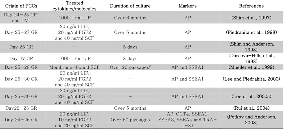
Table 4. List of embryonic germ cells and in vitro-cultured PGCs in pigs ... 58
Table 5. List of transgenic/cloned pigs using pluripotent stem cells ... 69
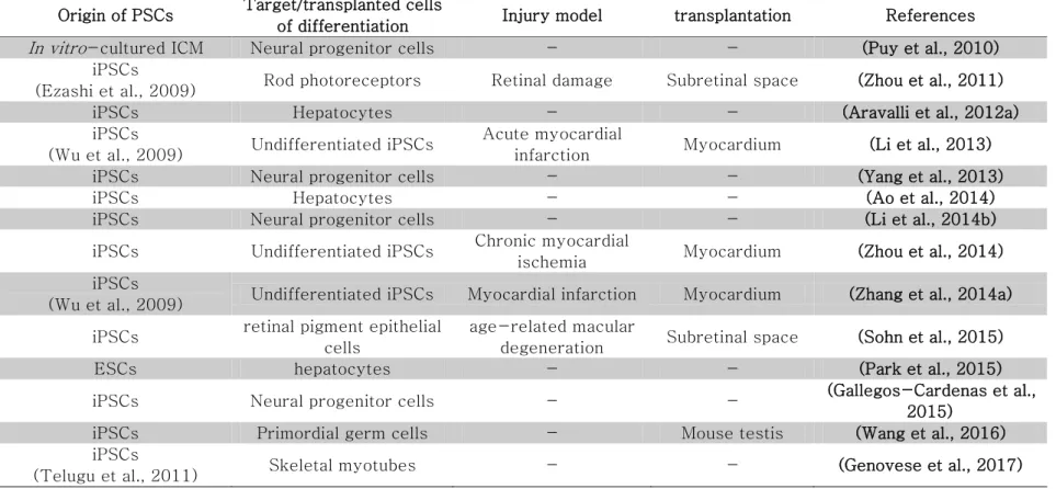
Table 6. List of differentiation studies using pig pluripotent stem cells ... 74
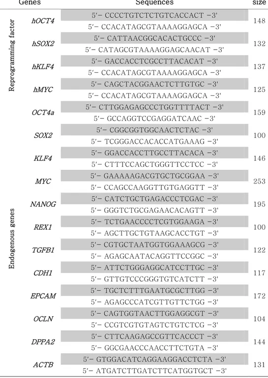
Table 7. RT-PCR primer sets for analyzing pluripotency and differentiation in pig ESCs ... 84
Table 8. Primer sets for detecting pluripotent and germ cell markers ... 86
x
IGF2/H19 DMR3 ... 90
Table 10. Primers for the detection of transgene insertion in gDNA ... 144
Table 11. Primers for detecting pluripotent and reprogramming markers ... 146
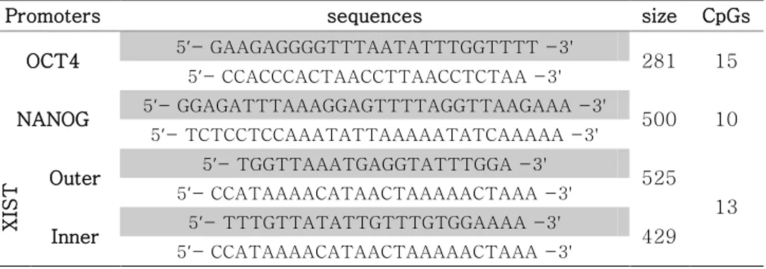
Table 12. Primers of bisulfite sequencing for promoters of OCT4,
NANOG and XIST ... 148
Table 13. Effects of serum replacements on outgrowth of primary pig ICM colonies ... 194
Table 14. Effects of cytokines on outgrowth of primary pig ICM colonies in KSR-supplemented media ... 196
Table 15. Effects of cytokines on outgrowth of primary pig ICM colonies in KSR+LC-supplemented media ... 198
Table 16. RT-PCR primer sets for pluripotent and differentiation markers in EGFP-transduced pig ESCs ... 220
Table 17. Primer sets of bisulfite sequencing for CMV promoter and EGFP ... 224
xi
xii
LIST OF FIGURES
Figure 1. Derivation and characterization of pig ESCs ... 95
Figure 2. Derivation of pig EGCs from fetal gonads ... 99
Figure 3. Characterization of pig EGCs by immunostaining ... 101
Figure 4. In vitro-differentiation ability of pig EGCs ... 103
Figure 5. Bisulfite sequencing at DMR region of IGF2/H19 and promoter region of XIST ... 105
Figure 6. Expression of SSEA4 in LIF-EGCs ... 108
Figure 7. Expression of germ cell markers during EGC culture ... 110
Figure 8. Expression of pluripotent genes and DNMTs during EGC culture ... 112
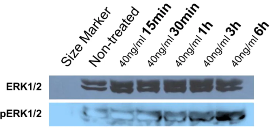
Figure 9. Analysis of ERK signaling in FGF2-EGCs ... 113
Figure 10. Analysis for plating efficiency of pig EGCs ... 117
Figure 11. Changes of gene expression during differentiation of LIF-EGCs ... 119
xiii
Figure 12. Candidate model for in vitro-reprogramming of pig primordial germ cells ... 131
Figure 13. Generation of pig induced pluripotent stem cells using drug-inducible vectors ... 153
Figure 14. Characterization of the AP-negative cell line ... 155
Figure 15. Characterization of three selected iPSC lines ... 157
Figure 16. Morphological changes of pig iPSCs in response to culture conditions ... 161
Figure 17. Expression of pluripotent markers in piPSCs ... 163
Figure 18. Expression of pluripotent and MET-related genes as measured by qPCR ... 167
Figure 19. Bisulfite sequencing at promoter regions of OCT4, NANOG, and XIST ... 171
Figure 20. Expression of SOX2 in primary porcine ICM outgrowths cultured with various serum replacements ... 195
xiv
cultured with KSR-supplemented media containing various
cytokines. ... 197
Figure 22. Expression of SOX2 in primary porcine ICM outgrowths cultured with KSR+LC-supplemented media containing various cytokines. ... 199
Figure 23. Long term culture of pig ICM with various combinations of culture media. ... 201
Figure 24. Morphology of pig embryonic stem cells derived by chemically defined media. ... 204
Figure 25. Expression of pluripotent genes in pig ESCs ... 206
Figure 26. Lentiviral transduction of pig ESCs ... 231
Figure 27. Characterization of EGFP-transduced pig ESCs ... 233
Figure 28. Change in EGFP expression during extended culture and in vitro differentiation ... 237
Figure 29. Lentiviral transduction efficiency of MEFs and PEFs at various MOIs ... 238
xv
Figure 30. Methylation level of the CMV promoter region in MEFs and PEFs ... 239
Figure 31. Silenced transgenes can be reactivated by treatment with the DNA methylase inhibitor, 5’-aza-2’-deoxycitidine (5-AzadC) ... 242
Figure 32. Doubling time of porcine embryonic stem cells ... 245
Figure 33. Expression level of PSIP1 in MEFs, PEFs, and pig ESCs ... 253
Figure 34. Correlation between the copy number of inserted transgenes and the EGFP expression level ... 254
Figure 35. Effects of culture conditions on neural differentiation in pig EGCs ... 268
Figure 36. Effects of RA concentration on neural differentiation in pig EGCs ... 271
Figure 37. Effects of FGF2 on growth of neural progenitor cells . 272
Figure 38. Expression of neural markers as determined by immunostaining ... 273
xvi
LIST OF ABBREVIATIONS
2i Two inhibitors; ERK and GSKb inhibitors ActA ACTIVIN A
AMD Age-related macular degeneration α-MEM α-Minimum Essential Medium ANOVA Analysis of variance
AP Alkaline phosphatase ATP Adenosine triphosphate BMP Bone Morphogenetic Proteins
cDNA Complementary deoxyribonucleic acid CDs Cluster of differentiation
CNTF Ciliary neurotrophic factor COCs cumulus oocyte complexes
DMEM Dulbecco's Modified Eagle's medium DNA Deoxyribonucleic acid
DNMT DNA methyltransferase Dox Doxycycline
DPBS Dulbecco's phosphate buffered saline dpc Days post coitus
E Embryonic day EBs Embryoid bodies
xvii
eCG Equine chorionic gonadotropin ECM Extracellular matrix
EGCs Embryonic germ cells EGF Epidermal growth factor
EGFP Enhanced green fluorescent protein
EpiSC Postimplantation epiblast-derived stem cells ESCs Embryonic stem cells
FBS Fetal bovine serum FGF Fibroblast growth factor Fsk Forskolin
gDNA Genomic deoxyribonucleic acid GFP Green fluorescent protein HAR Hyperacute rejection
hCG Human chorionic gonadotropin ICC Immunocytochemistry
ICM Inner cell mass
Id Inhibitor of differentiation IL Interleukin
iPSCs Induced pluripotent stem cells JAK Janus kinase
LIF Leukemia inhibitory factor
xviii LTR Long terminal repeat
MEFs Mouse embryonic fibroblasts
MET Mesenchymal-to-epithelial transition mRNA Messenger ribonucleic acid
OSKM OCT4, SOX2, KLF4 and cMYC OSM Oncostatin M
PCR Polymerase chain reaction pFF Pig follicular fluid
PGCs Primordial germ cells PI Propidium iodide
pre-iPSCs Partially reprogrammed-iPSCs PSCs Pluripotent stem cells
PZM Pig zygote media
qPCR Quantitative polymerase chain reaction RNA Ribonucleic acid
RPE Retinal pigment epithelium
RT-PCR Reverse transcription polymerase chain reaction SCF Stem cell factor
SCNT Somatic cell nuclear transfer SSEA Stage-specific embryonic antigen
STAT Signal transducer and activator of transcription STO Shimm's Thioguanine Ouabain resistant
xix TE Trophectoderm
TRA Tumor related antigen XCI X chromosome inactivation
1
CHAPTER 1
2
Stem cells indicates progenitor cells that are capable of self-renewal and differentiation into several cells. Especially, pluripotent stem cells (PSCs) have in vivo and in vitro differentiation potentials into three germ layers and can proliferate infinitely. First PSCs, known as embryonic stem cells (ESCs), were derived from preimplantation mouse blastocysts (Evans and Kaufman, 1981; Martin, 1981). Subsequently, another plutipotent cell lines, called embryonic germ cells (EGCs), were obtained though in vitro-culture of primordial germ cells (PGCs) (Matsui et al., 1992; Resnick et al., 1992). Recently, mouse epiblast stem cells (EpiSCs) and induced pluripotent stem cells (iPSCs) were derived from postimplantation embryos and somatic cells, respectively (Takahashi and Yamanaka, 2006; Tesar et al., 2007). Pluripotent states are divided into “naïve” and “primed” states depending on developmental competency of PSCs (Nichols and Smith, 2009). Naïve PSCs, represented by mouse ESCs and EGCs, are developmental ground state similar with early epiblasts of preimplantation embryos. On the other hand, primed PSCs, including EpiSCs and human ESCs, possess more differentiated pluripotency than naïve cells showing features of late epiblasts in postimplantation embryos. In permissive lines, both state of PSCs can be derived from embryos. However, in non-permissive lines such as human, only primed PSCs are derived in
3
absence of additional treatment such as genetic manipulation and chemicals (Buecker et al., 2010; Hanna et al., 2009; Park et al., 2013a)).
Because PSCs not only can differentiate into various type of cells and tissues, but also can produce germline-chimera by blastocyst injection (Bradley et al., 1984; Doetschman et al., 1985), PSCs have been considered as cell sources for cell therapy and producing transgenic animals. After establishment of human ESCs (Thomson et al., 1998), various researches for regenerative medicine by tissue engineering have been studied. Degenerative diseases could be treated by replacement of damaged tissues or cells with undamaged normal tissues or cells differentiated from PSCs (Tabar and Studer, 2014). And establishment of induced pluripotent stem cells (iPSCs) and cloned human ESCs allowed patient-specific cell therapies with PSCs (Tachibana et al., 2013; Takahashi and Yamanaka, 2006). In addition, transgenic animals could be generated by blastocyst injection and nuclear transfer using genetically modified PSCs (Capecchi, 2005; Robertson et al., 1986; Thomas and Capecchi, 1986). And, PSCs are more suitable for reprogramming within enucleated oocyte than differentiated cells (Hochedlinger and
4
Jaenisch, 2007), can provide an indefinite cell source for nuclear transfer.
In domestic animals, the aim of stem cell research is to create an indefinite cell source for transgenic animals used as bioreactors and tissue engineering materials as well as preliminary studies for human research (Keefer et al., 2007). To apply human PSCs as tools for regenerative medicine, preclinical studies with animal models are essential. Several animals such as pig, horse, cow and sheep etc. have been used for PSC researches (Ogorevc et al., 2016). Especially, pigs have been identified as an ideal animal model to study human disease, because of similarities between physiological and immunological features in humans and pigs, together with organ size (Brevini et al., 2007; Hall, 2008). Therefore, many research groups have attempted to derive pig PSCs including ESCs, EGCs and iPSCs, however, authentic pig ESCs have not yet been established (Ezashi et al., 2009; Kues et al., 2013; Park et al., 2013a; Piedrahita et al., 1990b; Son et al., 2009; Yang et al., 2009). Failures of deriving faithful pig PSCs are due to lack of understanding for mechanisms of pluripotency in pigs.
5
Because it was verified that pig has a different pluripotent networks in early embryos (Liu et al., 2015), it is essential to unveil mechanisms for establishing and maintaining pluripotency for deriving pig PSCs and optimizing pig-specific reprogramming methods. For these reasons, firstly, I attempted to analyze pig PSCs derived from various origins including embryos and somatic cells. In chapter 3 and 4, ESCs and EGCs were derived from pig in vitro -produced embryos and fetal gonads, respectively, and iPSCs were generated from pig somatic cells by ectopic expression of Yamanaka’s factors. Through an analysis of various PSC lines, it was explored which cell signaling are involved in supporting pluripotency of pig. Then, based on previous chapter, optimizing culture media for pig ESCs was performed by using various metabolic components and signaling molecules. Secondly, for applications of pig PSCs in preclinical study and production of transgenic pig, I tried to develop transgenensis and differentiation methods in pig PSCs. In chapter 6, lentiviral vectors carrying were examined to introduce transgene into pig pluripotent cell lines including ESCs and EGCs. In chapter 7, differentiation ability of pig EGCs into neuronal progenitor cells were investigated.
6
CHAPTER 2
7
1. Pluripotent stem cells (PSCs)
Researches on pluripotent stem cells (PSCs) were begun by embryonal carcinoma cells (EC cells) derived from murine gonadal teratomas and teratocarcinoma. Teratomas (benign) and teratocarcinomas (malignant) are tumors which are spontaneously formed in gonad, they are composed of several adult tissues including tooth, bone, muscle, skin and hair etc. When cells from teratocarcinomas were injected intraperitoneally into strain 129 mice, they re-formed teratocarcinomas (Kleinsmith and Pierce, 1964). This findings showed that stem cells which can proliferate indefinitely with differentiation potential into several tissues were existed in teratocarcinomas. In subsequent research, teratocarcinomas were artificially generated by injecting pre- and post-implantation embryos into testis in mouse, which indicated that stem cells in teratocarcinomas are similar with undifferentiated early embryos (Solter et al., 1970; Stevens, 1970) and Stevens developed 129/sv strain which has increased formation rate of gonadal tumor (Stevens, 1981).
8
from teratocarcinomas, so called EC cells, by co-culture with feeder cells made of mitotically-inactivated mouse embryonic fibroblasts in vitro (Martin and Evans, 1974). Subsequently, many scientists analyzed EC cells to elucidate their characteristics, which paved the way for researches on PSCs. EC cells possessed differentiation ability into three germ layers, so called pluripotency, and could proliferate indefinitely through symmetric division, so called self-renewal. Similar to teratocarcinomas, when injected subcutaneously into mouse, EC cells could produce teratocarcinomas differentiating into several tissues. When cultured in suspension, the cells aggregated and formed embryo-like structure with cavity, known as cystic embryoid bodies (Martin and Evans, 1974, 1975a; Martin and Evans, 1975b). In 1977, human EC cells were derived from malignant testicular teratomas and, as monoclonal antibodies were developed to detect specific marker protein, it was proven that mouse and human EC cells expressed SSEA1 and SSEA4 respectively (Fenderson et al., 1987; Hogan et al., 1977; Kannagi et al., 1983a; Kannagi et al., 1983b; Stern et al., 1978). Based on their features recapitulating embryonic development, early PSC researches had focused on cellular differentiation and embryology (Martin, 1975).
9
Although EC cells were potential materials to study pluripotent cells and embryogenesis, they have limitations to study embryology. They have abnormal karyotype and, when micro-injected into recipient blastocysts, couldn’t generate germline-chimeras showing restricted differentiation ability (Blelloch et al., 2004; Papaioannou et al., 1978; Papaioannou et al., 1975; Rossant and McBurney, 1982). In 1981, two groups, independently of each other, solved the problems through establishing embryonic stem cells (ESCs) derived by direct seeding of early blastocyst onto feeder cells, not by subcutaneous transplantation into mouse (Evans and Kaufman, 1981; Martin, 1981). ESCs not only possessed similar feature with EC cells in terms of cellular physiology, pluripotency and marker expression, but also could produce germline-chimeras with normal karyotype via blastocyst injection (Bradley et al., 1984; Doetschman et al., 1985). Especially, as gene targeting technologies were developed, transgenic mouse could be generated from genetically modified ESCs, which accelerated studies on gene functions during embryogenesis (Capecchi, 2005; Robertson et al., 1986; Thomas and Capecchi, 1986). On the other hand, because teratocarcinomas are spontaneously derived from germ cells in testis, this concept led to establish pluripotent cell lines from germ cells, as ESCs were derived from embryos (Dolci et al., 1991; Godin et al.,
10
1991; Matsui et al., 1991). In 1992, these efforts resulted in establishing pluripotent cell lines, so called embryonic germ cells (EGCs) from primordial germ cells (PGCs) (Matsui et al., 1992; Resnick et al., 1992).
In 1998, PSC researches entered on a new phase by establishment of human ESCs (Thomson et al., 1998). The derived human ESCs made people think that pluripotent cells could be used for regenerative medicine by tissue engineering. To take care of degenerative diseases including Parkinson`s disease, Alzheimer's disease, diabetes and retinitis pigmentosa etc., damaged tissues or cells should be replaced with undamaged normal tissues or cells, but it is hard to prepare the cells to care patients. However, if PSCs can be differentiated into specific cell lineage, human ESCs could supply normal cells or tissues for transplantation without limitations (Tabar and Studer, 2014). For these reason, PSC researches begun to focus on cell therapy, and were accelerated by establishment of induced pluripotent stem cells (iPSCs) and cloned human ESCs in 2006 and 2013, respectively (Tachibana et al., 2013; Takahashi and Yamanaka, 2006). So, in this chapter, it will be discussed that characteristics of PSCs derived from various origins and recent progress in PSC
11 research area.
1.1 Embryonic stem cells (ESCs)
To retain characteristics of ESCs, self-renewal and pluripotency, various extrinsic and intrinsic factors have to be delicately regulated. Distinct features of ESCs compared with somatic cells are used to identify them. Lack of extrinsic or intrinsic determinants induce differentiation of ESCs losing pluripotency. So, it is important to understand and investigate genes and culture conditions involved in pluripotent circuit.
Microenvironmental surrounding of ESCs, known as stem cell niche, including cytokines, extracellular matrix and metabolic sources is important extrinsic factors for supporting pluripotency. Viability and self-renewal of ESCs in in vitro-culture are dependent on stem cell niche. First mouse ESCs were obtained by co-culturing with mitotically-inactivated mouse embryonic fibroblasts (MEFs), so called feeder layer, and EC cell-derived conditioned media (Evans and Kaufman, 1981; Martin, 1981). Main function of feeder layer is
12
providing physical environment through extracellular matrix (ECM), which affects proliferation and survival rate by activating Cadherin and Integrin signaling in ESCs (Guilak et al., 2009; Nagaoka et al., 2006). And feeder cells activate intrinsic pathways of pluripotency by paracrine effects of several cytokines including LIF, FGF2, BMP4, ACTIVIN A and WNT etc (Eiselleova et al., 2008). Various type of cells including MEFs (Evans and Kaufman, 1981; Martin, 1981), STO cells (Park et al., 2003), and SNL 76/7 cells (Williams et al., 1988) have been used as a feeder cells. Recently, in order to reduce cell-to-cell variation and xeno-contamination occurred by feeder layer, mixture of ECM proteins and synthetic peptides are being used instead of feeder cells (Lee et al., 2010; Mei et al., 2010; Meng et al., 2010).
Together with physical environment, activation of cellular signaling by cytokines is also important to keep pluripotency. Because signaling molecules specifically support pluripotency of ESCs were not known, EC cell-derived conditioned media was used at the beginning of ESC culture (Martin, 1981). Later, it was verified that LIF and BMP4 signaling pathways have a crucial role in supporting pluripotency of ESCs (Smith et al., 1988; Williams et al.,
13
1988; Ying et al., 2003). Firstly, leukemia inhibitory factor (LIF) is a member of interleukin-6 family. If LIF bound extracellular domain of LIF receptor (LIFr) in plasma membrane, LIFr forms heterodimer with gp130. LIFr/gp130 heterodimer activates receptor-associated Janus kinase (JAK), which results in phosphorylation of STAT3. Phosphorylated STAT3 molecules act as a homodimer in nucleus, which facilitates transcription of pluripotency-related genes. The up-regulated genes prevent differentiation and support self-renewal in mouse ESCs (Boeuf et al., 1997). However, although LIF treatment is sufficient to maintain pluripotency in serum-supplemented media, ESCs differentiated into neural lineage in serum-free media, suggesting unknown factors of fetal bovine serum involved in sustaining pluripotency. Ying and colleagues found that BMP4 support pluripotency with LIF in serum-free media preventing neural differentiation (Ying et al., 2003). BMP4 facilitates expression of inhibitor of differentiation, Id, protein through SMAD signaling and these proteins inhibit neural differentiation. Recently, various small molecules which inhibit cell signaling of differentiation and sustain pluripotent gene networks are used in stem cell culture (reviewed by Ma et al., 2013).
14
If stem cell niche as mentioned above is prepared in in vitro -culture environment, intrinsic determinants of pluripotency are activated in ESCs. As transcription factors, the intrinsic determinants important roles in regulation and maintenance of pluripotent gene networks. Most important genes in regulating pluripotency are Oct4,
Sox2 and Nanog, which also are crucial for establishing pluripotency and lineage segregation in early embryo. Oct4 and Sox2 are starting to express from early blastomere stage and, after cavitation of blastocyst, exclusively express in inner cell mass (ICM) (Avilion et al., 2003; Nichols et al., 1998; Scholer et al., 1989). These two factors form transcriptional complex with various enhancer proteins and work as a complex (Ng and Surani, 2011). Balanced expression of Oct4 and Sox2 is important to maintain pluripotency (Radzisheuskaya and Silva, 2014). Overexpression of Oct4 induce mesodermal differentiation and, in case of Sox2, ESCs differentiate into ectodermal lineage. In addition, along with Oct4 and Sox2, Nanog
as a transcription factor have a crucial role in supporting stemness (Chambers et al., 2003; Loh et al., 2006; Mitsui et al., 2003).
Distinctive epigenetic feature of ESCs is that two X chromosomes are activated in female ESCs. Because, unlikely male
15
cells, female mammalian cells have two X chromosomes, one X chromosome is randomly inactivated for compensation of genetic materials in female cells (Okamoto and Heard, 2009). As ICM which have two activated X chromosomes undergoes gastrulation, X chromosome inactivation (XCI) is occurred by non-coding RNA, Xist
(Heard, 2004; Lee and Bartolomei, 2013). In this reason, ESCs as an
in vitro-counterpart of ICM have two-activated X chromosomes (Nichols and Smith, 2009). Recent researches indicated that pluripotent genes such as Oct4, Sox2 and Nanog, have a vital role in sustaining activated status of X chromosomes through regulation of
Xist and Tsix, known as negative regulator of Xist during murine preimplantation development(Donohoe et al., 2009; Navarro et al., 2008). Accordingly, several researches are being performed to verify relationship between pluripotency and XCI (reviewed in Minkovsky et al., 2012; Navarro and Avner, 2009).
Feeder cells are also required for culture of human ESCs, similar with mouse (Thomson et al., 1998). In addition, OCT4, SOX2
and NANOG play vital roles in establishing and maintaining of pluripotency in human embryos and ESCs (Boyer et al., 2005). However, there are some physiological differences between human
16
and mouse ESCs. Unlikely mouse in which LIF/BMP4 are involved, pluripotency of human ESCs is sustained through ERK and ACTIVIN/NODAL signal pathway activated by FGF2 and TGF-β (Pera and Tam, 2010). In female human ESCs, X-linked genes are monoallelic expressed because of inactivated X chromosome by expression of XIST in atmospheric O2 concentrations (Lengner et al.,
2010; Shen et al., 2008). These distinguishable features had been considered as species-specific characteristics for a long time. However, series of experiments discovered that the differences among PSCs are originated by two distinct grades of pluripotency, including“naïve” and “primed” states, based on developmental competence (Hanna et al., 2010b; Nichols and Smith, 2009; discussed in 1.5).
1.2 Epiblast stem cells (EpiSCs)
In 2007, two research groups, established novel type of pluripotent stem cells, named postimplantation epiblast-derived stem cells (shortly, epiblast stem cells or EpiSCs), from postimplantation embryos (Brons et al., 2007; Tesar et al., 2007). ICM of blastocyst divide into early epiblast and hypoblast through the
17
lineage segregation. After implantation, epiblast finally develop fetus through a gastrulation, hypoblast differentiate into extraembryonic tissues including visceral and parietal endoderm (reviewed in (Rossant and Tam, 2009)). To derive EpiSCs, late epiblasts were isolated at embryonic day 5.5 before gastrulation and seeded onto feeder cells in media supplemented with FGF2 and ACTIVIN A. After 3-5 days, epiblast explants rapidly grew and from OCT4-expressed compact colonies. Established EpiSCs expressed pluripotent genes such as Oct4, Sox2 and Nanog, and teratomas formed when the cells were grafted in immune-deficiency mice. However, EpiSCs showed distinct characteristics compared with ESCs derived from early epiblasts. Expression of ICM-specific genes which are up-regulated in ESCs, such as Pecam1, Tbx3 and Gbx2, were decreased in EpiSCs. On the other hand, epiblast and early germ layers-specific genes such as Otx2, Eomes, Foxa2, T, Gata4, Sox17 and Cer1 were highly expressed in EpiSCs. This cell line grew as a monolayer having flattened morphology and relied on ERK and ACTIVIN/NODAL signaling pathway to maintain pluripotency instead of LIF and BMP4 signaling. In epigenetic and developmental aspects, female EpiSCs possessed inactivated X chromosome similar with their in vivo -counterpart, late epiblasts. In the same manner, when assessed developmental stage by chimeric assay, although they could not
18
generate chimeric embryos with morula and early blastocyst, EpiSCs were incorporated within embryos and developed chimeric fetus when engrafted into postimplantation embryos (Huang et al., 2012; Kojima et al., 2014). While murine ESCs could be converted into EpiSCs by treatment of FGF2 and LIF antibody, EpiSC couldn’t be converted into ESCs without genetic manipulations, which means steps of mammalian development are irreversible (Bao et al., 2009; Guo et al., 2009; Hanna et al., 2009). Overall, EpiSCs shared defining similarities with human ESCs in terms of gene expression and cellular signaling.
Before derivation of this new type of cell line, it was thought that pluripotent state is fixed. However, this interesting discovery compelled to think that pluripotent states are flexibly altered dependent on culture environments and developmental origins of PSCs. Finally, all these results were sufficient to change paradigm of pluripotency and provoke a new hypothesis/theory for pluripotency status (discussed in 1.5).
19
1.3 Embryonic germ cells (EGCs)
PGCs as an alternative cell source can be used for deriving pluripotent stem cells. When cultured with feeder cells and adequate cytokines, PGCs can be reprogrammed into pluripotent stem cells, named EGCs. As ESCs were established from ICM via in vitro -culture, it had been attempted that in vitro culture of PGCs, origin of spontaneous teratocarcinomas in vivo, for deriving novel pluripotent stem cell line (Dolci et al., 1991; Donovan et al., 1986; Godin et al., 1991; Matsui et al., 1991).
In murine post-implantation embryo at dpc 5.5, PGCs arise from proximal posterior epiblast by stimulation of BMP4, BMP8b and BMP2 from extraembryonic ectoderm and visceral endoderm (Lawson et al., 1999; Ying et al., 2000; Ying and Zhao, 2001). In precursor of PGCs, BMP signaling stimulates expression of Blimp1
known as key transcription factors for specification of germline, which induces repression of somatic program and facilitates expression of pluripotent genes and epigenetic reprogramming (Durcova-Hills et al., 2008; Ohinata et al., 2005). Resulting PGCs migrate from posterior endoderm of the yolk sac at dpc 7.5 via
20
hindgut and mesentery to genital ridge at dpc 12.5, and primitive gonads and fetal ducts in turn develop into sex organs (Molyneaux et al., 2001). Apposing migration, dozens of cells actively proliferate into thousands and epigenetic remodeling including reconstruction of DNA methylation, imprint erasure and X-chromosome reactivation are occurred (reviewed in (Ewen and Koopman, 2010)). And after arrival at genital ridges, gender-specific imprinting patterns are re-establishment (Lees-Murdock and Walsh, 2008).
When cultured with feeder cells made of STO cell line and cytokines including LIF, FGF2 and SCF, mouse migrating PGCs isolated at dpc 8.5 were reprogrammed and converted into EGCs (Matsui et al., 1992; Resnick et al., 1992). In culture, roles of cytokines are as follows. Stem cell factor (SCF, also known as c-Kit ligand or Steel factor), although PGCs undergo apoptosis in a few days during in vitro culture, prevents apoptosis and promotes viability and proliferation rate of germ cells together with LIF (Dolci et al., 1991; Godin et al., 1991; Matsui et al., 1991). FGF2 up-regulates expression of Dhx38, Myc and Klf4 known as targets of BLIMP1 via down-regulation of Blimp1, facilitates reprogramming of PGCs into EGCs (Durcova-Hills et al., 2008). Finally,
fully-21
reprogrammed mouse EGCs cultured with only LIF.
Established mouse EGCs possess similar features with embryonic stem cells in physiological and developmental aspects. They express pluripotent marker genes such as OCT4, SOX2, NANOG and SSEA1, and have developmental competency in vitro
(embryoid body formation) as well as in vivo (teratoma and chimera formation) (Matsui et al., 1992; Resnick et al., 1992). In addition, recent data indicated that there’s close similarities between EGCs and ESCs in terms of genetics and epigenetics (Choi et al., 2017). In human, EGCs could be derived from gonad of aborted fetuses culturing with feeder cells and LIF, FGF2 and Fsk (Liu et al., 2004; Shamblott et al., 1998; Turnpenny et al., 2003). Human EGCs expressed several pluripotent markers such as OCT4, SSEA1 and SSEA3/4 and have in vitro developmental competency. However, because of ethical problems for using human fetuses, research on human germ cells is not as well performed (Turnpenny et al., 2006).
22
Developmental biologist Conrad H. Waddington, as known as pioneer of epigenetics, addressed that, because differentiation of cells is like a rolling stones through a downhill, stones reached at bottom of hill are in stable state and cannot move to anywhere (Sieweke, 2015). For a long time, developmental biologists had believed that developmental process of organism are irreversible event, and completely lineage-committed cells cannot be converted into the other type of cells including progenitor cells. In 1962, Sir John B. Gurdon generated tadpoles by nuclear transfer of somatic cells into enucleated eggs (Gurdon, 1962) and, after three decades, Sir Ian Wilmut and colleagues produced first cloned mammal, Dolly the sheep, with same technique (Wilmut et al., 1997). In addition, Japanese research group reported that epigenome of thymocyte is reconstructed into ESCs-like state by fusing with ESCs (Tada et al., 2001). In thymocyte-ESC hybrids, Oct4-GFP transgene was expressed and an inactivated X chromosome was reactivated. These serial experiments showed that cellular reprogramming of committed cells into a pluripotent state can be induced by unknown factors in oocytes and ESCs, which means that it could be possible to put stones at the bottom on top of the hill.
23
In 2006, inspired by previous studies, Yamanaka and colleagues developed new method to easily reprogram somatic cells (Takahashi and Yamanaka, 2006). New PSCs, so called iPSCs, were generated from adult somatic cells by nuclear reprogramming via introducing four genes such as Oct4, Sox2, Klf4 and cMyc. Comparison of global gene expression between ESCs and iPSCs showed that gene expression pattern of two cell lines was very close. The cells could stably be maintained having normal karyotype and differentiate into three germ layers in vitro and in vivo. In 2007, human induced pluripotent stem cells were derived by two independent groups (Takahashi et al., 2007; Yu et al., 2007). Yamanaka and colleagues generated human iPSCs by Yamanaka’s factors, the other groups used different combinations including OCT4,
SOX2, NANOG and LIN28. In addition, it was verified that nuclear reprogramming could be achieved by micro RNA and combination of chemicals (Anokye-Danso et al., 2011; Hou et al., 2013a; Miyoshi et al., 2011). The iPS technique was easy to use compared with prior methods (SCNT and cell fusion) and could be apply to adult cells, which accelerated stem cell researches for regenerative medicine and cell therapy using patient-specific PSCs.
24
However, iPS technique has problems for applying to therapeutic usages. First of all, abnormal features could be occurred by integration of transgenes in genome of cells during maintenance and differentiation of iPSCs. Especially, one of Yamanaka’s factors,
cMyc as a proto-oncogene induced tumor formation, when iPSC/iPSC-derived cells are transplanted in vivo (Nakagawa et al., 2008; Nakagawa et al., 2010). And also, because epigenetic memory derived from origin of somatic cells were remained in iPSCs after reprogramming, differentiation of iPSCs were biasedly progressed (Kim et al., 2010a; Polo et al., 2010). And this deficiency of epigenetic reprogramming caused differences between ESCs and iPSCs (Chin et al., 2009; Chin et al., 2010). These problems were solved with integration-free gene delivery systems such as plasmid vector (Okita et al., 2008), episomal vector (Yu et al., 2009), PiggyBack transposon system (Woltjen et al., 2009), adenovirus vector (Stadtfeld et al., 2008) and sendai virus vector (Fusaki et al., 2009), and transgene-free methods such as protein- and chemical-mediated systems (Hou et al., 2013a; Zhou et al., 2009). And recent study showed that such variations between ESCs and iPSCs were mainly originated by different genetic background and genetically matched human ESCs and iPSCs were molecularly and functionally equivalent (Choi et al., 2015).
25
The acquisition of pluripotency in fibroblasts is accomplished by genetic and epigenetic events termed initiation, maturation, and stabilization (Samavarchi-Tehrani et al., 2010). The initiation of reprogramming is defined by mesenchymal-to-epithelial transition (MET), in which epithelial-specific genes are up-regulated and
Tgfb1 (which blocks nuclear reprogramming) is downregulated by expression of reprogramming factors and BMP signaling (Li et al., 2010; Samavarchi-Tehrani et al., 2010). The cells that convert to iPSCs express predictive markers, such as ESRRB, UTF1, LIN28, and DPPA2. Subsequently, NANOG and SALL4 induced by SOX2 activate endogenous pluripotent networks, and pluripotent circuitry is stabilized via epigenetic remodeling such as DNA methylation, histone modification, and X chromosome reactivation (in the case of female cells) (Buganim et al., 2012; Polo et al., 2012). Since Nanog
as a gateway to pluripotency plays pivotal roles in epigenetic remodeling and X chromosome reactivation, it is important to reactivate Nanog in the late stages of reprogramming in mouse (Chambers et al., 2007; Silva et al., 2009). In human as a non-permissive species, NANOG plays an important role in reprogramming and maintaining pluripotency (Hyslop et al., 2005; Yu et al., 2007). Finally, the reprogrammed cells can be maintained
26
without ectopic expression of transgenes, which indicates that endogenous pluripotent network are fully activated and stabilized (Maherali et al., 2007; Wernig et al., 2007). Because the defective silencing of transgenes after reprogramming affects stability, carcinogenesis, and differentiation ability of iPSCs, silencing or eliminating transgene expression is one of the most important step for fully reprogrammed iPSCs (Okada and Yoneda, 2011; Okita et al., 2007).
As described above, several genetic and epigenetic changes occur during nuclear reprogramming from somatic cells to iPSCs. Achieving faithful pluripotency is required to overcome epigenetic and physiological obstacles such as the epigenetic memory of somatic cells (Kim et al., 2010a; Polo et al., 2010), MET (Li et al., 2010), repressive chromatin (Huangfu et al., 2008), and apoptosis and cell cycle arrest (Kawamura et al., 2009; Li et al., 2009). However, if these barriers are not overcome, silencing of transgenes, epigenetic remodeling, and lack of Nanog expression occur, resulting in partial reprogramming of iPSCs (pre-iPSCs) (Okita et al., 2007; Silva et al., 2008). The pre-iPSC lines share several common characteristics, such as incomplete expression of pluripotent genes, inactive X
27
chromosomes in female cells, and inability to generate germline chimeras, have been observed (Li et al., 2010; Okita et al., 2007; Silva et al., 2008; Silva et al., 2009).
To overcome the barriers of reprogramming, various studies have been performed using small molecules, another reprogramming factor, and nutrient supplements. At this time, drug-inducible vector systems, which can easily turn transgenes on and off, have been used in various studies involving the iPSC generation and the elucidation of reprogramming mechanisms (Buganim et al., 2012; Polo et al., 2012; Wernig et al., 2008). Chromatin remodeling and the erasing of epigenetic memory in somatic cells have been accomplished by inhibiting DNA methylation and using chromatin modifiers (Huangfu et al., 2008). Pre-iPSCs lacking Nanog expression were converted into fully reprogrammed iPSCs by inhibiting FGF2 signaling and
Nanog overexpression (Silva et al., 2008; Theunissen et al., 2011). In addition, serum free media and vitamin C could be used to overcome hurdles and increase reprogramming efficiency (Chen et al., 2010; Esteban et al., 2010). In addition, suppression of TGF signaling, apoptosis, senescence, and cell-cycle arrest are considered useful tools for reducing reprogramming barriers (Banito
28
et al., 2009; Ichida et al., 2009; Kawamura et al., 2009; Li et al., 2009). Recently, it has been shown that several factors including
Nanog, Lin28, Nr5a2, and Glis1 could be used for nuclear reprogramming instead of Yamanaka’s factors (Heng et al., 2010; Maekawa et al., 2011; Yu et al., 2007).
1.5 Two pluripotent states based on developmental
potency
Human and mouse ESCs have different features as described in Table 1. When human ESCs were firstly derived, it was thought that distinguished features of human ESCs were caused by basic developmental differences between human and mouse (Thomson et al., 1998). However, EpiSCs derived in FGF2/ACTIVIN A-culture media resembled human ESCs, which indicated that PSC could be categorized based on their pluripotent state and developmental potency. Recently, several studies have suggested that the states of PSCs are divided into two categories: naïve and primed (reviewed in (Hanna et al., 2010b; Nichols and Smith, 2009)).
29
Naïve PSCs derived from early epiblasts in pre-implantation blastocysts as a developmental ground state can generate the chimeric fetus when micro-injected into recipient blastocysts. In addition, primed PSCs derived from late epiblasts in post-implantation blastocyst possess more differentiated pluripotency than naïve cells in terms of developmental capacity, gene expression, and epigenetic signatures. Naïve PSCs, represented by mouse ESCs and EGCs, are characterized by dome-shaped colony morphologies, activation of LIF signaling, and two active X chromosomes in females. By contrast, primed PSCs, including EpiSCs and human ESCs, are defined by flattened colony morphologies and activated FGF signaling pathways. Compared with the primed state, naïve PSCs have developmental and functional ground states showing contributions to blastocyst chimeras and higher transgenic efficiency (Buecker et al., 2010; Hanna et al., 2010a). In permissive lines, both pluripotent states of PSCs can be derived from embryos. Only certain mouse strains, such as 129, C57BL/6 and BALB/C, were categorized as a permissive-line. (Nichols and Smith, 2012; Smith, 2001). However, in non-permissive lines, the stem cells cannot be stabilized in the naïve state, and are instead differentiated and stabilized at the primed state during the establishment process if no additional treatments (including genetic manipulation and chemicals) are performed (Hanna
30 et al., 2010b; Nichols and Smith, 2009).
Several studies have attempted to establish naïve-state PSCs from non-permissive species such as rats and humans (Buehr et al., 2008; Li et al., 2008). The first case of human naïve PSCs reported that derivation of naïve PSCs were accomplished via ectopic expression of OCT4 and KLF4 supplemented with LIF and two inhibitors for GSK and ERK1/2 signaling (Hanna et al., 2010a). However, these cell lines could not be maintained without transgene expression. Recent studies have reported that generation of transgene-free human naïve-like PSCs from primed PSCs could be achieved by several small molecules in addition to 2i (GSK and ERK inhibitors) (Chan et al., 2013; Gafni et al., 2013; Takashima et al., 2014; Theunissen et al., 2014; Valamehr et al., 2014; Ware et al., 2014). And Nichols and colleagues successfully derived naïve human ESCs directly from human early embryos by inhibiting PKC and ROCK signaling (Guo et al., 2016b). These cells expressed markers of naïve pluripotency and resembled mouse ESCS in terms of mitochondrial respiration, global gene expression and genome-wide methylation pattern. When transferred into FGF2-supplemented media, naïve cells were converted into conventional human ESCs.
31
These results demonstrated that in vivo counterpart of naïve cells exist in early human embryos and modulating signaling pathways is required for the maintenance of naïve state PSCs in non-permissive species.
32 Table 1. Characteristics of pluripotent stem cells.
Species Mouse Human
Permissive species (129, C57BL/6 and BALB/C) Non-permissive species Stem cell types Embryonic stem
cells Embryonic germ cells Epiblast stem cells Embryonic stem cells Embryonic germ cells Origins Preimplantation ICM Primordial germ cells Postimplantation epiblast Preimplantation ICM Primordial germ cells
Pluripotent states Naïve Primed -
Colony morphology Domed Flattened Domed
Pluripotent signaling LIF, BMP4 and WNT FGF2, ACTIVIN and WNT
FGF2, LIF and Fsk Pluripotent markers OCT4, SOX2, NANOG, SSEA1 OCT4, SOX2, NANOG and SSEA4 OCT4, SSEA1,
SSEA3/4 Chimeric assay Pre-implantation embryos Post-implantation embryos -
X chromosome inactivation in female
cells
Two active X chromosomes One inactive X chromosome and
33
2.
Large animal models in biotechnology/stem cell biology:
A pig review
Domestic animals such as pigs, cows and chickens have been considered as an ideal model for bioreactor producing medical proteins in by-product form such as milk, urine and egg (Keefer et al., 2007). For these reasons, many research groups have focused on production of transgenic farm animals for bioreactors, disease model and xenotransplantation for a long time (Houdebine, 2009; Pierson et al., 2009). Especially, pigs have a great potential in their application in xenotransplantation and disease model for human and in biopharming, because of its anatomical and physiological similarities to human (Brevini et al., 2007; Hall, 2008).
In order to produce transgenic animals, the somatic cell nuclear transfer (SCNT) and transgenic techniques are essential process. From the first cloned sheep named Dolly in 1997 (Wilmut et al., 1997), a large number of cloned animals including mice in 1998 (Wakayama et al., 1998), goat in 1999 (Baguisi et al., 1999), pig and cattle in 2000 (Kato et al., 2000; Onishi et al., 2000; Polejaeva et al., 2000), cat and rabbit in 2002 (Chesne et al., 2002; Shin et al., 2002),
34
horse, mule and rat in 2003 (Galli et al., 2003; Woods et al., 2003; Zhou et al., 2003), dog in 2005 (Lee et al., 2005), ferret in 2006 (Li et al., 2006), deer, buffalo and wolf in 2007 (Berg et al., 2007; Kim et al., 2007b; Shi et al., 2007), and camel in 2010 (Wani et al., 2010) have been produced. Together with improvement of cloning technique, the way to produce transgenic animals have been also advanced. First generated livestock animal to produce pharmaceutical proteins was transgenic sheep, named Molly and Polly (Schnieke et al., 1997). They were produced by somatic cell nuclear transfer. Transfected fetal fibroblasts carrying human clotting factor IX linked to the ovine β-lactoglobulin gene promoter known as mammary gland-specific promoter were used as donor cells. Lots of transgenic animals have been generated since then, and, in 2009, human antithrombin isolated from transgenic goat milk, named ATryn, was firstly approved by Food and Drug Administration of U.S. (2009; Edmunds et al., 1998; Konkle et al., 2003).
Along with production of bioreactors, development of animal models for human disease have been progressed for a long time. As pig genome project was completed, studies of transgenic pig models for human disease were accelerated. While some of human diseases
35
are caused by simple genetic mutations, pig genome project verified that pigs and humans share 112 DNA mutations implicated with human diseases (Groenen et al., 2012). So, scientists have made the transgenic pigs carrying human disease including Huntington’s disease (Uchida et al., 2001), retinitis pigmentosa (Ghosh et al., 2007), cystic fibrosis (Rogers et al., 2008), diabetes (Renner et al., 2010) and Alzheimer’s disease (Kragh et al., 2009; Søndergaard et al., 2012) through gene knockout technique. And porcine organs and tissues anatomically similar with human can be used for transplantation into human body in order to replace a malfunctioning organ. Because of problems caused by increasing imbalance between organ donors and recipients, various research groups are focusing on the xenotransplantation of human organs with those of pigs. Pig heart valves and skins have been transplanted into heart disease patients and burn victims respectively from 30 years ago (reviewed by Schuurman and Pierson, 2008). Moreover, attempt to treat diabetes though a transplantation of porcine pancreatic islet to human has been made by several groups. However, there is an obstacle to overcome such as an immune rejection in order to successful transplantation. Although immunosuppressant drugs are used, inhibition of immune rejection via genetic manipulations is necessary for minimizing side effects. When foreign tissues from other species are grafted into
36
human body, immune reactions including hyperacute rejection, acute vascular rejection, cellular rejection and chronic rejection are triggered. Hyperacute rejection (HAR) is immediately induced within minutes after the transplantation, and acute vascular rejection and cellular rejection occurred after the HAR (reviewed in (Yang and Sykes, 2007)). To avoid the immune rejection, transgenic pigs for xenotransplantation have been produced by modification of genes such as α1,3-galactosyltransferase, CAMH, heme oxygenase and human CDs (reviewed in (Niemann and Petersen, 2016)). It was proven that rat organs were successfully generated in mice by interspecies-chimera formation (Isotani et al., 2011; Kobayashi et al., 2010). When rat wild-type pluripotent stem cells (PSCs) were injected into blastocyst of transgenic mouse in which thymus and pancreas could be developed, rat PSCs were developed into various organs including thymus and pancreas while displaying chimerism. Belmonte and colleagues verified that human PSCs could generate chimeric fetuses incorporated with pig embryos (Wu et al., 2017). Accordingly, interspecies chimera technique might be an another source for providing therapeutic tissues and organs, if we consider consensus on ethical problems.
37
PSCs of domestic animals have been identified as promising tools for generating transgenic animals and preclinical researches. In making transgenic animals, because of unique epigenetic milieus, PSCs as a donor cell are more suitable for reprogramming within enucleated oocyte than differentiated cells (Hochedlinger and Jaenisch, 2007). Several studies showed that ESCs were more effective for cloning of mouse than differentiated cells such as immune cells, neuron, and fibroblasts (Blelloch et al., 2006; Blelloch et al., 2004; Eggan et al., 2004; Hochedlinger and Jaenisch, 2002; Inoue et al., 2005). And PSCs have another advantage in application to nuclear transfer. PSCs can proliferate indefinitely maintaining cellular characteristics including karyotype and genomic stability. This property, so called self-renewal, provides an indefinite cell source for nuclear transfer. With genetic manipulations, they can offer stable transgenic cell source for generation of transgenic animals for bioreactors, xenotransplantation and disease models. And also, as PSCs could be incorporated in development of early embryos, blastocyst injection could be apply for generation of transgenic animals. In these reasons, various transgenic piglets and embryos have been produced by using pig PSCs (Ahn et al., 2007; Fan et al., 2013; West et al., 2010; Zhang et al., 2015a).
38
In 2009, a clinical trial of human ES cell therapy for repairing spinal cord damages was approved by Food and Drug Administration (Alper, 2009). In 2014, first clinical test were operated in Japan (Sugita et al., 2016). Woman who suffered from aged-macular degeneration (AMD) received retinal pigment epithelium (RPE) differentiated from iPSCs, and her vision were partially recovered. However, because spinal cords and eyes are immune-privileged area without immune cells and are not affected to life, clinical tests for organs and tissues which are strongly involved in life, such as heart, brain and pancreas, need strict preclinical test using animal disease models. So, it makes the porcine study involving differentiation and transplantation of stem cells as a preliminary study more important. As a preliminary study for human cell therapy, many researchers are trying to convert the porcine PSCs into several differentiated cells including rod receptors, hepatocytes, endothelium and cardiomyocytes apposing transplantation of the differentiated cells into the pigs (Aravalli et al., 2012b; Gu et al., 2012; Kawamura et al., 2012; Zhou et al., 2011). In these reasons, in this chapter, it will be discussed history, features and research state of pig PSCs.
39
2.1 Pig embryonic stem cells
Because pluripotent cells have great potential as a cell source, research in this area has focused on embryonic carcinoma cells to iPSCs (Martin and Evans, 1974; Takahashi and Yamanaka, 2006). ESC research began in 1981 by the establishment of mouse ESCs and was accelerated by the establishment of human ESCs and iPSCs in 1998 and 2006, respectively (Evans and Kaufman, 1981; Takahashi and Yamanaka, 2006; Thomson et al., 1998). The purpose of PSC research in humans and mice includes the elucidation of basic cellular mechanisms contributing to the maintenance of pluripotency as well as applications in human cell therapies. However, in domestic animals, the research aim is to create an indefinite cell source for transgenic animals used as bioreactors and tissue engineering materials as well as preliminary studies for human research (Keefer et al., 2007). Because of the physiological and immunological similarities that exist between pigs and humans, porcine pluripotent cell lines have been identified as important candidates for preliminary studies on human disease (Brevini et al., 2007; Hall, 2008; Houdebine, 2009). Therefore, many research groups have attempted to derive pig PSCs from early embryos for a long time (Ezashi et al., 2009; Kues et al.,
40
2013; Park et al., 2013a; Piedrahita et al., 1990b; Son et al., 2009; Yang et al., 2009).
In early studies of pig embryonic stem cells (ESCs), researchers tried to establish stem cells based on teratocacinoma culture methods as mouse studies did. Various culture materials including serums (fetal bovine serum and calf serum), feeder cells (buffalo liver cells, mouse embryonic fibroblasts, pig embryonic fibroblasts and pig uterine epithelial cells) and conditioned media were tested for maintaining pluripotent inner cell mass (ICM) in vitro
(Anderson et al., 1994; Piedrahita et al., 1990a, b; Talbot et al., 1993b). Nonetheless, it was hard to derive pig stem cells due to lack of knowledge about cell signaling involved in early embryo development and maintaining pluripotency. When it was verified that LIF and FGF2 are responsible for maintaining mouse and human ESCs respectively (Amit et al., 2000; Smith et al., 1988; Williams et al., 1988), many cytokines like interleukins (IL), oncostatin M (OSM), ciliary neurotrophic factor (CNTF), epidermal growth factor (EGF), activin A (ActA) and stem cell factor (SCF) as well as LIF and FGF2 have been used for in vitro culture of pig ICM with various combinations and concentrations as summarized in Table 2. Beside,
41
as culture media were developed for culture of mouse and human ESCs, Knockout-DMEM, F10, F12 and αMEM have been used for basal media (Table 2). However, although some culture conditions could support in vitro-culture of pig ICM for a short term (Alberio et al., 2010; Puy et al., 2010; Vassiliev et al., 2010), cultured ICM lost their own features and in turn differentiated during prolonged culture. Instead of pluripotent cells, during in vitro-culture, multipotent stem cells, so called ES-like cells, have been spontaneously obtained by several groups (Brevini et al., 2010; Cheong et al., 2015; Kim et al., 2010b; Uh et al., 2014). To date, it is still endeavoring to establish authentic pig ESCs which have in vivo and in vitro-developmental competency.
Pig ES-like cells have been derived from various embryo origins as described in Table 2. In early studies, hatched blastocysts were collected from pregnant sow at 6-11 days after artificial insemination. These experiments showed that in vitro-survival rate of ICM was increased in older blastocysts (day 10 and 11) but spontaneous differentiation was frequently observed. Although freshly isolated ICM could be formed chimeric piglets after blastocyst injection, cultured ICM lost their developmental competency
42
(Anderson et al., 1994). Recent studies addressed that FGF2 and LIF supported in vitro-culture of ICM (Brevini et al., 2010; Li et al., 2004; Li et al., 2003) and ES-like cells could be derived in FGF2-supplemented media (Alberio et al., 2010; Cheong et al., 2015; Kim et al., 2016; Kim et al., 2010b; Park et al., 2013a). As summarized in Table 2, putative stem cell lines were derived from several in vitro-produced embryos including in vitro-fertilized, parthenogenetic and cloned embryos. Established pig ES-like cell lines have shown some common features. They express pluripotent markers such as AP, OCT4, SOX2 and NANOG, and have in vitro-differentiation ability, but not in vivo-developmental competency (chimera and teratoma formation ability). Since restricted differentiation is considered as obstacle for applying degenerative medicine, generating pig ESCs having faithful pluripotency remains ongoing missions in stem cell biology.
43 Table 2. List of embryonic/epiblast stem cell lines in pigs.
Origin of
ICM/epiblasts Basal media Serum sources Cytokines Markers References Day 8-9 in vivo blastocysts DMEM 10% FBSa + 10% CSb - - (Piedrahita et al., 1990a, b) Day 7-8 in vivo blastocysts DMEM 10% FBS - AP c (Talbot et al., 1993a; Talbot et al., 1993b) Day 6-10 in vivo blastocysts DMEM 5% FBS + 10% CS - - (Anderson et al., 1994) Day 7-11 in vivo
blastocysts DMEM 10% FBS 5000 U/ml LIF
d SSEA1, Cytokeratin 18 and Laminin (Wianny et al., 1997) Day 7 in vivo blastocysts DMEM/F10 15% FBS 1000 U/ml LIF, 100 ng/ml IL-6e, 2.5 μg/ml IL soluble receptor, 100 ng/ml OSMf and 100 ng/ml CNTFg - (Moore and Piedrahita, 1996) Day 7 in vivo
blastocysts DMEM/F10 15% FBS 1000 U/ml LIF AP
(Boeuf et al., 1997) Day 6-8 in vivo
44
(Table 2-continued 1) Origin of
ICM/epiblasts Basal media Serum sources Cytokines Markers References Day 7-9 in vivo
blastocysts DMEM 16% FBS
20 ng/ml FGF2h
and 40 ng/ml LIF AP (Li et al., 2003) Day 3-5 in
vitro-fertilized (IVF) blastocysts
DMEM 16% FBS 20 ng/ml FGF2
and 40 ng/ml LIF AP (Li et al., 2004) Day 5.5 in vivo
blastocysts DMEM 16% FBS - AP (Shiue et al., 2006)
Day 7-9 IVF, parthenogenetic, and NT blastocyst DMEM, DMEM/F10 and DMEM/NCSU23
15% FBS 1000 U/ml LIF AP (Kim et al., 2007a)
Day 8 in vivo blastocysts DMEM/M199 10% FBS - OCT4, SOX2, TDGF1, REX1 and NOG (Blomberg et al., 2008) Day 6.5-7.5 in vivo blastocysts Knockout (KO)-DMEM 10% KSRi + 10% human plasmanate 10-20 ng/ml FGF2 OCT4, SOX2,
NANOG and PGK1 (Puy et al., 2010) Day 10.5-12 in vivo blastocyst DMEM/F12 or conditioned DMEM/F12 20% KSR or 20% FBS 5-20 ng/ml FGF2 and ActAj AP, OCT4, NANOG, SSEA1 and NODAL (Alberio et al., 2010)
45
(Table 2-continued 2) Origin of
ICM/epiblasts Basal media Serum sources Cytokines Markers References Day 7 IVF & day 6
parthenogenetic blastocysts DMEM, KO-DMEM or DMEM/F10 15% KSR or 10% KSR +5% FBS 1000 U/ml LIF and 5ng/ml FGF2 OCT4, SOX2, NANOG, REX1 and
SSEA4
(Brevini et al., 2010)
Day 7 cloned
blastocyst DMEM/F12 20% KSR 20 ng/ml FGF2
AP, OCT4, SOX2, NANOG, SSEA1, SSEA4, TRA-1-60 and TRA-1-81
(Kim et al., 2010b)
Day 7 IVF blastocysts and day
6 in vivo blastocysts αMEM 10% KSR 20 ng/ml FGF2, 20 ng/ml EGFk, 10 ng/ml ActA and 10 ng/ml LIF
OCT4, NANOG and SSEA1 (Vassiliev et al., 2010) Day 7 parthenogenetic blastocysts DMEM/F10 15% FBS 40 ng/ml SCFl and 20 ng/ml FGF2
AP, OCT4, SOX2 and NANOG, SSEA1, TRA-1-60 and TRA-1-81
(Uh et al., 2014)
Day 7 IVF & parthenogenetic
blastocysts
DMEM/F10 10% FBS 4 ng/ml FGF2 AP and NANOG (Cheong et al., 2015) Day 7 aggregated parthenogenetic blastocysts DMEM, DMEM/F12 or αMEM 15% FBS or 20% KSR 5 ng/ml FGF2, 20 ng/ml LIF and 10μM ROCK inhibitor
AP, OCT4, SOX2, NANOG and REX1
(Siriboon et al., 2015)