저작자표시 2.0 대한민국 이용자는 아래의 조건을 따르는 경우에 한하여 자유롭게 l 이 저작물을 복제, 배포, 전송, 전시, 공연 및 방송할 수 있습니다. l 이차적 저작물을 작성할 수 있습니다. l 이 저작물을 영리 목적으로 이용할 수 있습니다. 다음과 같은 조건을 따라야 합니다: l 귀하는, 이 저작물의 재이용이나 배포의 경우, 이 저작물에 적용된 이용허락조건 을 명확하게 나타내어야 합니다. l 저작권자로부터 별도의 허가를 받으면 이러한 조건들은 적용되지 않습니다. 저작권법에 따른 이용자의 권리는 위의 내용에 의하여 영향을 받지 않습니다. 이것은 이용허락규약(Legal Code)을 이해하기 쉽게 요약한 것입니다. Disclaimer 저작자표시. 귀하는 원저작자를 표시하여야 합니다.
A DISSERTATION
FOR THE DEGREE OF DOCTOR OF PHILOSOPHY
Isolation of cellulolytic Bacillus species
indigenous to Jeju Island, comparison of
their cellulase genes and application to
cucumber seedling growth
YuKyoung Kim
Department of Food Science and Engineering
GRADUATE SCHOOL
JEJU NATIONAL UNIVERSITY
A DISSERTATION
FOR THE DEGREE OF DOCTOR OF PHILOSOPHY
Isolation of cellulolytic Bacillus species
indigenous to Jeju Island, comparison of
their cellulase genes and application to
cucumber seedling growth
YuKyoung Kim
(Supervised by Professor Young Hwan Ko)
Department of Food Science and Engineering
GRADUATE SCHOOL
JEJU NATIONAL UNIVERSITY
제주지역 토착 섬유소 분해성
바실러스 균주의 분리,섬유소
분해효소 유전자의 비교 및
오이 육묘에의 응용
지도교수 고 영
환
김
유
경
이 논문을 공학 박사학위 논문으로 제출함
20
1
3
년 2
월
김유경의 공학 박사학위 논문을 인준함
심사위원장 __
_
__
_
__
_
__
_
__
_
__
_
_
위
원 __
_
__
_
__
_
__
_
__
_
__
_
_
위
원 __
_
__
_
__
_
__
_
__
_
__
_
_
위
원 __
_
__
_
__
_
__
_
__
_
__
_
_
위
원 __
_
__
_
__
_
__
_
__
_
__
_
_
제주대학교 대학원
2
0
13
년 2
월
印 印 印 印 印Isolation of cellulolytic Bacillus species indigenous to
Jeju Island, comparison of their cellulase genes and
application to cucumber seedling growth
YuKyoung Kim
(Supervised by Professor Young Hwan Ko)
A dissertation submitted in partial fulfillment of the requirements for
the degree of Doctor of Philosophy
February, 2013
This dissertation has been examined and approved by
Chairperson of the supervising committee
Date
Department of Food Science and Engineering
GRADUATE SCHOOL
JEJU NATIONAL UNIVERSITY
i
-ABSTRACT
The bioconversion of cellulose and hemicellulose to soluble sugars is important for global stabilization and a sustainable human society. Here, hundreds of cellulolytic bacteria were screened and isolated from soil, compost, and animal waste slurry. Among the isolates, three strains, SL9-9, C5-16, and S52-2, showing higher potential for practical uses, were purified on carboxymethylcellulose (CMC) agar plates and identified as Bacillus subtilis strains by morphological, physiological, and biochemical characterization and 16S rRNA gene analysis. The production patterns of cellulose or hemicellulose-degrading enzymes were investigated during cell culture. All three isolated strains produced carboxymethylcellulase (CMCase), avicelase, β-glucosidase, and xylanase enzymes, which suggested synergic cellulolytic systems in
Bacillus subtilis. The enzymes showing CMCase, avicelase, and xylanase activities
existed in cell-free culture supernatant, meanwhile β-glucosidase activity was detected in cell debris suggesting that three of the enzymes, including CMCase, avicelase, and xylanase, were extracellular, and β-glucosidase was cell membrane-bound. The three isolates, SL9-9, C5-16, and S52-2, were not the same strains, presenting slight differences in biochemical characteristics, 16S rRNA gene sequences, and enzyme activities. The CMCase of three isolates were characterized and Bacillus subtilis KACC10111 was used as a reference for enzyme activity comparisons. Therefore, both of the enzyme of SL9-9, S52-2 and KACC10111 were most active at pH 5 and 60℃. The enzyme of C5-16 was most active at pH 5 and 50℃ and the optimum reaction time of CMCase is considered between 15 to 20 min. Also, studies on the optimization of conditions for CMCase production of three Bacillus subtilis strains were examined and the optimal culture conditions were determined as follows: as substrate medium containing 1% CMC, 0.4% soytone and 0.4% yeast extract; and initial medium pH of 5.0, temperature of 30~35℃, shaking speed of 150 rpm and
ii
-aeration of 1vvm. Furthermore, the effect of the addition of various agricultural wastes as a carbon source to the growth medium on cellulolytic enzymes production of B. subtilis SL9-9 was studied. We selected the rice bran as a carbon source because it was a more economical and easily available material for mass culture among various agricultural wastes. As a result, 0.5% rice bran was optimal for the production of cellulase from B. subtilis SL9-9.
Three B. subtilis strains were isolated from soil, compost, and animal waste slurry in Jeju Island, Korea and their endo-β-1,4-glucanase genes cloned. Cellulase genes from B. subtilis SL9-9, C5-16, and S52-2 encoded proteins of 480, 470, and 499 amino acid residues, respectively. DNA sequences of the genes were compared with those of other known cellulase genes. The deduced amino acid sequences of the cellulase genes from the isolates matched quite well the modern concepts of multidomain cellulolytic enzymes. The enzymes were composed of three discrete domains: catalytic domains (CD) of glucosyl hydrolase family 5/A2 and interdomain linker and cellulose-binding domains (CBD) of family IIIa. Similar to the modular organization of many Bacillus endoglucanases, the CDs of these enzymes were located in the N-terminal region and CBDs in the C-terminal region.
The effects of exogenous cellulase/cellulolytic bacteria application to bedsoils incorporated with or without organic liquid manure as a nurient source (especially in N, P, and K) on bedsoil fertility and seedling growth of cucumber were determined. The inoculation of B. subtilis SL9-9 had significantly greater impact on seedling growth of cucumber. Supplementation of Bacillus cells increased shoot dry weight (up to 79%), root dry weight (up to 71%) and leaf area (up to 108%), respectively compared to control (without inoculation of Bacillus cells). And, the supplementation of cellulolytic bacteria, B. subtilis SL9-9, promoted the bedsoil fertility and microbial activity. As a result, we supposed that these results came from mainly the acceleration in decomposition of organic matter i.e. organic liquid manure, peatmoss and cocopeat by B. subtilis SL9-9. Also, B. subtilis isolates had various plant growth promoting activities as well as antimicrobial activity against various pathogens. And
iii
-thus, we supposed that these activitises had a good impact on growth of plants. Therefore, the cellulolytic bacteria application has the potential to be an environment-friendly approach to manage crops and soils.
iv
-CONTENTS
ABSTRACT
--- ⅰCONTENTS
--- ⅳABBREVIATION
--- ⅶLIST OF FIGURES
--- ⅸLIST OF TABLES
--- ⅹⅲLITERATURE REVIEWS
--- 1PART I. Isolation and identification of cellulolytic microorganisms
I. Introduction --- 34
II. Materials and Methods
--- 361. Bacterial strains --- 36
2. Isolation of cellulolytic microorganisms --- 36
3. Identification of bacterial isolates --- 37
4. Enzyme activity assay --- 38
5. Characterization of cellulase --- 40
6. Optimization of culture conditions for cellulase production --- 41
7. Scale-up conditions for cellulase production --- 44
III. Results
--- 471. Screening of cellulolytic microorganisms --- 47
2. Identification of isolated cellulolytic bacteria --- 68
3. Production of cellulolytic enzymes by isolated B. subtilis strains --- 80
4. Characterization of cellulase from isolated B. subtilis strains --- 90
5. Optimization of culture conditions for cellulase production --- 96
6. Scale-up conditions for cellulase production by B. subtilis SL9-9 --- 119
IV. Discussion
--- 128v
-PART Ⅲ. Evaluation of Bacillus subtilis SL9-9 as a biofertilizer
in cucumber seedling growth
I. Introduction
--- 156II. Materials and Methods
--- 1591. Bacterial strain --- 159
2. Inoculation of B. subtilis SL9-9 --- 159
3. Preparation of bedsoils --- 159
4. Plant growth promotion --- 160
5. Field trial --- 160
6. Analysis of chemical properties of bedsoils --- 161
7. Analysis of dehydrogenase activity of bedsoils --- 162
8. Plant growth-promoting activity of B. subtilis strains --- 162
III. Results
--- 1661. Effect of B. subtilis SL9-9 on cucumber seedling growth --- 166
PART Ⅱ. Comparison of cellulase genes from isolated
Bacillus subtilis strains
I. Introduction
--- 138II. Materials and Methods
--- 1401. Bacterial strains and media --- 140
2. Cloning and DNA sequence analysis of cellulase genes --- 140
III. Results
--- 1411. Isolation of cellulase gene of B. subtilis SL9-9 --- 141
2. Isolation of cellulase gene of B. subtilis C5-16 --- 143
3. Isolation of cellulase gene of B. subtilis S52-2 --- 145
4. Comparison of cellulase genes of B. subtilis strains --- 147
IV. Discussion
--- 151vi
-2. Effect of B. subtilis SL9-9 on the chemical properties of bedsoils --- 188
3. Survival of B. subtilis SL9-9 during seedling growth --- 195
4. Dehydrogenase activity of bedsoils --- 197
5. Field trial --- 201
6. Plant growth-promoting activity of B. subtilis isolates --- 203
IV. Discussion
--- 209Ⅴ
. Conclusion
--- 215REFERENCES
--- 216국문요약
(ABSTRACT IN KOREAN)
--- 239vii -ACC 1-aminocyclopropane-1-carboxylate B1,3G Endo-β-1,3-glucanase B1,6G Endo-β-1,6-glucanase BCA 2,2'-Bicinchroninate BG β-glucosidase BTB Bromothymol blue C Carbon CB Commercial bedsoil CBD Cellulose-binding domain CBH Cellobiohydrolase CBM Carbohydrate-binding module CD Catalytic domain CD Cell debris CEL Cellulase
CFS Cell-free culture supernatant
CHI Chitinase
CMC Carboxymethylcellulose
CMCase Carboxymethylcellulase
CrI Crystallinity index
DHA Dehydrogenase
DNS Dinitrosalicylic acid
DOB Developed organic bedsoil
DP Degree of polymerization
EG Endo-β-1,4-glucanase
EXP Expansin
FFLM Fish-fermented liquid manure
FPA Filter paper assay
HC Hemicellulase
HCA Hydrophobic cluster analysis
HEC Hydroxyethyl cellulose
INT 2-(p-iodophenyl)-3-(pnitrophenyl)-5-phenyltetrazoliumchloride
K Potassium
KACC Korean Agricultural Culture Collection
KCTC Korean Collection for Type Cultures
viii -LM Liquid manure N Nitrogen NAGLU N-Acetylglucoaminidase ρNPG ρ-nitrophenyl-β-D-glucopyranoside P Phosphorus PAHBAH 4-hydroxybenzoylhydrazine
PCR Polymerase chain reaction
PT Pectinase
R Root
S Shoot
TEM Transmission electron microscope
TNP-CMC Trinitrophenyl-carboxymethylcellulose
XET Xyloglucan endotransglycosylase
ix
-Fig. 1. Molecular structure of cellulose. --- 17
Fig. 2. Most probable bond pattern of cellulose. --- 18
Fig. 3. A simplified model of enzymatic hydrolysis of cellulose. --- 20
Fig. 4. Distribution of microbes selected in the second round. --- 51
Fig. 5. Carboxymethylcellulase (CMCase) activities of isolated cellulolytic bacteria. --- 67
Fig. 6. TEM photos of isolated cellulolytic bacteria. --- 70
Fig. 7. Nucleotide sequence of 16S rRNA gene of SL9-9. --- 74
Fig. 8. Nucleotide sequence of 16S rRNA gene of C5-16. --- 75
Fig. 9. Nucleotide sequence of 16S rRNA gene of S52-2. --- 76
Fig. 10. Comparison of carboxymethylcellulase (CMCase) activity of Bacillus species at various cultivation pH. --- 79
Fig. 11. Total reducing sugar in cell-free culture supernatant of isolated B. subtilis strains. --- 81
Fig. 12. Carboxymethylcellulase (CMCase) activity in cell-free culture supernatant (A) and cell debris (B) of isolated B. subtilis strains. -- 83
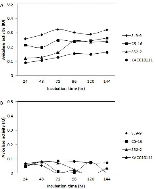
Fig. 13. Avicelase activity in cell-free culture supernatant (A) and cell debris (B) of isolated B. subtilis strains. --- 85
Fig. 14. β-Glucosidase activity in cell-free culture supernatant (A) and cell debris (B) of isolated B. subtilis strains. --- 87
Fig. 15. Xylanase activity in cell-free culture supernatant of isolated B. subtilis strains. --- 89
Fig. 16. Effect of pH on carboxymethylcellulase (CMCase) activity of B. subtilis strains. --- 91
Fig. 17. Effect of temperature on carboxymethylcellulase (CMCase) activity of B. subtilis strains. --- 93
Fig. 18. Effect of reaction time on carboxymethylcellulase (CMCase) activity of B. subtilis strains. --- 95
Fig. 19. Microbial growth of isolated B. subtilis strains at 15℃ (A), 25℃ (B), and 35℃ (C) for 5 days. --- 98
Fig. 20. Carboxymethylcellulase (CMCase) activity of isolated B. subtilis strains at 15℃ (A), 25℃ (B), and 35℃ (C) for 5 days. --- 99
x
-Fig. 21. Effect of initial pH on cell growth (A) and relative activity of CMCase (B) of isolated B. subtilis strains. --- 101 Fig. 22. Change of diameter of clear halos by B. subtilis. SL9-9 on CMC
agar medium adjusted initial pH in the range of 4 to 10. --- 102 Fig. 23. Change of diameter of clear halos by B. subtilis C5-16 on CMC
agar medium adjusted initial pH in the range of 4 to 10. --- 103 Fig. 24. Change of diameter of clear halos by B. subtilis S52-2 on CMC
agar medium adjusted initial pH in the range of 4 to 10. --- 104 Fig. 25. Change of diameter of clear halos by B. subtilis KACC10111 on
CMC agar medium adjusted initial pH in the range of 4 to 10. --- 105 Fig. 26. Effect of nitrogen sources on cell growth and CMCase activity of
isolated B. subtilis strains. --- 107 Fig. 27. Effect of carbon sources on cell growth and CMCase activity of
isolated B. subtilis strains. --- 109 Fig. 28. Effect of CMC concentration on cell growth and CMCase activity
of isolated B. subtilis strains. --- 111 Fig. 29. Effect of shaking speed on cell growth of isolated B. subtilis
strains. --- 113 Fig. 30. Cultural properties of B. subtilis SL9-9 under optimized conditions. 118 Fig. 31. CMCase and avicelase activities of B. subtilis SL9-9 when using
rice bran. --- 122 Fig. 32. CMCase activity during growth of B. subtilis SL9-9 under 1vvm-
aeration or none-aeration condition in 5 L bioreactor. --- 124 Fig. 33. CMCase activity of B. subtilis SL9-9 during cultivation in liquid
and solid media. --- 126 Fig. 34. Nucleotide and deduced amino acid sequences of cellulase gene of
B. subtilis SL9-9. --- 142 Fig. 35. Nucleotide and deduced amino acid sequences of cellulase gene of
B. subtilis C5-16. --- 144 Fig. 36. Nucleotide and deduced amino acid sequences of cellulase gene of
B. subtilis S52-2. --- 146 Fig. 37. Phylogenetic tree resulting from complete sequencing of cellulase
genes. --- 149 Fig. 38. Alignment of deduced amino acid sequences of endo-β-1,4-
xi
-Fig. 39. Shoot length of cucumber in organic bedsoils with different ratios of B. subtilis SL9-9 and organic liquid manure in cucumber seedlings for 30 days. --- 169 Fig. 40. Root length of cucumber in organic bedsoils with different ratios
of B. subtilis SL9-9 and organic liquid manure in cucumber seedlings for 30 days. --- 171 Fig. 41. Stem diameter of cucumber in organic bedsoils with different ratios
of B. subtilis SL9-9 and organic liquid manure in cucumber seedlings for 30 days. --- 173 Fig. 42. Shoot fresh weight of cucumber in organic bedsoils with different
ratios of B. subtilis SL9-9 and organic liquid manure in cucumber seedlings for 30 days. --- 175 Fig. 43. Root fresh weight of cucumber in organic bedsoils with different
ratios of B. subtilis SL9-9 and organic liquid manure in cucumber seedlings for 30 days. --- 177 Fig. 44. Shoot dry weight of cucumber in organic bedsoils with different
ratios of B. subtilis SL9-9 and organic liquid manure in cucumber seedlings for 30 days. --- 179 Fig. 45. Root dry weight of cucumber in organic bedsoils with different
ratios of B. subtilis SL9-9 and organic liquid manure in cucumber seedlings for 30 days. --- 181 Fig. 46. Leaf area of cucumber in organic bedsoils with different ratios of
B. subtilis SL9-9 and organic liquid manure in cucumber seedlings
for 30 days. --- 183 Fig. 47. Chlorophyll content of cucumber in organic bedsoils with different
ratios of B. subtilis SL9-9 and organic liquid manure in cucumber seedlings for 30 days. --- 185 Fig. 48. S/R ratio of cucumber in organic bedsoils with different ratios of
B. subtilis SL9-9 and organic liquid manure in cucumber seedlings
for 30 days. --- 187 Fig. 49. pH of organic bedsoils with different ratios of B. subtilis SL9-9
and organic liquid manure. --- 189 Fig. 50. Electronic conductivity (EC) of organic bedsoils with different
ratios of B. subtilis SL9-9 and organic liquid manure. --- 190 Fig. 51. Available phosphate of organic bedsoils with different ratios of B.
xii
-Fig. 52 Exchangeable potassium of organic bedsoils with different ratios of
B. subtilis SL9-9 and organic liquid manure. --- 192
Fig. 53. Exchangeable calcium of organic bedsoils with different ratios of B. subtilis SL9-9 and organic liquid manure. --- 193
Fig. 54. Exchangeable magnesium of organic bedsoils with different ratios of B. subtilis SL9-9 and organic liquid manure. --- 194
Fig. 55. Population of B. subtilis SL9-9 in bedsoils during cucumber seedling for 33 days. --- 196
Fig. 56. Dehydrogenase (DHA) activity of bedsoils with B. subtilis SL9-9 and organic liquid manure. --- 198
Fig. 57. Survival of B. subtilis SL9-9 after seedling for 33 days. --- 199
Fig. 58. Photo of cucumber seedling. --- 200
Fig. 59. Yield of cucumber in field trial. --- 202
Fig. 60. Nitrogen fixation of isolated B. subtilis strains. --- 205
Fig. 61. Phosphate solubilization of isolated B. subtilis strains. --- 206
Fig. 62. ACC deaminase activity of isolated B. subtilis strains. --- 207
xiii
-Table 1. Chemical composition of some typical cellulose-containing materials --- 19
Table 2. Systematic positions of the enzymes capable of splitting 1,4-β -glucosidic bonds in the general structural classification of glucosyl hydrolases --- 21
Table 3. Classification of cellulose binding domains --- 23
Table 4. Linker sequences of some cellulolytic enzymes and their comparison with central parts of family I CBD sequences --- 26
Table 5. Microorganisms having cellulolytic activities --- 28
Table 6. Substrates containing β-1,4-glucosidic bonds hydrolyzed by cellulases and their detections --- 29
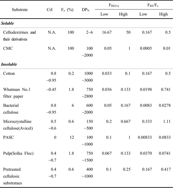
Table 7. Summary of typical values of model celluloses for crystallinity index(CrI), the fraction of β-glucosidic bond accessible to cellulase(Fa), which is estimated by maximum cellulase adsorption capacity(Zhang and Lynd, 2004), the number average of degree of polymerization(DPN), the fraction of reducing ends(FRE), and relative ratio of FRE/Fa --- 30
Table 8. Potential applications of CELs and related enzymes and/or microorganisms in agriculture, biotechnology and bioenergy --- 31
Table 9. CMC basal medium --- 37
Table 10. Some nitrogen sources --- 43
Table 11. Additive concentration of metal ions --- 44
Table 12. Conditions of mass culture medium --- 45
Table 13. Samples collected for screening of cellulolytic microbes --- 48
Table 14. The cellulolytic clones detected by clear halos in the first round --- 49
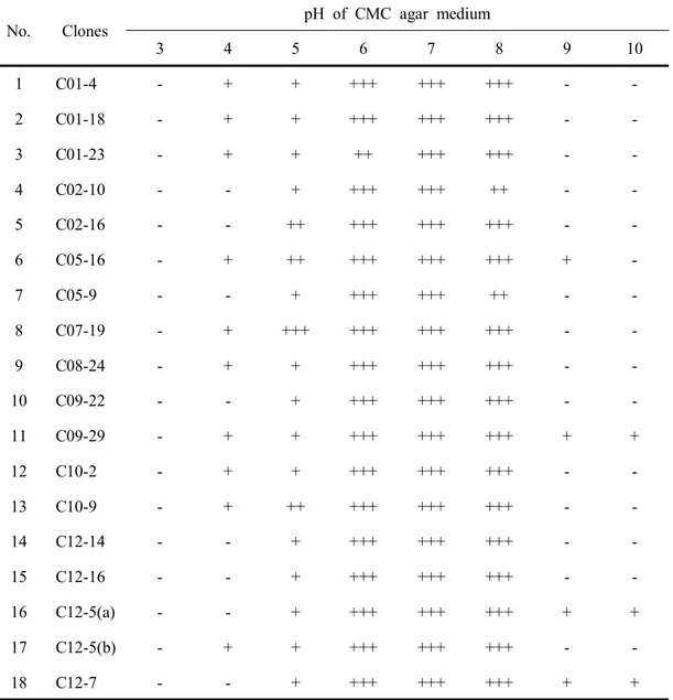
Table 15. Selection of some clones from soils based on clear zone diameters of cellulolytic activities on trypan blue agar plates --- 52
Table 16. Selection of some clones from composts based on clear zone diameters of cellulolytic activities on trypan blue agar plates --- 53
Table 17. Selection of some clones from slurry based on clear zone diameters of cellulolytic activities on trypan blue agar plates at neural pH --- 55
xiv
-Table 18. Selection of some clones from slurry based on clear zone diameters of cellulolytic activities on trypan blue agar plates at
alkali pH --- 58
Table 19. Selection of some clones from cultured native microbes based on clear zone diameters of cellulolytic activities on trypan blue agar plates --- 61
Table 20. Cell growth of the clones selected from soils in the pH range of 3-10 63 Table 21. Cell growth of the clones selected from composts in the pH range of 3-10 64 Table 22. Cell growth of the clones selected from cultured native microbes in the pH range of 3-10 --- 65
Table 23. Cell growth of the clones selected from animal waste slurry in the pH range of 3-10 --- 66
Table 24. Morphological and physiological properties of isolated cellulolytic bacteria --- 69
Table 25. Biochemical properties of isolated cellulolytic bacteria. --- 71
Table 26. Comparison of 16S rRNA gene sequences with those of other bacteria --- 77
Table 27. Effect of incubation temperature on cell growth of isolated B. subtilis strains --- 97
Table 28. Effect of metal ions on cell growth of isolated B. subtilis strains -- 115
Table 29. Effect of metal ions on CMCase activity of isolated B. subtilis strains --- 116
Table 30. Production of cellulase from different agricultural wastes by Bacillus isolates --- 120
Table 31. Scale-up culture condition for cellulase production by B. subtilis SL9-9 --- 127
Table 32. Homology among nucleotide sequences of cellulase genes from Bacillus strains --- 148
Table 33. Nfb medium contents --- 163
Table 34. DF minimal salts media --- 164
Table 35. Test pathogens and culture conditions --- 165
Table 36. Effect of organic bedsoils with different ratios of B. subtilis SL9-9 and organic liquid manure on the germination of cucumber seedlings 167 Table 37. Plant growth-promoting activities of B. subtilis isolates --- 204
1
-LITERATURE REVIEWS
1. Cellulose
Cellulose is the primary product of photosynthesis in terrestrial environments, and the most abundant renewable bioresource produced in the biosphere (~100 billion dry tons year-1) (Holtzapple, 1993; Jarvis, 2003; Zhang and Lynd, 2004). Of great scientific importance is access to cellulose using enzymatic and chemical methods, respectively, developed during the last decade.
Cellulose is a linear condensation polymer consisting of D-anhydro glucopyranose joined together by β-1,4-glycosidic bonds with a degree of polymerization (DP) from 100 to 20,000 (O'Sullivan, 1997; Zhang and Lynd, 2004). Anhydrocellobiose is the repeating unit of cellulose (Fig. 1). Coupling of adjacent cellulose chains and sheets of cellulose by hydrogen bonds and van der Waals forces results in a parallel alignment and a crystalline structure with straight, stable supra-molecular fibers of great tensile strength and low accessibility (Notley et al., 2004; Zhbankov, 1992) as shown in Fig. 2. The cellulose molecule is very stable, with a half life of 5~8 million years for β-glucosidic bond cleavage at 25℃ (Wolfenden and Snider, 2001), while the much faster enzyme-driven cellulose biodegradation process is vital to return the carbon in sediments to the atmosphere (Berner, 2003; Cox et al., 2000). The main source of cellulose is the occurrence of this polysaccharide in different types of plants often combined with other biopolymers. The primary occurrence of cellulose is the existing lignocellulosic material in forests, with wood as the most important source. Other cellulose-containing materials include agriculture residues, water plants, grasses and other plant substances. Besides cellulose, they contain hemicelluloses, lignin and a comparably small amount of extractives (Klemm, 2005). Commercial cellulose production concentrates on harvested sources such as wood or on naturally highly pure sources such as cotton (Table 1).
2
Cellulose biodegradation by cellulases (CELs) and cellulosomes, produced by numerous microorganisms, represents a major carbon flow from fixed carbon sinks to atmospheric CO2 (Berner, 2003; Falkowski et al., 2000; Melillo et al., 2002), is very important in several agricultural and waste treatment processes (Angenent et al., 2004; Das and Singh, 2004; Haight, 2005; Hamer, 2003; Schloss et al., 2005), and could be widely used to produce sustainable biobased products and bioenergy to replace depleting fossil fuels (Demain et al., 2005; Galbe and Zacchi, 2002; Hoffert
et al., 2002; Kamm and Kamm, 2004; Moreira, 2005; Reddy and Yang, 2005;
Wyman, 2003).
2. Cellulase
Cellulase (CELs) is a collective term referring to enzymes able to hydrolyze cellulose (Bhat and Bhat, 1997). Although cellulose is a homopolymer of repeated units of cellobiose, the β-1,4-glycosidic linkages make the structural organization highly ordered and tightly packed (crystallinity), with few amorphous regions. To achieve complete hydrolysis of cellulose, three categories of CELs are required. Firstly, endoglucanases (EG; endo-1,4-β-D-glucanase, EC 3.2.1.4), preferably, attack amorphous regions and randomly cleave the internal bonds of the glycan chains, thus providing reducing or nonreducing ends of cellooligosaccharides for cellobiohydrolases (CBH; or exoglucanase, 1,4-β-D-glucan-cellobiohydrolase, EC 3.2.1.91) to attack. CBH then hydrolyzes those chain ends in the processive manner, yielding cellobiose as the major product. Lastly, β-glucosidase (BG; cellobiase, β-D-glucoside -glucanohydrolase, EC 3.2.1.21) further hydrolyzes cellobiose to glucose and also releases glucose from the nonreducing ends of soluble cellooligosaccharides (Fig. 3) (Jørgensen et al., 2007; Lynd et al., 2002).
Unlike soluble substrates that can diffuse the active sites of enzymes, cellulose is insoluble; thus, CELs, on the contrary, have to diffuse, attach, and move the segment of the cellulose polymer to their active sites (Wilson, 2011). Most CELs are modular
3
-proteins comprising discrete catalytic modules that typically appended one or more carbohydrate-binding modules (CBMs) joined by a flexible linker (Shoseyov et al., 2006). The CBM functions as a cellulose probe, in which the main responsibility is binding the enzyme to the cellulose and increasing the effective concentration of enzymes on the surface of the cellulose (Araki et al., 2010). In addition, some CBMs are known to possess the ability to disrupt crystalline cellulose (Shoseyov et
al., 2006). Therefore, the presence of CBMs appears to be important in enhancing
the enzymatic activity toward insoluble polysaccharides, as well as crystalline cellulose.
Classification of cellulases and hemicellulases according to the structural features of their CDs was first introduced in the end of the 1980s and beginning of the 1990s (Gilkes et al., 1991). It was based on hydrophobic cluster analysis (HCA) and later spread to all glycosyl hydrolases. According to HCA methodology, protein amino acid sequence is represented as two-dimensional longitudinal section of a cylinder formed by a hypothetical helical folding of polypeptide chain. Current classification of cellulase-hemicellulase enzymes is partially reproduced in the Table 2. Corresponding CDs are grouped in 15 families: 5/А, 6/B,7/C, 8/D, 9/E, 10/F, 11/G, 12/H, 26/I, 44/J, 45/K, 48/L, 51, 60, and 61, three of which (5/A, 9/E, and 45/K) being additionally divided in subfamilies. Separated by the slash lettering designations correspond to the initial structural nomenclature of cellulases introduced in 1991. Besides cellulases, other types of glycosyl hydrolases involved in plant cell wall polysaccharide degradation are also included (Rabinovich et al., 2002).
CBD is the second important and the most wide spread element of cellulase structure involved in cellulose transformation. In fact, CBD often plays the role of a recognition factor, which is used by enzyme producing microorganism to address secreted polysaccharide hydrolases to the plant cell wall to be decomposed. The first evidence of their existence was obtained more than two decades ago (Rabinovich, 1977). Since then, the number of CBDs identified as the elements of cellulases, xylanases, mannanases, and other enzymes now exceeds 150 and constantly increases.
4
-Current structural classification of CBDs includes at least thirteen different families (Mattinen et al., 1997). Variable localization of CBDs (at the С or N terminus, or in the middle of polypeptide chain) as well as the presence of some identical or even different CBDs in one protein molecule, and finally, the existence of non hydrolytic proteins containing only CBD repeats suggest an independent function of these structural modules and high level of their autonomy. Therefore, their coupling with the enzyme CDs may be a recent evolutionary event. Table 3 illustrates the CBD classification developed by the scientists of the University of British Columbia (Vancouver). It aims to overview the spread of CBDs among different species as well as the diversity of their structural forms and localization in the molecules of different proteins. As seen from this table, bacterial CBDs significantly differ in their polypeptide chain length and form nearly a decade of families (II to X), large families II and III being additionally divided in subfamilies (a) and (b). Contrary to that, known eucaryotic CBDs are (with minor exceptions) highly homologous and are grouped in one family I. Apart from CBDs originating from the enzymes of basidiomycetous, filamentous, and anaerobic fungi, this family also includes CBDs of the polysaccharide binding protein from the alga Porphyra purpurea, which is, in fact, the fourfold repeat of fungal CBD. However, family does not include any procaryotic CBDs (Tomme et al., 1995).
In most cases CDs and CBDs are separated in the molecules of cellulases and other enzymes by linker sequences. Usually, linkers comprise flexible disordered chains rich in proline and hydroxy amino acid residues (serine and threonine), as well as glycine and alanine (so-called PT-linkers similar with extensins of plant cell walls) (Table 4) (Gilkes et al., 1991). Their length can vary from 5~6 to 100 residues, although most often is limited by the range 20 to 50 residues. In many linkers repeated motifs of 4~7 residues can be identified, where some positively, or negatively charged, or hydrophobic residues are inserted within PTS-rich sequence. Periodicity with high proline content can result in collagen-like secondary structure typical for extensins (Tomme et al., 1995). However, their crystal structure
5
-cannot be obtained because numerous threonines serve as O-glycosylation sites, while glycines provide increased flexibility. Because of this, linker sequences along with attached CBDs are usually removed by specific proteolysis before crystallization. Proteolysis occurs in the most flexible part of linker usually adjacent to one the functional domains, where G and Р residues are localized. More rigid linker parts containing numerous hydroxyamino acids are supposed to be protected from proteolysis by glycosylation (Rabinovich et al., 2002).
Linkers are believed to provide spatial separation of CDs from CBDs to allow their autonomous function on the surface of insoluble substrate. Rigid glycosylated part is responsible for spatial separation, whereas flexible part provides autonomous domain function and is supposed to play a role of a hinge. Reduction of linker’s length has almost no effect on binding and within the certain limits only slightly decreases enzyme activity towards insoluble substrate. However, mutant fungal cellobiohydrolases I with deleted hinge and rigid parts of linker demonstrated reduced activity towards ordered cellulose while had almost the same affinity for the insoluble substrate (Srisodsuk et al., 1993). Therefore, the role of linkers may not be restricted by the solely spatial separation of functional domains; rather, they provide a concerted action of all parts of the enzymatic molecular machine on the cellulose surface. It is interesting to note in this respect, that extensins, the proteoglycans of primary cell walls of higher plants, whose amino acid sequence reveals similarity with linkers, play, according to one of hypotheses, a principal role in the shift of protein matrix along cellulose fibers during expansion growth of plant cells (Rabinovich et al., 2002).
Rabinovich et al. (1984) suggested that It was obvious that the mode of action of a certain enzyme on cellulose surface was defined by a number of parameters: length and structure of its active site, the strength of fixation of the polypeptide loops forming active site, the probability of free rotation of the segments of polymeric substrate molecule within the active site, affinities of different subsites for monomeric units of polymer, location of CBD with respect to CD and its affinity for cellulose
6
-surface, length, and degree of conformational elasticity of the interdomain linker, the presence of other domains in the enzyme structure, which can form protein-protein aggregate on cellulose surface. Because of strictly topochemical character of the overall process, structural data should be considered in the context of heterogenous kinetics and thermodynamic, taking into account evolution of the enzyme-substrate interactions during hydrolysis.
3. Cellulolytic microorganisms
Cellulolytic microorganisms are found among extremely variegated taxonomic groups as shown in Table 5. Most belong to eubacteria and fungi, but anaerobic, cellulose-degradation protozoa have also been identified in the rumen (Coleman, 1978). Cellulolytic microorganisms can be found in all biota where cellulosic waste accumulates. They usually occur in mixed populations comprising cellulolytic and non cellulolytic species, which often interest synergistically. These interactions lead to the complete degradation of cellulose. which is ultimately converted into carbon dioxide and water under aerobic conditions, and into carbon dioxide, methane, and water under anaerobic conditions (Zhang et al., 2005).
Unlike non-complexed fungal cellulase, anaerobic microorganisms possess complexed cellulase systems, called cellulosomes (Bayer et al., 2004; Demain et al., 2005; Doi and Kosugi, 2004; Schwarz, 2001). Leschine (1995) estimated that anaerobic cellulose degradation could account for only 5~10% of total cellulose biodegradation, but it could be underestimated because anaerobic cellulose hydrolysis is responsible for considerable carbon recycling in the anoxic zones of ponds, lakes, oceans, and intestines of ruminants and guts of termites.
7
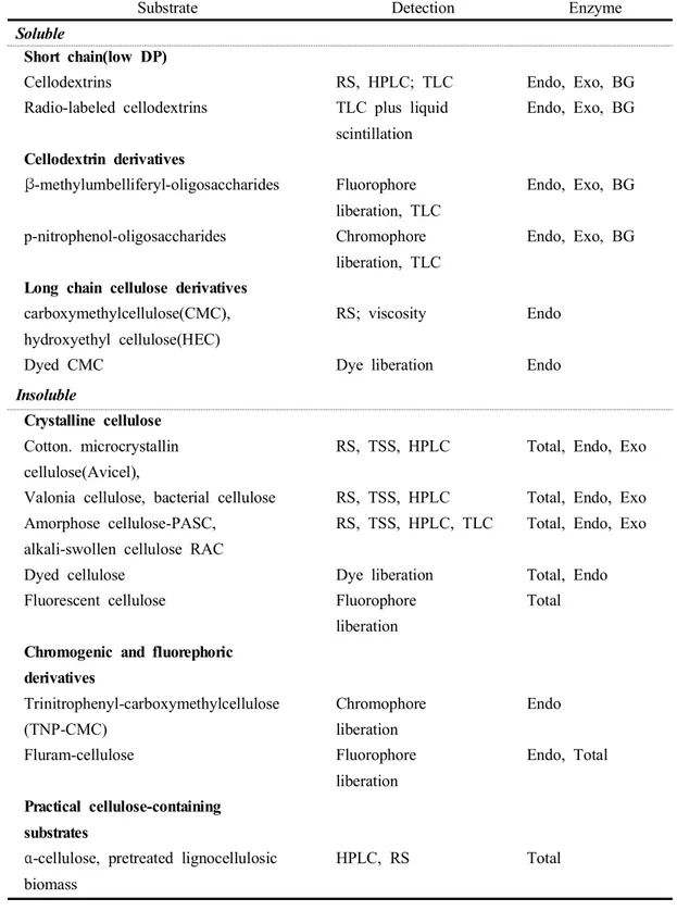
Substrates for cellulase activity assays can be divided into two categories, based on their solubility in water (Table 6).
Soluble substrates. Soluble substrates include low DP cellodextrins from 2 to 6 sugar units and their derivatives, as well as long DP cellulose derivatives (ca. several hundreds of sugar units). They are often used for measuring individual cellulase component activity. Long DP cellulose derivatives can be dissolved in water because of their chemical substitutions. Ionic substituted carboxymethylcellulose (CMC) is often used for determining endoglucanase activity, called CMCase, because endoglucanases cleave intramolecular β-1,4-glucosidic bonds randomly, resulting in a dramatic reduction in the DP (ie, specific viscosity) of CMC. Also, the viscosity of ionic CMC is influenced by pH, ionic strength, and polyvalent cation concentration. Therefore, it is recommended to use nonionic substituted celluloses, such as hydroxyethylcellulose (HEC), for determining endoglucanase activity (Wood and Bhat, 1988).
Insoluble substrates. Insoluble cellulose-containing substrates for cellulase activity assays include nearly pure celluloses (cotton linter, Whatman No. 1 filter paper, bacterial cellulose, microcrystalline cellulose, and amorphous cellulose) and impure cellulose-containing substrates (dyed cellulose, α-cellulose, and pretreated lignocellulose). Native cellulose, referred to as cellulose I, has two distinct crystallite forms, Iα which is dominant in bacterial and algal cellulose, and Iβ, which is dominant in higher plants (Atalla and Vanderhart, 1984). The crystallinity index (CrI) of cellulose, quantitatively measured from its wide range X-ray diffraction pattern (Ramos et al., 2005; Zhang and Lynd, 2004), is not strongly associated with hydrolysis rates (Mansfield et al., 1999; Zhang and Lynd, 2004). Nevertheless, it is still a convenient indicator representing the change in cellulose characteristics for one material before and after treatment. Cotton, bacterial cellulose, and the Valonia ventricosa algal cellulose are examples of highly crystalline cellulose (Fierobe et al., 2002), whereas amorphous cellulose is at the other extreme. Microcrystalline cellulose, filter paper, α -cellulose, and pretreated cellulosic substrates have modest CrI values, and can be
8
-regarded as a combination of crystalline fraction and amorphous fraction, but there is no clear borderline between two fractions. Whatman No.1 filter paper is made from long fiber cotton pulp with a low CrI=∼45% (Dong et al., 1998). Microcrystalline cellulose, called hydrocellulose or avicel (the commercial name), can be purchased from several companies, such as FMC, Merck, and Sigma. Avicel is a good substrate for exoglucanase activity assay, because it has a low DP and relatively low accessibility (i.e., the highest ratio of FRE/Fa) (Table 7). Therefore, some researchers feel that "avicelase" activity is equivalent to exoglucanase activity (Wood and Bhat, 1988). However, some endoglucanases can release considerable reducing sugars from avicel (Zhang and Lynd, 2004). α-Cellulose contains major cellulose and a smallamount of hemicellulose. The commercial Sigma α-cellulose is often used as a reference cellulosic material to evaluate the hydrolysis ability of total cellulase (Kim
et al., 2003).
5. Quantitative assays
All existing cellulase activity assays can be divided into three types: (1) the accumulation of products after hydrolysis, (2) the reduction in substrate quantity, and (3) the change in the physical properties of substrates (Zhang et al., 2006).
The majority of assays involve the accumulation of hydrolysis products, including reducing sugars, total sugars, and chromophores. The most common reducing sugar assays include the dinitrosalicyclic acid (DNS) method (Ghose, 1987; Miller, 1959), the Nelson-Somogyi method (Breuil et al., 1985a; Nelson, 1944; Somogyi, 1952), the 2,2′-bicinchroninate (BCA) method (Waffenschmidt and Janeicke, 1987), the 4-hydroxybenzoylhydrazine (PAHBAH) method (Lever et al., 1973), and the ferricyanide methods (Kidby and Davidson, 1973). Total soluble sugars, regardless of their chain lengths, can be measured directly by the phenol-H2SO4 method (Zhang and Lynd, 2005) or the anthrone-H2SO4 method (Roe, 1955). Glucose can be measured by an enzymatic glucose kit using coupled hexokinase and
9
-glucose-6-phosphate dehydrogenase (Zhang and Lynd, 2004), or HPLC after post-hydrolysis conversion to glucose.
The DNS and Nelson-Somogyi methods are two of the most common assays for measuring reducing sugars for cellulase activity assays because of their relatively high sugar detection range (i.e., no sample dilution required) and low interference from cellulase (i.e., no protein removal required). However, the primary drawback for this method is the poor stoichiometric relationship between cellodextrins and the glucose standard (Coward-Kelly et al., 2003). For example, the results may suffer from an underestimation of cellulase activity when glucose is used as the standard and β -glucosidase is not in excess (Schwarz et al., 1988). The ferricyanide, PAHBAH, and BCA methods, having higher sensitivity to reducing sugar, can detect as little as several micrograms per sample, but suffer from non-specific interference from protein. Total carbohydrate assays, including the phenol-H2SO4 method and the anthrone-H2SO4 method, offer two obvious advantages as compared with reducing sugar assays: a strict stoichiometric relationship between cellodextrins (glucose equivalent) and the glucose standard, and little or no interference from protein. But they are limited for application to pure celluloses, because any carbohydrates and their derivatives can have strong interference readings. Using an enzymatic glucose assay kit or HPLC can overcome nonspecific readings from other sugars, but this requires an extra step—conversion of longer cellodextrins to glucose (Zhang et al., 2006).
The two basic approaches to measuring cellulase activity are (1) measuring the individual cellulase (endoglucanases, exoglucanases, and β-glucosidases) activities, and (2) measuring the total cellulase activity. In general, hydrolase enzyme activities are expressed in the form of the initial hydrolysis rate for the individual enzyme component within a short time, or the end-point hydrolysis for the total enzyme mixture to achieve a fixed hydrolysis degree within a given time. For cellulase activity assays, there is always a gap between initial cellulase activity assays and final hydrolysis measurement (Sheehan and Himmel, 1999). To be most meaningful,
10
-individual cellulase component assays must also be based on a reliable estimation of the amount of individual enzyme component present in the assay. This information permits the calculation of specific activity, i.e., bonds broken per milligram enzyme per unit time.
Endoglucanases. Endoglucanases cleave intramolecular β-1,4-glucosidic linkages randomly, and their activities are often measured on a soluble high DP cellulose derivative, such as CMC with the lowest ratio of FRN/Fa (Table 7). The modes of actions of endoglucanases and exoglucanases differ in that endoglucanases decrease the specific viscosity of CMC significantly with little hydrolysis due to intramolecular cleavages, whereas exoglucanases hydrolyze long chains from the ends in a processive process (Teeri, 1997; Zhang and Lynd, 2004). Endoglucanase activities can be measured based on a reduction in substrate viscosity and/or an increase in reducing ends determined by a reducing sugar assay. Because exoglucanases also increase the number of reducing ends, it is strongly recommended that endoglucanase activities be measured by both methods (viscosity and reducing ends). Because the carboxymethyl substitutions on CMC make some glucosidic bonds less susceptible to enzyme action, a linear relationship between initial hydrolysis rates and serially diluted enzyme solutions requires (1) dilute enzyme preparation, (2) a short incubation period (e.g., 2~4min) or a very low enzyme loading, (3) a low DS CMC, and (4) a sensitive reducing sugar assay. Many workers agree that the BCA method for reducing sugar assay is superior to the DNS method (Carcia et al., 1993). For example, the modified BCA method, which is conducted at 75°C to avoid β -glucosidic bond cleavage during the assay, delivers a strict stoichiometry for the reducing ends of cellodextrins regardless of sugar chain lengths and offers a much higher sensitivity as shown in Table 6 (Zhang and Lynd, 2005). Soluble oligosaccharides and their chromophore-substituted substrates, such as p-nitrophenyl glucosides and methylumbelliferyl-β-D-glucosides, are also used to measure endoglucanase activities based on the release of chromophores or the formation of shorter oligosaccharide fragments, which are measured by HPLC or TLC (Zverlov et
11
-al., 2005). Endoglucanase activities can also be easily detected on agar plates by
staining residual polysaccharides (CMC, cellulose) with various dyes because these dyes are adsorbed only by long chains of polysaccharides (Jang et al., 2003; Kim et
al., 2000; Ten et al., 2004). These methods are semi-quantitative, and are well suited
to monitoring large numbers of samples. Precision is limited because of the relationship between the cleared zone diameters and the logarithm of enzyme activities. For example, differences in enzyme activity levels less than 2-fold are difficult to detect by eye (Sharrock, 1988). Unfortunately, most exoglucanase activities are not detected by these methods, since the processive action of exoglucanases is blocked by carboxymethyl substitutions, which prohibits cellulose chain from shortening. The lack of efficient exoglucanase plate screening method explains some of the difficulty in detecting exoglucanase genes cloned from C.
thermocellum (Demain et al., 2005).
Exoglucanases. Exoglucanases cleave the accessible ends of cellulose molecules to liberate glucose and cellobiose. T. reesei cellobiohydrolase (CBH) I and II act on the reducing and non-reducing cellulose chain ends, respectively (Teeri et al., 1998; Zhang and Lynd, 2004). Avicel has been used for measuring exoglucanase activity because it has the highest ratio of FNR/Fa among insoluble cellulosic substrates (Table 7).
During chromatographic fractionation of cellulase mixtures, enzymes with little activity on soluble CMC, but showing relatively high activity on avicel, are usually identified as exoglucanases. Unfortunately, amorphous cellulose and soluble cellodextrins are substrates for both purified exoglucanases and endoglucanases. Therefore, unlike endoglucanases and β-glucosidases, there are no substrates specific for exoglucanases within the cellulase mixtures (Sharrock, 1988; Wood and Bhat, 1988). Claeyssens and his coworkers (van Tilbeurgh et al., 1982) found that 4-methylumbelliferyl-β-D-lactoside was an effective substrate for T. reesei CBH I, yielding lactose and phenol as reaction products, but it was not a substrate for T.
12
-Tilbeurgh et al., 1982). T. reesei EG I, structurally homologous to CBH I, also cleaves 4-methylumbelliferyl-β-D-lactoside, yet these enzymes can be differentiated by adding cellobiose, an inhibitor that strongly suppresses cellobiohydrolase activity (Claeyssens and Aerts, 1992). T. reesei CBH II does not hydrolyze 4-methylumbelliferyl-β-D-aglycones of either glucose or cellobiose units, but does cleave 4-methylumbelliferyl-β-D-glycosides with longer glucose chains (van Tilbeurgh
et al., 1985). Deshpande et al. (1984) reported a selective assay for exoglucanases in
the presence of endoglucanases and β-glucosidases. This assay is based on the following: (1) exoglucanase specifically hydrolyzes the aglyconic bond of p-nitrophenyl-β-D-cellobioside to yield cellobiose and p-nitrophenol, (2) β-glucosidase activity is inhibited by D-glucono-1,5-δ-lactone (Holtzapple et al., 1990) and (3) the influence of exoglucanase hydrolysis activities must be quantified in the assay procedure in the presence of added purified endoglucanases. However, this technique has its own limitations: (1) CBH II activity cannot be measured using p-nitrophenyl-β-D-cellobioside, (2) the specific activity of the available purified endoglucanases may not be representative of all existing endoglucanases in the mixture, and (3) the product ratio from endoglucanase actions may be influenced by the presence of exoglucanases.
β-D-Glucosidases. β-D-Glucosidases hydrolyze soluble cellobiose and other cellodextrins with a DP up to 6 to produce glucose in the aqueous phase. The hydrolysis rates decrease markedly as the substrate DPs increase (Zhang and Lynd, 2004). The term "cellobiase" is often misleading due to this key enzyme's broad substrate specificity beyond a DP of 2. β-D-glucosidases are very amenable to a wide range of simple sensitive assay methods, based on colored or fluorescent products released from p-nitrophenyl-β-D-1,4-glucopyranoside (Strobel and Russell, 1987), β-naphthyl-β-Dglucopyranoside, 6-bromo-2-naphthyl-β-D-glucopyranoside (Polacheck et al., 1987), and 4-methylumbelliferyl-β-D-glucopyranoside (Setlow et al., 2004). Also, β-D-glucosidase activities can be measured using cellobiose, which is not hydrolyzed by endoglucanases and exoglucanases, and using longer cellodextrins,
13
-which are hydrolyzed by endoglucanases and exoglucanases (McCarthy et al., 2004). Total cellulase. The total cellulase system consists of endoglucanases, exoglucanases, and β-D-glucosidases, all of which hydrolyze crystalline cellulose synergically. Total cellulase activity assays are always measured using insoluble substrates, including pure cellulosic substrates such as Whatman No. 1 filter paper, cotton linter, microcrystalline cellulose, bacterial cellulose, algal cellulose; and cellulose-containing substrates such as dyed cellulose, α-cellulose, and pretreated lignocellulose. The heterogeneity of insoluble cellulose and the complexity of the cellulase system cause formidable problems in measuring total cellulase activity. Experimental results show that the heterogeneous structure of cellulose (filter paper and bacterial cellulose) gives rise to a rapid decrease in the hydrolysis rate within a short time (less than an hour), even when the effects of cellulase deactivation and product inhibition are taken into account (Valjamae et al., 1998; Zhang et al., 1999). In an attempt to clarify this situation, a functionally based model has been developed to demonstrate that the degree of synergism between endoglucanase and exoglucanase is influenced by substrate characteristics, experimental conditions, and enzyme loading/composition ratio. This model clearly suggests the complexity of total cellulase activity assays and infers that it is nearly impossible to apply the results of the total cellulase activity assay measured on one solid substrate to a different solid substrate. The most common total cellulase activity assay is the filter paper assay (FPA) using Whatman No. 1 filter paper as the substrate, which was established and published by the International Union of Pure and Applied Chemistry (IUPAC) (Ghose, 1987). This assay requires a fixed amount (2 mg) of glucose released from a 50 mg sample of filter paper (i.e., 3.6% hydrolysis of the substrate), which ensures that both amorphous and crystalline fractions of the substrate are hydrolyzed. A series of enzyme dilution solutions is required to achieve the fixed degree of hydrolysis. The strong points of this assay are (1) it is based on a widely available substrate, (2) it uses a substrate that is moderately susceptible to cellulases, and (3) it is based on a simple procedure (the removal of residual substrate is not necessary
14
-prior to the addition of the DNS reagent). However, the FPA is reproduced in most laboratories with some considerable effort and it has long been recognized for its complexity and susceptibility to operators' errors (Coward-Kelly et al., 2003; Decker
et al., 2003). Reliability of results could be influenced by (1) the β-D-glucosidase
level present in the cellulase mixture (Schwarz et al., 1988; Sharrock, 1988), because the DNS readings are strongly influenced by the reducing end ratio of glucose, cellobiose, and longer cellodextrins (Kongruang et al., 2004; Zhang and Lynd, 2005); (2) the freshness of the DNS reagent, which is often ignored (Miller, 1959); (3) the DNS reaction conditions, such as boiling severity, heat transfer, and reaction time (Coward-Kelly et al., 2003); (4) the variations in substrate weight based on the area size (1×6 cm a strip), because this method does not require substrate excess (i.e., substrate amounts strongly influence enzyme activity) (Griffin, 1973); and (5) filter paper cutting methods, because the different papercutting methods such as paper punching, razoring, or scissoring could lead to different accessible reducing ends of the substrate (Zhang and Lynd, 2005). Cotton fiber, microcrystalline cellulose, bacterial cellulose, and algal cellulose are several other common pure cellulosic substrates. Powder microcrystalline cellulose could become a preferred substrate to replace filter paper because (1) it can be rapidly dispensed volumetrically as a slurry and thus permits robotics methods; (2) it can be easily pelleted by centrifugation, and the total sugars released are measured more exactly by the phenol-H2SO4 method than by the DNS assay; (3) it is a more recalcitrant substrate, yielding a more stringent substrate for total cellulase activity than does filter paper; and (4) activities measured on microcrystalline cellulose could more accurately represent hydrolysis ability on pretreated lignocellulose, because its characteristics are closer to those of pretreated lignocelluloses, based on cellulose accessibility to cellulase and the degree of polymerization (Zhang and Lynd, 2004). Sigmacell-20, a readily available microcrystalline cellulose powder, could also be a good alternative substrate for a total cellulase activity assay, replacing Whatman No. 1 filter paper. Keep that in mind, some of the pretreated lignocellulose still contains significant amounts of
15
-hemicellulose and lignin, while microcrystalline cellulose does not contain hemicellulose and lignin. α-Cellulose and pretreated lignocellulose are often used to evaluate the digestibility of commercial cellulase or of a reconstituted cellulase mixture for a prolonged reaction. The primary difference, as compared to cellulase activity assays using model cellulosic substrates, is the time required for assays, which ranges from several minutes to hours for model substrates (initial hydrolysis rate) to several days for pretreated lignocellulose to obtain the final digestibility (cellulose conversion). Clearly, the presence of hemicellulose and even lignin results in more complexity. Again, the desired outcome of the experiment must indicate the substrate chosen, especially in the case of total cellulase performance.
In conclusion, the measurement of isolated individual cellulase activity is relatively easy, but it is still challenging to measure T. reesei CBH I and CBH II activities specifically in the presence of endoglucanases. There is no clear relationship between the hydrolysis rates obtained on soluble substrates and those on insoluble substrates, mainly because of huge differences in substrate accessibility and DP. For insoluble cellulose, it is highly unlikely that any substantial solubilization of crystalline or semicrystalline cellulose will proceed linearly with time, due to varying β-glucosidic bond accessibilities and chain end availability for different regions of fibers. Researchers must state clearly all parameters of their assay conditions, and resist temptation to compare their results to those of other researchers using different substrates, assay methods, etc.
6. Application of cellulase and/or microorganisms
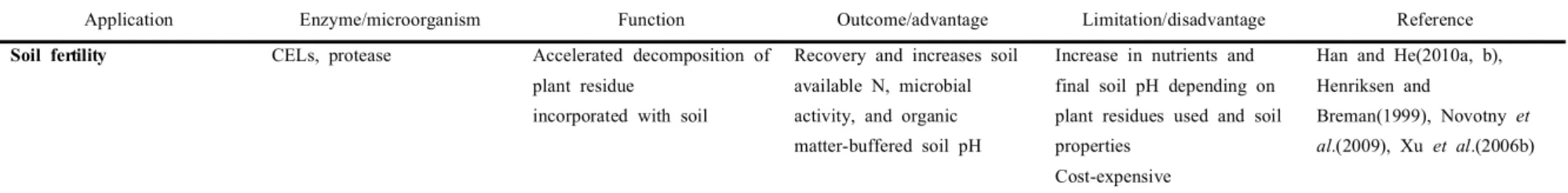
Recently, many researchers have discussed the potential applications of CELs and related enzymes and/or microorganisms in agriculture, biotechnology, and bioenergy as shown in Table 8. Supplementation of CELs to accelerate decomposition of plant residues in soil results in improved soil fertility (Han and He, 2010a, b; Henriksen and Breman, 1999; Novotny et al., 2009; Xu et al., 2006b). So far, applying
16
-CELs/antagonistic cellulolytic fungi to crops has shown to promote plant growth performance, including enhanced seed germination and protective effects (Brummel and Harpster, 2001; Catala et al., 2000; Cosgrove, 2005; Ding et al., 2008b; Payasi
et al., 2009). Their actions are believed mainly to trigger plant defense mechanisms
and/or to act as biocontrol agents that mediate disease suppression (Cotes et al., 1996; Inbar et al., 1994; Moreno et al., 2009). However, the exact interaction between the enzymes/fungi and plants has not been clearly elucidated. CELs have recently shown great potential in enzyme aid extraction of bioactive compounds from plant materials before selective extraction through enhancing release of target molecules, especially those associated with the wall matrix (Barzana et al., 2002; Ishida and Chapman, 2009; Kapasakalidis et al., 2009; Kim et al., 2005; Sun et al., 2005). To date, attempts have been made to formulate CEL preparation for cellulosic-based bioethanol production (Lim and Lian, 2001). The high cost of CELs has created a bottleneck, resulting in an uneconomic production process. The utilization of low-cost carbohydrates, strain improvement, and gene manipulations has been alternatively aimed at reducing the cost of CEL production.
17
18
19
-Table 1. Chemical composition of some typical cellulose-containing materialsa
Source Composition (%)
Cellulose Hemicellulose Lignin Extract
Hard wood 43~47 25~35 16~24 2~8 Soft wood 40~44 25~29 25~31 1~5 Bagasse 40 30 20 10 Coir 32~43 10~20 43~49 4 Corn cobs 45 35 15 5 Corn stalks 35 25 35 5 Cotton 95 2 1 0.4 Flax(retted) 71 21 2 6 Flax(unretted) 63 12 3 13 Hemp 70 22 6 2 Henequen 78 4~8 13 4 Istle 73 4~8 17 2 Jute 71 14 13 2 Kenaf 36 21 18 2 Ramie 76 17 1 6 Sisal 73 14 11 2 Wheat straw 30 50 15 5
20
-Fig. 3. A simplified model of enzymatic hydrolysis of cellulose.
EGs are presumed to first cleave amorphous regions of the cellulose polymer, thus providing reducing or nonreducing ends of cellooligosaccharides for CBHs to attack and processively hydrolyze those chain ends. BGs further hydrolyze the resulting products, cellobiose, to glucose and also release glucose from the nonreducing ends of the higher oligomers (Redrawn from Lynd et al., 2002).
21
-Table 2. Systematic positions of the enzymes capable of splitting 1,4-β-glucosidic
bonds in the general structural classification of glucosyl hydrolasesa
Family Type and origin of the enzyme EC Clan Fold Type of catalysis
1 2 3 4 5 6
5/A1 fungal endo-1,4-β-mannanases; endo glucanases of aerobic and anaerobic bacteria
3.2.1.78 3.2.1.4
GHA TIM-barrel = (α/β)8 barrel = (β/α)8 barrel; active site includes nucleophilic glutamate at the C- terminus of β-strand 7 and general acidbase asparagine-glutamate at C-terminus of β-strand 4 retain configuration of the anomeric carbon in the reaction products
5/A2 endoglucanases of ctinomycetes and aerobic and anaerobic bacteria, as well as animals (nematodes)
3.2.1.4 same same same
5/A3 exo-1,3-β-glucanases; endoglucanases/1,3-1,4-β -glucanases; 1,3-βglucanases (yeast, Clostridium) 3.2.1.58 (cryst.) 3.2.1.4/73 3.2.1.39
same same; insertion of an additional helical subdomain near active site
same
5/A4 endoglucanases and mannanases of actinomycetes, aerobic and anaerobic bacteria, and anaerobic fungi
3.2.1.4 3.2.1.78
same same same
5/A5 endoglucanases of filamentous fungi and aerobic bacteria
3.2.1.4 same same same
5 endo-1,6-β-glucanases 3.2.1.75 3.2.1.123
same same same
6/B endoglucanases and cellobiohydrolases of aerobic bacteria, actinomycetes, and anaerobic and filamentous fungi 3.2.1.91 (cryst.) 3.2.1.4 (cryst.) incomplete TIM barrel inverse configuration
7/С cellobiohydrolases and endoglucanases of filamentous fungi 3.2.1.91 (cryst.) 3.2.1.4 GHB concanavalin A type β-sandwich retain configuration
8/D 1,31,4βglucanases and endoglu canases of aerobic and anaerobic bacteria 3.2.1.73 3.2.1.4 (cryst.) not defined (α,α)6-barrel inverse
9/E1 endoglucanases of actinomycetes and aerobic and anaerobic bacteria; cellobiohydrolases of anaerobic bacteria
3.2.1.4 (cryst.) 3.2.1.91
same same same
9/E2 endoglucanases of aerobic and anaerobic bacteria, plants, and insects (termites); endo/exoglucanase of actinomycetes
3.2.1.4 (cryst.)
3.2.1.4/91
same same; additional strongly fixed type III CBD
22 -Table 2. (contd.)
1 2 3 4 5 6
10/F xylanases and
xylanases/endoglucanases of actinomycetes, aerobic bacteria, anaerobic archaeand eubacteria, and aerobic and anaerobic fungi; bacterial xylanase /exoglucanase
3.2.1.4/8 3.2.1.8/91 (cryst.) 3.2.1.8 (cryst.)
GHA TIM-barrel retain
11/G strictly specific xylanases of aerobic and anaerobic fungi, aerobic and anaerobic bacteria, and actinomycetes 3.2.1.8 (cryst.) GHC “ribcage”like β-sandwich same
12/H endoglucanases of aerobic fungi, actinomycetes, aerobic bacteria, and anaerobic archaebacteria
3.2.1.4 (cryst.)
same same; two β-sheets of six and nine strands, and α-helix across
same
26/I endoglucanases; endoglucanases /xylanases; endomannanases of aerobic and anaerobic bacteria
3.2.1.78 3.2.1.4 3.2.1.4/8
GHA TIM-barrel same
44/J endoglucanases of aerobic and anaerobic bacteria
3.2.1.4 not defined
not defined inverse
45/K1 endoglucanases of filamentous and anaerobic fungi and aerobic bacteria 3.2.1.4 not defined 6-β-barrel same 45/K2 endoglucanase of mollusks 48/L endoglucanases; cellobiohydrolases; endoglucanases/cellobiohydrolases of actinomycetes and aerobic and anaerobic bacteria 3.2.1.4 3.2.1.91 3.2.1.4/91 not defined (α,α)6-barrel same 51 α-L-arabinofuranosidases of ascomycetes and actinomycetes; endoglucanase of F. succinogenes
3.2.1.55 3.2.1.4
GHA TIM-barrel retain
60 Clostridium endoglucanases 3.2.1.4 not defined
not defined not defined
61 endoglucanases of ascoand basidiomycetes
3.2.1.4 not defined
not defined same
Note: EC, enzyme classification according to IUB; (cryst.), proteins with resolved crystalline structure.