Nucl Med Mol Imaging Vol. 43, No. 6, Dec 2009
509
다발성 골수종에서의
18
F-FDG PET의 임상이용
성균관대학교 의과대학 삼성서울병원 핵의학과
이수진․최준영
Clinical Application of
18F-FDG PET in Multiple Myeloma
Su Jin Lee, M.D. and Joon Young Choi, M.D.
Department of Nuclear Medicine, Samsung Medical Center, Sungkyunkwan University School of Medicine, Seoul, Korea
This review focuses on the clinical use of 18F-FDG PET to evaluate multiple myeloma. 18F-FDG PET is useful for diagnosis, staging of multiple myeloma and differential diagnosis of myeloma related disease such as monoclonal gammopathy of undetermined significance or plasmacytoma. For therapy response, 18F-FDG PET may be effective after chemotherapy for multiple myeloma and radiotherapy for plasmacytoma. (Nucl Med Mol Imaging 2009;43(6):509-512) Key Words: Multiple myeloma, 18F-FDG, PET, clinical application
∙Received: 2009. 9. 18. ∙Revised: 2009. 9. 25. ∙Accepted: 2009. 10. 1.
∙Address for reprints: Joon Young Choi, M.D., Ph.D., Department of Nuclear Medicine, Samsung Medical Center, Sungkyunkwan University School of Medicine, 50 Ilwon-dong, Gangnam-gu, Seoul, Korea
Tel: 82-2-3410-2648, Fax: 82-2-3410-2639 E-mail: jynm.choi@samsung.com ※이 연구는 보건복지부 인체구조 영상화 신기술 개발사업(02-PJ3- PG6-EV06-0002)의 지원으로 수행되었음.
서 론
다발성 골수종은 B림프구의 성숙형태인 형질세포가 증
식하여 생기는 암이다. 우리나라에서는 연간 약 500명 정
도 발병하는 드문 암이지만, 예후가 아주 나쁘며, 평균수명
의 증가와 함께 발병빈도가 꾸준히 늘어나고 있다.
1)다발
성 골수종의 진단은 미국의 Southwest Oncology Group
(SWOG)이 제시한 기준이 보편적으로 사용되고 있다. 진
단기준으로는 골수에서 비정상적인 형질세포가 10% 이상
이거나 또는 조직학적으로 진단된 형질세포종, 소변이나
혈청에서의 M단백의 검출, 그리고 용해성 뼈병변이나 병
적골절 등이 있다. 위 3가지 항목을 충족할 때 다발성 골수
종으로 진단을 내릴 수 있지만, 점진적인 M단백의 증가와
연관된 골수의 비정상적인 형질세포 증가의 경우나, 골수
외 형질세포종의 경우는 용해성 뼈병변이 없어도 진단을
내릴 수 있다.
2)다발성 골수종의 진단에 사용되는 영상법으로는 단순
X-선 촬영, MRI 등이 주로 사용되어 왔다. 단순 X-선 촬
영이나 CT는 용해성 뼈병변의 발견에 유용하나, 뼈파괴를
일으키지 않은 뼈병변이나 골수외 병변의 진단이 어려우며,
병변의 활성도를 알 수 없어 치료 후 추적관찰에 제한적이
다.
3)MRI는 골수 병변의 진단에 유용하고 치료반응 평가와
예후예측에 이용된다.
4,5)그러나, 전신 MRI가 아닌 경우에
는 골수외 병변의 진단이 어렵다.
18F-Fluorodeoxyglucose
(
18F-FDG)를 사용한 positron emission tomography
(PET)는 많은 종류의 종양에서 임상적 유용성이 입증되
고 있다. 여기서는
18F-FDG PET의 다발성 골수종에서의
임상적 이용에 대하여 다루고자 한다.
문헌 검토
1. 진단 및 병기결정
다발성 골수종의 진단 및 병기결정에서 PET을 이용한
연구는 많지 않으나, 병의 범위를 정확하게 측정할 수 있다
는 점에서 병의 진단 및 병기결정에 유용하다고 보고되고
있다.
6-14)다발성 골수종과 감별을 요하는 미결정유의성 단
일클론성 감마글로불린혈증(monoclonal gammopathy of
undetermined significance)은
18F-FDG PET에서 유의한
18
F-FDG 섭취가 없고, 활성 골수종(active myeloma)에서
는
18F-FDG 섭취증가가 나타나므로 두 질환의 감별에 유
용하다.
10-12)다발성 골수종의 소수에서는 단일 뼈만을 침
범하는 고립성 형질세포종(solitary plasmacytoma)의 형태
로 나타나거나, 골수외 병변만 침범하는 골수외 형질세포종
(extramedullary plasmacytoma)의 형태로 나타날 수 있다.
Reference evidenceLevels of Type of study subjectsNo. of Characteristics of subjects interpretationsPET images & Diagnostic value of PET Other diagnostic tools Influence on subjects
6 2- Retrospective 6
Diagnosis & staging (n = 3), therapy response (n = 3)
Nonattenuation-cor rected PET, visual analysis
X-ray, CT, MRI, bone scan for lesion verification
Useful to diagnose initial active lesions and to evaluate therapy response
7 2+ Retrospective 13
Diagnosis & staging (n = 4), therapy response (n = 9) Attenuation-correct ed PET, visual analysis, SUV analysis Sensitivity 85% Specificity 92% MR, CT, X-ray, biopsy results for lesion verification
Useful to evaluate initial disease extent, staging and therapy response
8 2++ Prospective 46
Diagnosis & staging (44; multiple myeloma, 2; solitary plasmacytoma) Attenuation-correct ed PET/CT, osteolytic lesion on CT with SUV > 2.5 -> (+) Sensitivity 92% Sensitivity of X-ray 61%, Sensitivity of pelvis/spine MRI 92% Useful to evaluate whole body lesions and therapy response compared with pelvis/spine MRI
9 2++ Prospective 33 Diagnosis & staging
Attenuation-correct ed PET/CT, visual analysis, uptake pattern analysis
Sensitivity 97% Tc-99m MIBI scan, pelvis/spine MRI
PET/CT was superior in the detection of focal lesions, and useful to evaluate whole body.
10 2++ Prospective 43
Diagnosis & staging (28; multiple myeloma, 15; solitary plasmacytoma) Attenuation-correct ed PET, visual analysis, uptake pattern analysis Focal lesion: Sensitivity 92.7%, PPV 100% Diffuse lesion: Sensitivity 83.8-91.9%, Specificity 83.3-100% MR, CT, X-ray, biopsy results for lesion verification
PET showed high sensitivity & specificity.
11 2+ Retrospective 66
Diagnosis & staging (n = 30; 16 multiple myeloma, 14 MGUS a)), Tx response (n = 36; 10 CR, 26 Recur) Attenuation-correct ed PET, visual analysis (-) Useful in the differential diagnosis of myeloma-related diseases and prognosis evaluation
13 2+ Retrospective (45 scans)30 Diagnosis & staging (n = 12), therapy response (n = 33)
Attenuation-correct
ed PET/CT (-)
PEC/CT can detect extramedullary lesions (11%) adjacent to bone compared with PET.
14 2+ Retrospective 22 Diagnosis & staging
Attenuation-correct ed PET, visual & SUV analysis of spines Stage 1, 2; Sensitivity 78%, Stage 3; Sensitivity 80% Spine MRI: Stage 1-2; Sensitivity 86%, Stage 3; Sensitivity 92% In stage 1 and 2, MRI was comparable to PET, whereas in stage 3, MRI was superior to PET.
15 2+ Retrospective 14
Diagnosis & staging (n = 3), therapy response (n = 11) Attenuation-correct ed PET, visual analysis (-) Useful to detect unexpected sites of bone involvement in solitary plasmacytoma patients 16 2+ Retrospective 21
Diagnosis & staging (n = 17), restaging (n = 4)
Attenuation-correct ed PET, visual
analysis Tc-99m MIBI scan
Useful in staging, planning of radiation treatment and restaging in solitary plasmacytoma patients
17 2++ Prospective 24 Diagnosis & staging
Attenuation-correct ed PET, visual analysis, SUV analysis (cut-off = 2.5) Sensitivity 59%, Specificity 75%, PPV 81%, NPV 50% Whole-body MRI: Sensitivity 68%, Specificity 83%, PPV 88%, NPV 59% PET and whole-body MRI were found to have a specificity and positive predictive value of 100%.
a)monoclonal gammopathy of undetermined significance
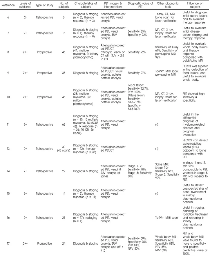
Table 1. Summary of Literature Dealing with the Utility of PET for Staging and Therapy Response of Multiple Myeloma Su Jin Lee, et al. Clinical Application of 18F-FDG PET in Multiple Myeloma
이수진 외. 다발성 골수종에서의 18F-FDG PET의 임상이용 511
이런 아형들의 경우는 전신적인 치료보다는 국소 방사선
치료가 효과적이므로 질병의 범위를 아는 것이 중요한데,
18F-FDG PET은 전신촬영을 한 번에 할 수 있어 위 질환
들의 감별에 유용하므로 치료계획 수립에 도움을 준
다.
10,11,15,16)임상적으로 다발성 골수종이 의심되나 단순
X-선 검사에서 분명하지 않은 환자에서
18F-FDG PET상 활
성 병변을 관찰하여 진단에 도움을 줄 수 있으며, 임상적으
로 생검이 필요한 경우라면 적절한 생검 부위를 제시할 수
있다. 특히, 단일클론성 감마글로불린 분비를 잘 안 하는
골수종의 진단에
18F-FDG PET이 도움이 된다.
11)현재까지 PET/CT를 이용한 다발성 골수종 연구는 적
으나, PET/CT에서 CT상 뼈파괴 병변을 관찰할 수 있고,
PET에서 활성 골수종 병변을 찾을 수 있으므로 다발성 골
수종 진단에 이상적인 진단 도구가 될 수 있다. 또한, 골수
외 병변을 발견하는 데에도 PET/CT가 PET 보다 유용하
다.
9,13,15)최근 한 연구에서는 전신 MRI와
18F-FDG PET의 진단
성적을 보고하였는데, PET은 질병의 범위와 활성도를 평
가하는데 유용하며 특히 국소병변 진단에 우수하다고 하였
다. 반면 전신 MRI는 미만성 골수 침범 진단에 우수하다고
하였다. 두 검사를 같이 시행하였을 경우 특이도와 양성예
측도는 100%였다.
17)2. 재발 평가, 치료효과 판정 및 재병기 설정
18F-FDG PET을 이용하여 재발 진단에 사용된 연구는
별로 없으나, 고립성 형질세포종의 상당수가 3-4년 내에
다발성 골수종으로 진행하게 되므로, 고립성 형질세포종의
환자의 추적검사에 사용할 수 있다. 고립성 형질세포종 환
자의 방사선치료 후 치료반응을 평가하는데도
18F-FDG
PET이 유용하다.
15,16)활성 다발성 골수종은 치료 후
18F-FDG 섭취가 감소되
므로 치료 효과 판정에 유용하다. 치료 후에도 지속적인
18F-FDG 섭취증가를 보이는 경우에는 조기 재발을 예측할
수 있다. 골수 이식 치료 후에도 지속적인
18F-FDG 섭취증
가를 보이는 경우는 예후가 나빠 6개월 이내에 재발을 잘
한다는 연구도 있다.
11.12)최근 한 연구에서는 치료받지 않은 활성 골수종 환자군,
미결정유의성 단일클론성 감마글로불린혈증군, 임상적 관
해를 보인 골수종 환자군, 재발한 골수종 환자군을 대상으
로
18F-FDG PET을 시행하여, 재발한 골수종 군에서 골수
외 침범이 있거나 치료 후에도 지속적인 FDG 섭취가 있으
면서 생존기간이 짧은 점을 보고하여
18F-FDG PET이 골
수종 고위험군을 분류하는데 유용하였다.
11)결 론
18F-FDG PET은 단순 X-선 촬영, MRI와 함께 상호보
완적으로 다발성 골수종의 진단 및 병기결정(권고등급 B),
치료효과 판정 및 재병기 설정(권고등급 C)에 유용한 것
으로 보인다. 다발성 골수종의 재발 진단에는 근거가 없으
므로 권고하지 않는다. 그러나, 아형인 고립성 형질세포종
인 경우에는 재발 진단을 위한 환자의 추적검사에 사용할
수 있다(권고등급 D).
References
1. Incidence of Multiple Myeloma (C90), 1999-2002 year. National Cancer Statistics of Ministry for Health, Welfare and Family Affairs, 2008. http://www.cancer.go.kr/candat/cms_renewal/statics/ stat/1193978 _1611.html
2. Munshi Nikhil C, Longo Dan L, Anderson Kenneth C, "Chapter 106. Plasma Cell Disorders" (Chapter). Fauci AS, Braunwald E, Kasper DL, Hauser SL, Longo DL, Jameson JL, Loscalzo J:
Harrison's Principles of Internal Medicine. 17th Edition:
http://www.accessmedicine.com/content.aspx?aID=2891791. 3. Angtuaco EJ, Fassas AB, Walker R, Sethi R, Barlogie B.
Multiple myeloma: clinical review and diagnostic imaging. Radiology 2004;231:11-23.
4. Van de Berg BC, Lecouvet FE, Michaux L, Labaisse M, Malghem J, Jamart J, et al. Stage I multiple myeloma: value of MR imaging of the bone marrow in the determination of prognosis. Radiology 1996;201:243-6.
5. Baur-Melnyk A, Buhmann S, Dürr HR, Reiser M. Role of MRI for the diagnosis and prognosis of multiple myeloma. Eur J Radiol 2005;55:56-63.
6. Jadvar H, Conti PS. Diagnostic utility of FDG PET in multiple myeloma. Skeletal Radiol 2002;31:690-4.
7. Bredella MA, Steinbach L, Caputo G, Segall G, Hawkins R. Value of FDG PET in the assessment of patients with multiple myeloma. AJR Am J Roentgenol 2005;184:1199-204.
8. Zamagni E, Nanni C, Patriarca F, Englaro E, Castellucci P, Geatti O, et al. A prospective comparison of 18 F-fluorodeoxy-glucose positron emission tomography-computed tomography, magnetic resonance imaging and whole-body planar radiographs in the assessment of bone disease in newly diagnosed multiple myeloma. Haematologica 2007;92:50-5.
9. Fonti R, Salvatore B, Quarantelli M, Sirignano C, Segreto S, Petruzziello F, et al. 18F-FDG PET/CT, 99mTc-MIBI, and MRI in evaluation of patients with multiple myeloma. J Nucl Med 2008; 49:195-200.
10. Schirrmeister H, Bommer M, Buck AK, Müller S, Messer P, Bunjes D, et al. Initial results in the assessment of multiple myeloma using 18F-FDG PET. Eur J Nucl Med Mol Imaging 2002;29:361-6.
11. Durie BG, Waxman AD, D'Agnolo A, Williams CM. Whole- body 18F-FDG PET identifies high-risk myeloma. J Nucl Med. 2002;43:1457-63.
12. Durie BG. The role of anatomic and functional staging in myeloma: description of Durie/Salmon plus staging system. Eur J Cancer 2006;42:1539-43.
Su Jin Lee, et al. Clinical Application of 18F-FDG PET in Multiple Myeloma
512
13. Lee SJ, Choi JY, Kim KH, Lee EJ, Cho YS, Hyun SH, et al.
18
F-FDG PET/CT in multiple myeloma: Is it necessary to include the skull and lower extremity distal to mid-thigh? Nucl Med Mol Imaging 2008;42:39-43.
14. Hur J, Yoon CS, Ryu YH, Yun MJ, Suh JS. Comparative study of fluorodeoxyglucose positron emission tomography and magnetic resonance imaging for the detection of spinal bone marrow infiltration in untreated patients with multiple myeloma. Acta Radiol 2008;49:427-35.
15. Nanni C, Rubello D, Zamagni E, Castellucci P, Ambrosini V, Montini G, et al. 18F-FDG PET/CT in myeloma with presumed
solitary plasmocytoma of bone. In Vivo 2008;22:513-7.
16. Kim PJ, Hicks RJ, Wirth A, Ryan G, Seymour JF, Prince HM, et al. Impact of 18F-fluorodeoxyglucose positron emission tomo-graphy before and after definitive radiation therapy in patients with apparently solitary plasmacytoma. Int J Radiat Oncol Biol Phys 2009;74:740-6.
17. Shortt CP, Gleeson TG, Breen KA, McHugh J, O'Connell MJ, O'Gorman PJ, et al. Whole-body MRI versus PET in assessment of multiple myeloma disease activity. AJR Am J Roentgenol 2009;192:980-6.