BTK functions as a corepressor of Nurr1
in human neural stem cells
by
Myung Sun Park
Major in Neuroscience
Department of Biomedical Sciences
The Graduate School, Ajou University
BTK functions as a corepressor of Nurr1
in human neural stem cells
by
Myung Sun Park
A Dissertation Submitted to The Graduate School of Ajou University
in Partial Fulfillment of the Requirements for the Degree of
Master of Biomedical Sciences
Supervised by
Myung Ae Lee, Ph.D.
Major in Neuroscience
Department of Biomedical Sciences
The Graduate School, Ajou University
This certifies that the dissertation
of Myung Sun Park is approved.
SUPERVISORY COMMITTEE
The Graduate School, Ajou University
June, 25th, 2010
ACKNOWLEDGENENT
2년이 넘는 시간을 보내고 이 논문을 쓰면서 감사를 보낼 사람이 너무 많습니다. 우선, 서른이라는 적지 않은 나이에 공부할 수 있는 기회를 주시고 부족한 저에 게 아낌없는 지도와 사랑을 주신 지도교수 이명애 교수님께 감사의 마음을 전합 니다. 더불어 이 논문을 심사해주신 김혜선, 이준규 교수님께도 감사 드립니다. 나이도 많고 투정도 심한 늙은 후배를 처음 파이펫 잡는 법부터 가르치느라 고 생이 많았을 수민이, 실험실의 영원한 선배이자 유일하게 나처럼 가정을 꾸려 이 런저런 소소한 얘기까지 통할 수 있었던 정용씨, 학기를 같이 시작하고 수업도 같이 듣고 실험도 같이 하며 많은 얘기를 나누었던 든든한 동기 동철이, 짧은 시 간이었지만 긴 시간을 만났던 것 같은 친구 용근이, 실험실이 아니면 만나보기 힘들었을 나이터울을 가진 귀엽고 싹싹한 은지, 처음 대학원에 들어와서 흔들리 는 마음을 다잡을 수 있도록 용기와 조언을 주셨던 감사한 안정희 박사님, 그리 고, 마지막으로 정말 친동생처럼 나의 갖은 구박을 견뎌내며 많이 싸우기도 하고 울기도 하고 웃기도 하며 티격태격 식구처럼 지낸 수연이까지 모두 감사합니다. 또 20년이 가까운 시간 동안 변함없이 나를 응원해주고 아껴주는 내 소중한 친 구 현진이, 려진이에게도 감사의 마음을 전합니다. 마지막으로 언제나 나의 가장 가까운 곳에서 힘과 사랑을 주는 우리 멋쟁이 신 랑님, 엄마의 늦은 공부를 위해 세상에 나오길 오랫동안 기다렸을 세상에서 가장 사랑하는 우리 아들 대현이, 그리고 큰며느리의 늦은 공부에 언제나 든든하게 응 원을 보내주신 우리 시부모님, 낳아주시고 길러주셨고 언제나 내편인 사랑하는 친정부모님께도 깊은 감사와 사랑을 보냅니다.ABSTRACT
BTK protein functions as a corepressor of Nurr1 in human
neural stem cells
Nurr1, a member of the orphan nuclear receptor superfamily, was demonstrated to be of critical importance in the developing central nervous system, where it is required for the generation of midbrain dopaminergic cell. Although its role during dopaminergic neurogenesis has been extensively studied, its transcriptional regulation of TH, the rate-limiting enzyme of dopamine biosynthesis, is not well understood.
In our previous study, we found that Nurr1 directly mediated transcriptional activation or repression of TH gene through NBRE-A site and a key regulating complex of TH expression, that may be formed in NBRE-A site during dopaminergic neurogenesis. In recent years, a growing number of cofactor has been discovered that participate in the regulation of the transcriptional activity of TH promoter. So, we present the identification of a cofactors binding in and around NBRE-A, which differentially regulates the transcriptional activity of the human TH promoter in human neural stem cell, F3, and human neuroblastoma cell, SH-SY5Y. In order to identify the transcription factors that mediate these contradictory functions in each of the cell types, we conducted biotin-labeled oligonucleotide of Nurr1-binding element (NBRE) and streptavidin-agarose beads pull-down assays. We identified three proteins, bruton tyrosine kinase-associated protein (BTK) and topoisomerase II β (TOPO II β) in F3 and poly (ADP-ribosyl) transferase (PARP) in SH-SY5Y. To know the interaction of
these candidate proteins with NBRE-A site, we performed by immunoprecipitation and chromatin immunoprecipitation. BTK and TOPOII interact specifically with Nurr1 in the F3 cells, PARP interacts with SH-SY5Y cells. And to know these proteins regulate TH promoter activity, we performed the Luciferase assays. We found BTK repressed TH promoter activities but TOPO and PARP did not. Also when BTK was knockdown, TH promoter activity in F3 cells was activated. These results showed that these Nurr1 binding proteins perform a crucial role expression of the TH gene
Keywords: tyrosine hydroxylase, Nurr1, doapminergic neuron, neural stem cell, neuroblastoma cell, bruton tyrosine kinase-associated protein (BTK) and topoisomerase II β (TOPO II β), poly (ADP-ribosyl) transferase (PARP)
TABLE OF CONTENTS
ACKNOWLEDGENENT ... i ABSTRACT ... ii TABLE OF CONTENTS ... iv LIST OF FIGURES ... vi I. INTRODUCTION ... 1II. MATERIAL AND METHOD ... 4
1. Cell culture ... 4
2. Plasmid constructs and mutagenesis. ... 4
3. Transient transfection ... 5
4. Luciferase Assay ... 6
5. Biotinylated Oligonucleotide-Streptavidin Pull-Down ... 6
6. Immunoprecipitation assay and Western blotting. ... 7
7. Chromatin immunoprecipitations (ChIP). ... 8
8. Construction of siBTK Duplexes and Transfection. ... 8
III. RESULT ... 10
1. Cell type-specific expression patterns of human TH promoter constructs through NBRE-A motif. ... 10
2. Purification of Nurr1 and recruitment cofactor complex in NBRE-A region. ... 11
3. Conformation BTK, TOPO IIβ and PARP co-immunoprecipitate with Nurr1 in F3 and SH-SY5Y cells. ... 13
4. Physical interaction both identified proteins and NERE-A motif of Nurr1. ... 14
5. BTK, TOPO II and PARP are co-regulators of Nurr1 in the F3 and SH-SY5Y cells. 15 IV. Discussion ... 22
V. CONCLUSION ... 24
VI. REFERENCE ... 25
LIST OF FIGURES
Figure. 1. Cell type-specific regulation of human TH promoter through NBRE -A site... 17
Figure. 2. Purification of NBRE- A
binding
protein by DNA pull-down assay. ... 18Fig. 3. Physical interaction between candidate proteins and Nurr1 ... 19
Fig. 4. Transcriptional activities of human TH promoter by BTK, PARP ... 20
I. INTRODUCTION
Tyrosine hydroxylase (TH) is the first and rate-limiting enzyme in the synthesis pathway of catecholamine in the central and peripheral nervous system (Mallet et al., 1996). TH is expressed in several cell groups, including the dopaminergic neurons of the substantia nigra, ventral tegmental area, hypothalamus, and olfactory bulb, the noradrenergic neurons of the locus coeruleus and lateral tegmental system, and the adrenergic neuron of the brainstem. Given this wide distribution, it is clear that different transcriptional regulatory mechanisms may account for regulated expression of the TH gene in different cell populations. Some positive and negative cis-regulatory elements within the TH gene have been associated with the control of cell type-specific expression (Yang et al., 1998), but the sequences and positions of these elements are not fully conserved across species (Gandelman et al., 1990). Thus, it is very likely that the TH gene relies on multiple regulatory elements to ensure the plasticity and diversity of its expression.
Nurr1 (NR4A2) is an orphan member of the nuclear receptor superfamily of transcriptionfactors critical for the generation of dopamine (DA) neurons of the substantia nigra (SN) andventral tegmental area (VTA) (Zetterstrom et al. 1997, Castillo et al. 1998, Saucedo-Cardenaset al. 1998). Precursor cells of DA neurons are born in Nurr1-null mice, however tyrosine hydroxylase (TH), the DA transporter (DAT) are not expressed in these precursor cells (Wallen et al. 2001, Smits et al. 2003, Perlmann & Wallen-Mackenzie 2004). Recently, it was demonstrated that Nurr1 is also essential to maintain midbrain DA neurons in the adult (Kadkhodaei et al. 2009). Indeed, specific ablation of Nurr1 in mature DA
neurons induces TH, VMAT2 and DAT loss, and neuronal degeneration (Kadkhodaei et al. 2009). Nurr1 continues to be expressed in mature DA cells, which are those cells that degenerate in patients with Parkinson’s disease. Overexpression of Nurr1 in another immortalized neural stem cell line has provided additional evidence that this approach can result in the expression of dopaminergic markers in cultured stem cells (Kim et al., 2003).
Members of the NR4A subgroup are known to bind to DNA as monomers, homodimers, and heterodimers. As monomers, the NR4A subgroup binds to the NGFI-B response element (NBRE; AAAGGTCA). This sequence consists of the canonical NR-binding motif (AGGTCA) proceeded by two adenines (Wilson et al., 1991; Wilson TE and Fahrner TJ, 1993). As homodimers and heterodimers with other NR4A members, all three members of the NR4A subgroup bind to the Nur-responsive element, which consists of everted repeats of the sequence AAAT(G/A)(C/T)CA similar to the monomeric NBRE motif (Philips A and Lesage S, 1997 and Maira M and Martens C, 1999) Nurr1 (but not Nor-1) can also heterodimerize with retinoid X receptor (Perlmann T and Jansson L, 1995; Zetterstro¨m RH and Solomin L, 1996). NR4A/ retinoid X receptor heterodimers bind to a combination of an NBRE and retinoic acid response element as direct repeats (Perlmann T and Jansson L, 1995).
Here we show that tyrosine hydroxylase (TH) expression is transcriptionally activated by Nurr1 through the NBRE site in dopaminergic neuron-like cells (SH-SY5Y cell) but is repressed by Nurr1 in human neural stem cells (NSCs, F3). We demonstrated this nurr1 dependant regulation of TH gene expression made by recruitment of cofactors and this recruitment is different with cell stage. To identify the novel cofactors, we tried to search for
other component that can bind to the NBRE region in TH gene using DNA pull-down assay. After that we have performed 1D: LC mass and 2D: proteomics. And we identified selected cofactoers biding with Nurr1and regulation of TH gene expression. Our results showed that Nurr1 and differential cofactor complexes may regulate differential transcriptional activity of the TH promoter in neural stem cells and dopaminergic neurons.
II. MATERIAL AND METHOD
1. Cell culture
Immortalized human neural stem cell (NSC) line, F3, was established as described previously (Kim, 2003), and were grown in Dulbecco's modified Eagle's medium (DMEM, GIBCO) containing 10% fetal bovine serum (FBS, Hyclone). SH-SY5Y (human dopaminergic neuroblastoma cell line) were grown in DMEM (sigma) supplemented with 10% FBS (Hyclone).
2. Plasmid constructs and mutagenesis.
A 3301-bp fragment containing sequence -3174 to +127 was directly subcloned upstream of the luciferase gene in order to assay TH promoter activity (phTH-3174-Luc). Within this region of the human TH promoter, 3 consensus elements for Nurr1 are located at positions -2413 to –2406, -1440 to -1433 and -833 to -824, and designated as NBRE-A, -B, and -C, respectively.
Site-directed mutagenesis of the Nurr1 binding sites (NBREs) within the human TH promoter was conducted using the QuickChange Mutagenesis kit (Stratagene) in accordance with the manufacturer’s instructions. In brief, pGL3-basic-hTH expression vector was utilized as a template. Oligonucleotides with 34-36 nucleotides harboring the desired point mutations were as follows:
mNBRE-B 5’-GAAGCAGTTTTAGGAAAAACAGCAGGGGCTATTGTTG-3’ mNBRE-C 5’-GAGGAGAAACTGCAAAAACAGCTCCAAGGGGAAGGC-3’ The base codings for mutated residues are underlined. The site-directed mutations were confirmed via sequence analysis.
The pLPCX-Nurr1, pLPCX-Nurr1-variant-A, and pLPCX-Nurr1-variant-B contain the coding cDNA sequences of human Nurr1, spliced variant type A and B .
3. Transient transfection
Two days prior to transfection, SH-SY5Y cells were cultured in Dulecco modified Eagle medium (Sigma) supplemented with 10% fetal bovine serum (Hyclone). Cell grown in 35mm culture dishes to 60~70% confluence were transiently transfected with 3 g of total plasmid DNA (pGL3-basic-hTH-Luc constructs and pGEM3zf(+)) complexed with 4㎕ of lipofectAMINE reagent and 6㎕ of PLUS reagent (Invitrogen), according to protocol provided by the manufacturer. The plasmid DNA pGEM3zf(+) was used as carrier to keep the total DNA amount constant between wells. The DNA, lipofectamine and plus mixture were added to the cells (SH-SY5Y). After incubated for 4 h transfection, 1ml of the medium containing 20% fetal bovine serum was added and then the transfected cells were replaced with fresh medium after a 16h incubation. Cells were also co-transfected with three plasmids mixed in the following amount: pLPCX-Nurr1(1.6 g/well) and various amounts of pGL3-basic-hTH constructs. The total amount of per well was adjust to 3 g by addition of the plasmid pGEM-3zf(+). Cells were treated with TSA (Tricostatin-A, sigma) 48 h after transfection. The cells were harvested apprximately 72 h later, after changing fresh medium,
and then the cell extracts were assayed luciferase activities by Single luciferase assay kit (Promega, Madison, WJ, USA). Each experiment normalized three sets of independent triplicates to overcome the variability inherent in transfection experiments.
4. Luciferase Assay
Promoter activity was determined using the Single-LuciferaseTM Reporter Assay System (Promega) in accordance with the manufacturer’s recommendations. Luciferase activities were normalized on the basis of pSV-β-galactosidase activity in each well. Statistical analysis of the results was conducted with GraphPAD Instat, version 1.13 (Graph Pad Software, San Diego, CA).
5. Biotinylated Oligonucleotide-Streptavidin Pull-Down
Binding of NBRE-A and NBRE-mA to the following 5’-biotinylated DNA oligonucleotides was tested using the biotin-streptavidin affinity system:
NBRE - A : AAGACATTTGCTGAAAGGTTAAATCCACAT NBRE - mA : AAGACATTTGCTGAAAcaTTAAATCCACAT The NBRE – A and NBRE – mA sites are underlined, and boldface.
After annealing, biotinylated oligonucleotides(1ug) were incubated with precleared whole cell lysates (0.5-1mg) derived from F3, and SH-SY5Y cell line, transfected Nurr1 or not as above, and 100㎕ streptavidin – agarose (Pirece, Rockford, IL) in a 2ml of incubation buffer [ Tris 10mM(pH 7.4), NaCl 50mM, glycerol 5%, EDTA 1mM, MgCl2 5mM, BSA 1㎍, poly-deoxyin-osine-deoxycytosine 20㎍, complete mini 1 tablet /10 ml(protease inhibitor cocktail
tablets, Roche) ] Incubation was carried out on a rotating wheel for 2hr. at 4℃. After centrifugation, the pellet containing the streptavidin-agarose was washed four times the washing buffer [ Tris 10mM (pH 7.4), EDTA 1mM, NaCl 100mM, complete mini 1 tablet/10 ml(protease inhibitor cocktail tablets, Roche) ] and the protein eluted from the resin with laemmil sample buffer were resolved on SDS-PAGE and examined by 2-dimentional PAGE with lysis buffer [ 8M urea, 2% CHAPS, 0.8%pharmalyte, 1% DTT ]. The quantitative binding of oligonucleotides to streptavidine-agarose was verified by the analysis of the supernatant in appropriate nondenaturing polyacrylamide gel or IEF strip.
6. Immunoprecipitation assay and Western blotting.
Cells grown in 100mm culture dishes and transiently transfected with the indicated plasmids were lysed in 1 ml lysis buffer (0.1M Tris-HCL, ph 7.4, 150 mM NaCl, 1 mM EDTA, 2 mM MgCl2, 1% trioX-100, 10% glycerol, 2 ㎎/ml phenylethylsulfonyl fluoride, and 1 mM Na3VO4). 1 mg protein aliquots of the extracts were pre-cleared after dilution with an equal quantity of lysis buffer, soluble proteins were immunoprecipitated at 4℃ for 4 h with 2 ㎍ anti-Nurr1, anti-BTK, anti-TOPO IIβ (Santacruz) and anti-PARP (BD) antibody cross-linked to protein A-rotation. The immunoprecipitated proteins were eluted with SDS sample buffer and resolved via SDS-PAGE. Thus, the proteins were electrophoretically transferred onto PVDF membranes (Millipore). The membranes were incubated with an anti-Nurr 1, anti-BTK, anti-TOPO IIβ, and anti-PARP with 5% BSA in TBS-T buffer overnight at 4 °C. The membrane was washed three times for 15 min each in TBS-T, and then incubated for 1 h with secondary antibody peroxidase-conjugated
anti-rabbit and anti-mouse (1: 2000, Zymed). Immunoblots were detected with an ECL (Amersham) kit and visualized after exposure to film.
7. Chromatin immunoprecipitations (ChIP).
ChIP assays were conducted with a ChIP kit (Upstate) in accordance with the manufacturer's recommendations. For each ChIP assay, 2 – 3 × 10⁷ cells (80 – 90% confluency) were crosslinked with 1% formaldehyde for 20 min at room temperature. The cells were washed twice in ice-cold PBS, scraped from the culture dish and resuspended in SDS lysis buffer (Upstate). Chromatin was fragmented via sonication (Labsonic U, B. Braun) in 20 sec pulses on ice to an average size of 300-400 bp. IP was conducted via the addition of 5 ㎍ anti-BTK, TOPO IIβ , PARP antibody , or 5 ㎍ unspecific rabbit IgG overnight at 4°C under rotation, and an aliquot was incubated without antibody. Immunocomplexes were precipitated via the addition of protein G plus protein A agarose for 1 h at 4°C. After interaction with protein G plus protein A beads and overnight incubation at 65°C to reverse the cross-links, the DNA was dissolved in Tris-EDTA buffer and PCR analyzed. Site-specific PCR was conducted using primer pairs located at the NBREA binding sites (374 bp). PCR products were run on 2% agarose gel and visualized via ethidium bromide staining. Each ChIP experiment was conducted at least three times with similar results.
8. Construction of siBTK Duplexes and Transfection.
The c DNA-targeted region and the sequences of the siRNA duplexes for BTK are as follows:
Btk siRNA-1 (accession no. NM_000061 and L29788);
targeted region (cDNA): 1682-TTGGTAAACGATCAAGGAG-1700; sense siRNA: 5′-UUGGUAAACGAUCAAGGAGUU;
antisense siRNA: UUAACCAUUUGCUAGUUCCUC-5′;
Btk siRNA-2; targeted region (cDNA): 895-GGGAAAGAAGGAGGTTTCA-913; sense siRNA: 5′-GGGAAAGAAGGAGGUUUCAUU;
antisense siRNA: UUCCCUUUCUUCCUCCAAAGU-5′;
Btk siRNA-3; targeted region (cDNA): 518-GAAGCTTAAAACCTGGGAG-536; sense siRNA: 5′-GAAGCUUAAAACCUGGGAGUU;
antisense siRNA: UUCUUCGAAUUUUCCACCCUC-5′.
The siRNA duplexes of BTK were transfected using LiofectAMINEPLUS reagent in accordance with the manufacturer's instructions.
III. RESULT
1. Cell type-specific regulation of human TH promoter constructs through NBRE -A motif.
In our previous study, deletion analysis indicated that the hTH promoter upstream -3174 to -2164 regions retain the majority of the responsiveness to Nurr1 transcriptional activation or repression in human neural stem and human neuroblastoma cells. We hypothesized that NBRE-A may be crucial cis-regulatory elements for the transcriptional regulation of the TH promoter activity by Nurr1 in differential cell lines. In order to assess these hypotheses, we mutagenized all or each of these three putative cis-elements in the context of the 3,174 kb upstream sequences.
As shown in Fig. 1A, series of luciferase reporter construct containing various fragments of point mutation of the human TH promoter, was cotransfected in both differential cell line such as, human neuroblastoma cells, SH-SY5Y, human neural stem cells, F3, A4, F5 (Fig. 1B) and in the presence or absence of Nurr1 addition. The point mutation of all three sites (i.e. mABC) almost completely not abolished transactivation of hTH promoter by Nurr1 in SH-SY5Y, F3, A4, F5 cell lines. This result thus strongly supports the idea that Nurr1 directly transcriptional regulated hTH transcription by interacting with not only some or all of these NBREs motif, but also 447 regions (k. s. kim et al. 2003). When the NBRE-A site or NBRE-B site or NBRE-C site mutated (mA, mB, mC), the hTH promoter was transcription repressed as efficiently by Nurr1 as the wild-type promoter in SH-SY5Y and F3 cell lines. Moreover, mutation of the NBRE-A (mA) site, caused an approximately 60% decreased in
hTH reporter gene expression by Nurr1 in SH-SY5Y cell (Fig. 1B). These results suggested that, The NBRE-A region important for mediated transactivation of the hTH by Nurr1 in SH-SY5Y cell line. In contrast, mutation of the NBRE-A (mA) site, bring out an approximately 130% increase in hTH reporter gene expression by Nurr1 in F3 cell (Fig. 1B). Therefore, NBRE-A was critical region for mediated transcriptional repression of the hTH in F3 cell line. also, in A4, F5 cell line, the similarly properties of F3 cell in human neural stem cell line, NBRE-A very critical region for transcriptional repression in hTH promoter. Based on previous TH promoter studies indicated that, Neuron-like cell-specific of the human TH gene promoter (Kim et al. 2003). To determine cell- specific regulation of human TH promoter activity by Nurr1, we performed cotransfection of these constructs with Nurr1 or without Nurr1. As shown in Fig. 1C, in SH-SY5Y cell, Neuron-like cell forced expression of Nurr1 caused an approximately 11-fold increase in each reporter expression. In contrast, human neural stem cell, F3, A4, F5 (Fig. 1C) activated only 0.7-fold increase in hTH reporter gene expression. According to results that, Neuron-like cell-specific of the human TH gene promoter by Nurr1.
2. Purification of Nurr1 and recruitment cofactor complex in NBRE-A region. We have demonstrated that Nurr1 directly mediates the transcriptional activation or repression of TH gene expression through the NBRE-A site and is a key regulating complex of TH expression, which may be formed in the NBRE-A site during dopaminergic neurogenesis. We also determined that Nurr1 transcriptional activity is cell-type-specific, thereby suggesting that the transcriptional cofactors of Nurr1-interacted might participate in
the regulation of promoter transcriptional activity. In order to evaluate Nurr1 and potential recruitment cofactor by Nurr1 in the NBRE-A region, we utilized the 5’- biotinylated oligonucleotide / streptavidin - agarose bead system in transfected Nurr1 or none in a differential cell line. Thus, we established a 30bp biotinylated oligonucleotide probe DNA affinity pull-down assay in order to identify NBRE-A motif-interacting proteins in the identification of cofactors that binding in and around the area (Fig. 3). The eluted sample from the pull-down assay (in Nurr1 transfected or non-transfected cells) was analyzed via SDS-PAGE followed by silver staining. As is shown in Fig. 3B, the eluted sample contained several protein bands, which were not present in the control sample (without the addition of biotinylated oligonucleotide NBRE-A probe in the DNA pull-down assay procedure, lanes 1, 2 and lanes 5, 6). Several different protein-Nurr1 or NBRE-A region complexes evidenced differently-sized patterns in each cell line, F3 and SH-SY5Y cells. Western blot analysis confirmed a high level of Nurr1 protein in the lanes 3, 4 in the F3 cell line, and lanes 7, 8 in the SH-SY5Y cell line, as compared with lanes 1, 2, 5, 6 in the controls (Fig. 3B). The results of the analysis showed that a total of 24 specific binding proteins evidenced significant alterations in their expression in F3 cells. The proteins were then categorized according to their functions, including transcription factors and metabolism-related proteins. These involved burton tyrosine kinase (BTK) associated protein, heterogeneous nuclear ribonucleoprotein family (A, H, K), topoisomerase II α, β (TOPO II β), DNA-3-methyadenine glycosylase, RUv B-like1, RCC1 like protein, ATP-dependent RNA helicase A, DEAD/H (Asp-Glu-Ala-Asp/His) box polypeptide 3, U5 snRNP-specific protein , PRP9 protein, and many others. However, 5 proteins that interact specifically with the NBRE site
were identified in SH-SY5Y cells, including single-stranded DNA-binding protein, protease serine 4 isoform, replication protein, core 2 beta-1,6-N- acetylglucosaminyltransferase and HSPC 255 protein(data was not show). We selected 3 candidate proteins which are BTK, TOPO II β and PARP.
These proteins were identified as new potential transcriptional partners of Nurr1. In order to confirm the specificity of the NBRE-A region-binding proteins, 3 proteins were selected randomly for quantitative Western blot analysis (2C). Thus, the specific proteins of BTK, TOPO IIβ and PARP in the NBRE-A region binding site were identified either in the respective cells or in the SH-SY5Y and F3 cells transfected transiently with Nurr1. The expression level of BTK increased to a greater degree in the F3 cells than in the SH-SY5Y cells, whereas the expression of TOPO II β was not significantly altered in SH-SY5Y cell. PARP expression increased in the Nurr1-overexpressed SH-SY5Y cells as compared to the control cells, whereas the results of Western blotting demonstrated that their expressions were not significantly altered in the Nurr1-overexpressed cells as compared to the respective controls group.
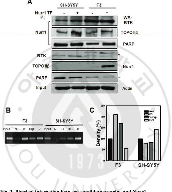
3. Conformation BTK, TOPO IIβ and PARP co-immunoprecipitate with Nurr1 in F3 and SH-SY5Y cells.
To corroborate the above-mentioned observations, we conducted immunoprecipitation analysis for each of the above-mentioned proteins or Nurr1, and conducted Western blotting for specific antigens in order to determine whether they associated in a complex in F3 and SH-SY5Y cells (Fig 3A). The results showed that Nurr1 associated with PARP, TOPO II β,
and BTK using a specific antibody for Nurr1 and Western blotting for each of the three candidate proteins. Importantly, in the reverse immunoprecipitation using specific antibodies against candidate proteins, Western blot analysis demonstrated that Nurr1 was detected. Thus, BTK is profoundly associated with Nurr1 in F3 cells, as compared to SH-SY5Y cells. Additionally, the interaction of PARP and Nurr1 was found to be increased in SH-SY5Y cells, but similar patterns were noted between the wild-type and Nurr1-transfected cell lines. By way of contrast, TOPO II β associated only with Nurr1 from F3 cells in the reverse immunoprecipitation analysis.
4. Physical interaction both identified proteins and NERE-A motif of Nurr1. In order to determine whether the candidate proteins are able to bind to the NBRE region, we assessed the interaction between candidate proteins and Nurr1 in F3 and SH-SY5Y cells via ChIP analysis (Fig 3B). The cross-linked chromatin from 5x10⁵ cells was immunoprecipitated with specific antibodies against BTK, TOPO II β, and PARP, and was amplified with primers that amplified a 374-bp amplicon within the NBRE-A elements. ChIP analysis demonstrated that three candidate proteins, PARP, TOPO II β, and BTK, were all associated with the NBRE-A element, with no detectable signals observed with the match antibody control. In particular, BTK was strongly bound at 210% at the NBRE-A element in F3 cells, whereas PARP was increased by 140% in the SH-SY5Y cells(3C). These results indicate that these proteins are associated with the NBRE elements, and their signals may also include association with Nurr1.
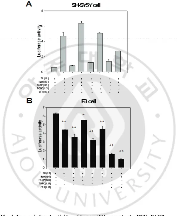
5. BTK, TOPO II and PARP are co-regulators of Nurr1 in the F3 and SH-SY5 Y cells.
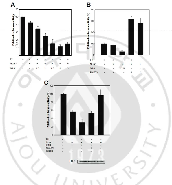
Our results identified proteins as a potential modulator of Nurr1-induced TH transcriptional activity. Co-immunoprecipitation and streptavidin pull-down experiments established an interaction between Nurr1 and PARP, TOPO II β, and BTK. In order to assess the functionality of the TH promoter and to test the cell specificity of TH gene expression, we introduced a series of TH promoter/luciferase reporter gene constructs into the F3 and SH-SY5Y cells (Fig. 4). The cells were cotransfected with TH and different combinations of Nurr1, PARP, TOPO II β, and BTK expression vectors. Importantly, the co-transfection of Nurr1-PARP and Nurr1-TOPO II β was enhanced significantly in TH gene luciferase levels in both cell lines. By way of contrast, BTK of hTH promoter activity repressed irrelevant Nurr1. These data show clearly that BTK may function as a repressor of the TH promoters in F3 cells. Also, these results show that Nurr1 directly activates the TH promoter in a cell-specific manner, and demonstrated that Nurr1 interacted directly to BTK, TOPO II β, and PARP, may regulate TH transcription activity via the identified proteins. In F3 cells, BTK repressed Nurr1-induced hTH promoter activity in a dose–dependent manner (Fig. 5A). Additionally, BTK profoundly repressed the transcriptional effects, inducing up to an 80% inhibition in F3 cells. In order to determine whether BTK influences Nurr1-induced transactivity, F3 cells that express neither Nurr1 nor BTK were co-transfected with mutant BTK and siBTK. Cotransfection of Nurr1 with BTK caused a significant repression of luciferase activity, whereas double tyrosine–mutant BTK significantly enhanced TH promoter activity (Fig. 5B). Surprisingly, siBTK was still found to be capable of enhancing
Nurr1-induced TH transcriptional activity (Fig 5C). As shown, the transfection of BTK siRNA duplexes to neural stem cells was quite effective, as BTK expression was strongly downregulated (Fig 5C). These findings show that Nurr1-dependent transcriptional repressor activity in neural stem cells depends upon the coexpression of the BTK. Our present data clearly demonstrate the requirement for BTK kinase at the repressor activity in TH function.
Figure. 1. Cell typespecific regulation of human TH promoter through NBRE
-A site.
(A) Schematic diagram of the TH promoter encompassing three putative NBREs
(NBRE-A, -B, -C). (B) Three of mutated TH constructs with or without Nurr1 were
influenced in the activity of TH in SH-SY5Y in human NSCs (Neural stem cells), F3
(C), A4 (D), and F5 (E) cells. Effect of mutations on transactivation of the TH
promoter activity by Nurr1 in SH-SY5Y (B’), and F3 (C’), A4 (D’), F5 (E’) cells.
Figure. 2. Purification of NBRE- A binding protein by DNA pull-down assay.
(A) Flow diagram for purification of the NBRE-A site binding protein. (B)
SDS-PAGE analysis of purified NBRE-A binding, in and around protein eluted from
streptavidin agarose bead affinity resin. (C) Western blotting of the purified NBRE-A
binding, in and around protein.
Fig. 3. Physical interaction between candidate proteins and Nurr1
(A) The interaction between BTK, TOPO II β or PARP and Nurr1 in F3 and
SH-SY5Y cells were confirmed by coimmunoprecipitation analysis. (B) ChIP assay. (C)
Quantification of the immmunoprecipitation based on the quantification of the
specific bands
Fig. 4. Transcriptional activities of human TH promoter by BTK, PARP
and TOPO.
(A) SH-SY5Y cell and (B) human Neural stem cell F3 *, p<0.01
compared with TH promoter group. **, p< 0.001.
Fig. 5. BTK functions as a repressor in human neural stem cells.
(A) TH promoter activity by Nurr1 with increasing amount of BTK expressio
n. (B) Expression of kinase-deficient BTK indicated that BTK functions as a t
ranscriptional co-repressor of Nurr1 on TH expression. (C) Transcriptional acti
vities of human TH promoter in BTK knockdown F3 cells.
IV. Discussion
Our previously report, we studied human TH sequence analysis to identify transcriptional regulatory element binding sites, which perform essential functions in dopaminergic neurogenesis (Kim et al., 2003). Also to check the cell type-specific and differentiation-related expression of TH, we performed many experiment. And we focus on Nurr1 and revealed Nurr1 direct regulation of TH gene expression. When Nurr1 regulate the TH gene expression, it is different with cell stage that repression in undifferentiated cell stage(neural stem cells) and activation in differentiated cell stage(neuroblastoma cells). We demonstrated this regulation of TH gene expression by Nurr1 may affect other cofactors. So we identified the protein that binds to Nurr1. Our human TH promoter has three Nurr1 binding sites which was mutated one amino acid with NBRE sequence, so we called NBRE-A,B,and C. We made variety structure with or without NBRE-A,B and C. After that check the TH promoter activities with or without Nurr1.In the results NBRE-A site is critical to TH promoter regulation(Fig.1) which repress in F3 cells and activate in SH-SY5Y cells. Therefore, we performed DNA pull-down assay to find cofactors binds to the NBRE-A site. In fact, in our assays we detected an interaction with the TH repressor Nurr1, which was further confirmed by streptavidin-agarose beads pull-down assays. Also LC/mass spectrophotometry analysis identifies new potential dimmer partners of nurr1. We identified three candidate proteins including BTK, TOPO II beta, and PARP.
BTK is a type of kinase enzyme implicated in the primary immunodeficiency disease X-linked agammaglobulinemia and plays a crucial role in B cell maturation. Also BTK
translocate to the plasma membrane upon stimulation with growth factor and expressed in a variety of mammalian neural cells. And TOPO II beta is critical acts for the cell proliferation and transaction including DNA replication, transcription, and recombination. Last, PARP is a protein involved in an number of cellular processes involving mainly DNA repair and programmed cell death.
Each protein expressed in experimental cells(Fig.2) and interacted with Nurr1(Fig3). To check the interaction we performed co-immunopricipitation and ChIP assay, BTK was strongly interacts with Nurr1 in F3 cells and PARP interacts in SH-SY5Y cells. Also BTK strongly repressed TH promoter activities with Nurr1 in F3 cells, PARP activated TH promoter activities in SH-SY5Y cells(Fig.4). At this result, we pointed BTK in F3 cells and so, performed further experiments to check BTK’s functions and effects in F3 cells. In the result, BTK repressed the TH promoter activities as dose dependent manner and when BTK was mutation or knockdown, this activation was disappeared. (Fig.5)
Our results indicate when Nurr1 repress the TH gene expression in immature cell stage(neural stem cell), BTK strongly repress by recruitment of Nurr1. After cell maturation(dopaminergic cell) Nurr1 may recruit PARP, the TH gene expression may restart.
V. CONCLUSION
The findings of our study demonstrate that Nurr1directly activates the TH promoter in a cell line-specific manner and cofactors are required for Nurr1-mediated TH gene induction. Additionally, these data indicate that these specifically interacting cofactors may perform a crucial role in the neuron cell-specific expression of the TH gene. Thus, these identified Nurr1 binding proteins provide a number of clues and potential links in understanding the mechanisms of the transcription of TH genes. Finally, these study of TH gene may offer Elucidation of etiology of neuronal disorders such as Parkinson’s disease.
VI. REFERENCE
1. Aarsland D., Tandberg E., Larsen J.P. and Cummings J.L. Frequency of dementia in Parkinson disease. Arch. Neurol. 53(6):538-542, 1996.
2. Berg L.J., Finkelstein L.D., Lucas J.A. and Schwartzberg P.L. Tec family kinases in T lymphocyte development and function. Annu. Rev. Immunol. 23:549-600, 2005.
3. Brunner C., Muller B. and Wirth T. Bruton's Tyrosine Kinase is involved in innate and adaptive immunity. Histol. Histopathol. 20(3):945-955, 2005.
4. Conley M.E., Mathias D., Treadaway J., Minegishi Y. and Rohrer J. Mutations in btk in patients with presumed X-linked agammaglobulinemia. Am J Hum Genet 62(5):1034-1043, 1998.
5. Ekman N., Arighi E., Rajantie I., Saharinen P., Ristimaki A., Silvennoinen O. and Alitalo K. The Bmx tyrosine kinase is activated by IL-3 and G-CSF in a PI-3K dependent manner. Oncogene 19(36):4151-4158, 2000.
6. Felices M. and Berg L.J. The Tec kinases Itk and Rlk regulate NKT cell maturation, cytokine production, and survival. J. Immunol. 180(5):3007-3018, 2008.
7. Felices M., Falk M., Kosaka Y. and Berg L.J. Tec kinases in T cell and mast cell signaling. Adv. Immunol. 93:145-184, 2007.
8. Freed C.R., Greene P.E., Breeze R.E., Tsai W.Y., DuMouchel W., Kao R., Dillon S., Winfield H., Culver S., Trojanowski J.Q., Eidelberg D. and Fahn S. Transplantation of embryonic dopamine neurons for severe Parkinson's disease. N. Engl. J. Med. 344(10):710-719, 2001.
9. Hasan M., Lopez-Herrera G., Blomberg K.E., Lindvall J.M., Berglof A., Smith C.I. and Vargas L. Defective Toll-like receptor 9-mediated cytokine production in B cells from Bruton's tyrosine kinase-deficient mice. Immunology 123(2):239-249, 2008.
10. Jui H.Y., Tseng R.J., Wen X., Fang H.I., Huang L.M., Chen K.Y., Kung H.J., Ann D.K. and Shih H.M. Protein-tyrosine phosphatase D1, a potential regulator and effector for Tec family kinases. J. Biol. Chem. 275(52):41124-41132, 2000.
11. Khan W.N., Alt F.W., Gerstein R.M., Malynn B.A., Larsson I., Rathbun G., Davidson L., Muller S., Kantor A.B., Herzenberg L.A. and et al. Defective B cell development and function in Btk-deficient mice. Immunity 3(3):283-299, 1995.
12. Kim T.E., Lee H.S., Lee Y.B., Hong S.H., Lee Y.S., Ichinose H., Kim S.U. and Lee M.A. Sonic hedgehog and FGF8 collaborate to induce dopaminergic phenotypes in
the Nurr1-overexpressing neural stem cell. Biochem. Biophys. Res. Commun. 305(4):1040-1048, 2003.
13. Kim T.E., Park M.J., Choi E.J., Lee H.S., Lee S.H., Yoon S.H., Oh C.K., Lee B.J., Kim S.U., Lee Y.S. and Lee M.A. Cloning and cell type-specific regulation of the human tyrosine hydroxylase gene promoter. Biochem. Biophys. Res. Commun. 312(4):1123-1131, 2003.
14. Kobayashi S., Kamo S., Ohmae A., Agui K., Li Y. and Anzai K. Identification of a negative regulatory DNA element for neuronal BC1 RNA expression by RNA polymerase III. Biochim. Biophys. Acta 1493(1-2):142-150, 2000.
15. Kuwahara K., Saito Y., Ogawa E., Takahashi N., Nakagawa Y., Naruse Y., Harada M., Hamanaka I., Izumi T., Miyamoto Y., Kishimoto I., Kawakami R., Nakanishi M., Mori N. and Nakao K. The neuron-restrictive silencer element-neuron-restrictive silencer factor system regulates basal and endothelin 1-inducible atrial natriuretic peptide gene expression in ventricular myocytes. Mol. Cell. Biol. 21(6):2085-2097, 2001.
16. Lawinger P., Venugopal R., Guo Z.S., Immaneni A., Sengupta D., Lu W., Rastelli L., Marin Dias Carneiro A., Levin V., Fuller G.N., Echelard Y. and Majumder S. The neuronal repressor REST/NRSF is an essential regulator in medulloblastoma cells.
Nat. Med. 6(7):826-831, 2000.
17. Lemonde S., Rogaeva A. and Albert P.R. Cell type-dependent recruitment of trichostatin A-sensitive repression of the human 5-HT1A receptor gene. J. Neurochem. 88(4):857-868, 2004.
18. Lewis C.M., Broussard C., Czar M.J. and Schwartzberg P.L. Tec kinases: modulators of lymphocyte signaling and development. Curr. Opin. Immunol. 13(3):317-325, 2001.
19. Lindvall J.M., Blomberg K.E., Valiaho J., Vargas L., Heinonen J.E., Berglof A., Mohamed A.J., Nore B.F., Vihinen M. and Smith C.I. Bruton's tyrosine kinase: cell biology, sequence conservation, mutation spectrum, siRNA modifications, and expression profiling. Immunol. Rev. 203:200-215, 2005.
20. Lunyak V.V., Burgess R., Prefontaine G.G., Nelson C., Sze S.H., Chenoweth J., Schwartz P., Pevzner P.A., Glass C., Mandel G. and Rosenfeld M.G. Corepressor-dependent silencing of chromosomal regions encoding neuronal genes. Science 298(5599):1747-1752, 2002.
21. Matise M.P., Epstein D.J., Park H.L., Platt K.A. and Joyner A.L. Gli2 is required for induction of floor plate and adjacent cells, but not most ventral neurons in the mouse
central nervous system. Development 125(15):2759-2770, 1998.
22. Murai K., Naruse Y., Shaul Y., Agata Y. and Mori N. Direct interaction of NRSF with TBP: chromatin reorganization and core promoter repression for neuron-specific gene transcription. Nucleic Acids Res 32(10):3180-3189, 2004.
23. Nagatsu T. The human tyrosine hydroxylase gene. Cell. Mol. Neurobiol. 9(3):313-321, 1989.
24. Naruse Y., Aoki T., Kojima T. and Mori N. Neural restrictive silencer factor recruits mSin3 and histone deacetylase complex to repress neuron-specific target genes. Proc. Natl. Acad. Sci. U. S. A. 96(24):13691-13696, 1999.
25. Nisitani S., Satterthwaite A.B., Akashi K., Weissman I.L., Witte O.N. and Wahl M.I. Posttranscriptional regulation of Bruton's tyrosine kinase expression in antigen receptor-stimulated splenic B cells. Proc. Natl. Acad. Sci. U. S. A. 97(6):2737-2742, 2000.
26. Nore B.F., Vargas L., Mohamed A.J., Branden L.J., Backesjo C.M., Islam T.C., Mattsson P.T., Hultenby K., Christensson B. and Smith C.I. Redistribution of Bruton's tyrosine kinase by activation of phosphatidylinositol 3-kinase and Rho-family GTPases. Eur. J. Immunol. 30(1):145-154, 2000.
27. Nunes I., Tovmasian L.T., Silva R.M., Burke R.E. and Goff S.P. Pitx3 is required for development of substantia nigra dopaminergic neurons. Proc. Natl. Acad. Sci. U. S. A. 100(7):4245-4250, 2003.
28. Oda A., Ikeda Y., Ochs H.D., Druker B.J., Ozaki K., Handa M., Ariga T., Sakiyama Y., Witte O.N. and Wahl M.I. Rapid tyrosine phosphorylation and activation of Bruton's tyrosine/Tec kinases in platelets induced by collagen binding or CD32 cross-linking. Blood 95(5):1663-1670, 2000.
29. Palm K., Belluardo N., Metsis M. and Timmusk T. Neuronal expression of zinc finger transcription factor REST/NRSF/XBR gene. J. Neurosci. 18(4):1280-1296, 1998.
30. Pelletier G., Stefanovsky V.Y., Faubladier M., Hirschler-Laszkiewicz I., Savard J., Rothblum L.I., Cote J. and Moss T. Competitive recruitment of CBP and Rb-HDAC regulates UBF acetylation and ribosomal transcription. Mol. Cell 6(5):1059-1066, 2000.
31. Roopra A., Sharling L., Wood I.C., Briggs T., Bachfischer U., Paquette A.J. and Buckley N.J. Transcriptional repression by neuron-restrictive silencer factor is mediated via the Sin3-histone deacetylase complex. Mol. Cell. Biol. 20(6):2147-2157, 2000.
32. Sacchetti P., Mitchell T.R., Granneman J.G. and Bannon M.J. Nurr1 enhances transcription of the human dopamine transporter gene through a novel mechanism. J. Neurochem. 76(5):1565-1572, 2001.
33. Schmidt U., Boucheron N., Unger B. and Ellmeier W. The role of Tec family kinases in myeloid cells. Int. Arch. Allergy Immunol. 134(1):65-78, 2004.
34. Schoenherr C.J., Paquette A.J. and Anderson D.J. Identification of potential target genes for the neuron-restrictive silencer factor. Proc. Natl. Acad. Sci. U. S. A. 93(18):9881-9886, 1996.
35. Schurer H., Lang K., Schuster J. and Morl M. A universal method to produce in vitro transcripts with homogeneous 3' ends. Nucleic Acids Res 30(12):e56, 2002.
36. Sediva A., Smith C.I., Asplund A.C., Hadac J., Janda A., Zeman J., Hansikova H., Dvorakova L., Mrazova L., Velbri S., Koehler C., Roesch K., Sullivan K.E., Futatani T. and Ochs H.D. Contiguous X-chromosome deletion syndrome encompassing the BTK, TIMM8A, TAF7L, and DRP2 genes. J. Clin. Immunol. 27(6):640-646, 2007.
37. Smidt M.P., Asbreuk C.H., Cox J.J., Chen H., Johnson R.L. and Burbach J.P. A second independent pathway for development of mesencephalic dopaminergic neurons requires Lmx1b. Nat. Neurosci. 3(4):337-341, 2000.
38. Storch A., Sabolek M., Milosevic J., Schwarz S.C. and Schwarz J. Midbrain-derived neural stem cells: from basic science to therapeutic approaches. Cell Tissue Res. 318(1):15-22, 2004.
39. Vihinen M., Kwan S.P., Lester T., Ochs H.D., Resnick I., Valiaho J., Conley M.E. and Smith C.I. Mutations of the human BTK gene coding for bruton tyrosine kinase in X-linked agammaglobulinemia. Hum. Mutat. 13(4):280-285, 1999.
40. Wallen A. and Perlmann T. Transcriptional control of dopamine neuron development. Ann. N. Y. Acad. Sci. 991:48-60, 2003.
−
국문요약
−미분화 신경줄기세포에서 BTK와 전사인자 Nurr1의
상호작용에 관한 연구
아주대학교 대학원 의생명과학과
박 명 선
(지도교수: 이 명 애)
파킨슨 병은 주로 중뇌의 substantia nigra에서 도파민 신경세포의 소멸로 인 해 발병된다. 카테콜아민 생합성 과정에서 속도 조절 효소인 tyrosine hydroxyl ase (TH) 유전자의 발현은 신경세포의 발달과정동안 전사과정과 다양한 환경적 인 자극에 의해 조절된다. 또한 Orphan nuclear receptor 중 하나인 Nurr1은 발달 과정 중 중뇌의 substantia nigra와 ventral tegmental 부위에서 발현되고, 도파민 신경 세포 전구체가 완전한 도파민 신경세포로 분화하는데 중요한 역할 을 한다. Nurr1은 발달 과정 동안 TH 유전자의 발현을 유도하는데 중요한 전사 인자라고 생각된다. 우리는 TH 유전자의 발현에서 세포특이적인 전사 조절을 하고, Nurr1에 의한 활성에 가장 크게 관여하는 NBRE-A 부분에서 Nurr1과 함께 복합체를 이루어 TH 발현을 위해 전사 조절에 관여하는 인자들을 찾아 동정하고자 하였다. 이에DNA pull-down assay를 이용하여 NBRE-A 부분에 결합하여 전사 조절 인 자들을 살펴보았다. 이 결과 Nurr1과 결합는 후보 단백질인 PARP, TOPO, BT K를 선택할 수 있었고, 이 각각의 단백질이 Nurr1과 결합하여 TH 유전자의 발 현을 조절하는지를 확인하였다. 이 중 BTK는 Nurr1과 결합하여 사람 줄기세포 주인 F3 세포에서 TH유전자의 발현을 억제시키는 것을 확인 할 수 있었다. 이 결과는 향후 TH 유전자의 발현 정도에 따라 나타나는 퇴행성 뇌질환의 연구 분야에 큰 공헌을 할 것으로 기대된다. 핵심어 : 파킨슨병, TH 유전자, Nurr1, NBRE-A, BTK, 퇴행성 뇌질환