Review Article
The Role of Protein Modifications of T-Bet in Cytokine
Production and Differentiation of T Helper Cells
Sera Oh and Eun Sook Hwang
College of Pharmacy, School of Pharmaceutical Sciences and Global Top 5 Research Program, Ewha Womans University, 52 Ewhayeodae-gil, Seodaemun-gu, C206 Science Building, Seoul 120-750, Republic of Korea
Correspondence should be addressed to Eun Sook Hwang; eshwang@ewha.ac.kr Received 14 March 2014; Accepted 15 April 2014; Published 13 May 2014 Academic Editor: Mizuko Mamura
Copyright © 2014 S. Oh and E. S. Hwang. This is an open access article distributed under the Creative Commons Attribution License, which permits unrestricted use, distribution, and reproduction in any medium, provided the original work is properly cited.
T-Bet (T-box protein expressed in T cells, also called as TBX21) was originally cloned as a key transcription factor involved in the commitment of T helper (Th) cells to the Th1 lineage. T-Bet directly activates IFN-𝛾 gene transcription and enhances development of Th1 cells. T-Bet simultaneously modulates IL-2 and Th2 cytokines in an IFN-𝛾-independent manner, resulting in an attenuation of Th2 cell development. Numerous studies have demonstrated that T-bet plays multiple roles in many subtypes of immune cells, including B cell, dendritic cells, natural killer (NK) cells, NK T cells, and innate lymphoid cells. Therefore, T-bet is crucial for the development and coordination of both innate and adaptive immune responses. To fulfill these multiple roles, T-bet undergoes several posttranslational protein modifications, such as phosphorylation at tyrosine, serine, and threonine residues, and ubiquitination at lysine residues, which affect lineage commitment during Th cell differentiation. This review presents a current overview of the progress made in understanding the roles of various types of T-bet protein modifications in the regulation of cytokine production during Th cell differentiation.
1. Introduction
T-Bet (T-box protein expressed in T cells, also called TBX21) was firstly described in 2000 in a report examining the effects of T-bet on the differentiation of T helper 1 (Th1)
cells [1]. For the past 15 years, many studies have examined
the functions of T-bet and have revealed multiple roles for this protein during Th cell differentiation, with a focus on the molecular mechanisms involved, the novel functions of this transcription factor in innate immune cells, and
T-bet-mediated modulation of inflammatory diseases [2–9].
It has been clarified that T-bet plays a critical role in the coordination of innate immunity and adaptive immunity and that it fulfills an important function in modulating chronic inflammatory diseases, including asthma and inflammatory bowel disease, by controlling a network of highly conserved
genetic programs [10–12]. Thus, optimal regulation of T-bet
expression and activity seems to be beneficial for preventing or treating chronic inflammation and autoimmune diseases.
Although attempts have been made at identifying the small molecules that control the expression and activity of
T-bet that affect the T cell-mediated immune response, little progress has been made on this to date. Given the importance of T-bet in the immune regulation, elucidating the functional mechanisms underlying the multiple roles of T-bet would facilitate the development of novel therapeutic interventions for treating chronic inflammatory and autoimmune diseases. This review summarizes the current state of knowledge about the molecular mechanisms underlying the multiple roles played by T-bet in Th cell development.
2. Structure of T-Bet
The T-bet contains an amino-terminus, a T-box domain, and a carboxyl-terminus, which show 82%, 100%, and 79% homology, respectively, between mice (530 amino-acid residue protein) and humans (535 amino-acid residue
pro-tein) (Figure 1). The T-box domain, located between residues
135 and 326 in mouse T-bet, is highly conserved in 18
mem-bers of the T-box protein (TBX) family [13, 14]. Common
features shared by T-box proteins include a capacity for DNA binding through the T-box domain and transcriptional http://dx.doi.org/10.1155/2014/589672
Transactivation domain 1 136 327 535 T-box 1 135 530 T-box 326 Mouse Human 100% identity T302 T 30 2 S508 Y304 K313 K 31 3 S508 Y525 Y219/Y265/Y304 82% identity 79% identity P P P P P P ubub ubub
Position Modification Physiological effect Reference
Y
219 Y265 Y304 Y525
c-Abl-mediated phosphorylation
Induction of Th1 cell development
Suppression of Th2 cell development [28] Phosphorylation Interaction with NFAT
Suppression of IL-2 and Th2 cytokines [29] Interaction with RUNX1
Inhibition of Th17 cell development [21] Ubiquitination Binding to DNA sequence
Control of protein stability [29] GSK-3-mediated phosphorylation Interaction with NF-𝜅B p65 Inhibition of IL-2 [19] ITK-induced phosphorylation
Interaction with GATA-3
Suppression of Th2 cytokines [5]
—
Figure 1: Structure and protein modification of T-bet. Mouse and human T-bet is 100% identical in the T-box domain. Several amino acid residues are conserved in mice and undergo posttranslational modifications, including phosphorylation at serine, threonine, and/or tyrosine residues, and ubiquitination at lysine residues.
regulatory activity, which plays a role in controlling the expression of developmental gene in all animal species.
The T-box domain is made up of about 180 amino-acid residues and is both sufficient and necessary for binding to the
consensus DNA sequence TCACACCT [13–15]. Brachyury
(T) was the first T-box protein to be identified and, in dimeric form, interacts with the major and the minor grooves of DNA through hydrophobic interactions and unusual
main-chain carbonyl contact with a guanine as a dimer [16]. TBX1
also binds to the DNA sequence as a dimer, whereas TBX2 appears to bind to the same DNA sequence as a monomer
[17]. Although TBX1 and TBX2 share 61% identity in the
T-box domain, the structure of the DNA-T-box binding complex appears to be different, because of the low homology among the amino- and carboxyl-terminal regions. The T-box domain in T-bet shows 50% homology with the correspond-ing domain in brachyury (T), TBX1, and TBX2; however, the crystal structure of T-bet bound to the DNA sequence remains to be characterized.
3. Regulation of Th Cell
Differentiation by T-Bet
3.1. Stimulation of Th1 Cell Differentiation by T-Bet. T-bet directly binds to the consensus DNA sequence within the
IFNG promoter and activates its transcription. The T-bet-induced expression of IFNG derives Th precursor cells to differentiate into Th1 effector cells. While exogenous T-bet overexpression in na¨ıve Th cells preferentially increases development of Th1 cells, T-bet deficiency leads to a failure to produce sufficient IFN-𝛾 and therefore reduces generation of
Th1 cells [1,18]. T-Bet expression is substantially increased by
stimulation of the T cell receptor (TCR) and is augmented by cotreatment with IFN-𝛾 and IL-12. IFN-𝛾 binds to its receptor and induces activation of signal transducer and activator of transcription (STAT) 1 and transcription of T-bet gene (TBX21). Subsequently, T-T-bet directly stimulates the transcription of IFNG as well as IL12RB2. Expression
of IL-12 receptor (IL-12R) 𝛽2 on the cell surface further
enhances IFN-𝛾 production through IL-12 and the STAT4 signaling pathway, thereby resulting in preferential Th1 cell differentiation. Interestingly, enforced T-bet expression can
also convert the differentiated Th2 cells into Th1 cells [1].
Therefore, T-bet is positioned at the crux of the regulatory pathways that induce IFN-𝛾 in Th cells.
3.2. Attenuation of IL-2 Production by T-Bet. In addition to IFN-𝛾 regulation, T-bet significantly suppresses IL-2 expres-sion. This cytokine, an early T cell growth factor, is essential for activation, proliferation, and differentiation of Th cells and
is abundantly produced upon TCR stimulation. Ectopically introduced T-bet significantly suppresses IL-2 production
through inhibition of nuclear factor𝜅B (NF-𝜅B) p65 activity,
under conditions of both Th1 and Th2 differentiation [19].
During Th1 cell differentiation, IL-2 transcription is also attenuated upon induction of T-bet. The T-bet-mediated IL-2 inhibition may affect Th cell expansion and exquisitely modulate the Th1-mediated immune response upon exposure to a pathogenic antigen.
3.3. Suppression of Th2 Cell Development by T-Bet. Further-more, exogenous T-bet introduction into Th cells suppresses the production of Th2 cytokines, such as 4, 5, and IL-13, via suppression of GATA-binding protein-3 (GATA-3). Accordingly, a lack of T-bet induces spontaneous Th2 cell
development in vitro and in vivo [1,18]. The Th2-suppressive
activity of T-bet was also confirmed in the absence of IFN-𝛾, indicating that T-bet has a discrete inhibitory function, independent of IFN-𝛾 stimulation.
3.4. Other Functions of T-Bet. Recently, many studies have reported that T-bet also modulates other Th cell lineages,
including Th17, Treg, and follicular Th (TFH) cells, in
coor-dination with many transcription factors, such as the retinoic acid-related orphan receptor-𝛾t (ROR𝛾t), runt-related tran-scription factor 3 (RUNX3), and B-cell lymphoma-6 (BCL6)
[20–25]. These findings suggest that T-bet is a transcription
factor that is critical for fine-tuning Th cell development.
4. Posttranslational Modification of T-Bet
T-Bet functions as a multitasking player in the regulation of Th cell differentiation. However, the molecular mechanisms that underlie the stimulatory and inhibitory activity of T-bet in regulating target gene expression remain to be clarified. Many multitasking proteins are known to undergo posttrans-lational protein modifications and to determine cell fates by exerting direct stimulatory and indirect inhibitory activity on
target gene expression [26,27].
4.1. Tyrosine Phosphorylation of T-Bet. Antibody-based detection of T-bet proteins in western blots results in multiple bands, suggesting the posttranslational modification of T-bet in TCR-triggered Th cells. Tyrosine phosphorylation of T-bet protein occurs primarily during the early stages (days 2 to 3) of Th cell development, upon TCR engagement, and declines afterwards. Treating Th cells with the tyrosine phosphatase inhibitor pervanadate enhances the tyrosine phosphorylation of T-bet. T-Bet is mainly localized in the nucleus, and the nuclear tyrosine kinase IL2-inducible tyrosine kinase (ITK) was identified as the responsible upstream tyrosine kinase. ITK deficiency prevents tyrosine phosphorylation of T-bet in Th cells after stimulation with TCR and IL-12. Mutational research has revealed that tyrosine residue 525 (Y525) is the relevant phosphorylation site and that phosphorylation at this site plays an important role in the interaction with GATA-3. Although T-box domain in T-bet is important for DNA and protein-protein interaction, tyrosine phosphorylation of
Y525 is prerequisite for the suppression of GATA-3-mediated Th2 cell differentiation. Blockade of Y525 phosphorylation abrogates the interaction with and suppression of GATA-3,
resulting in impairment of Th2 suppression [5].
Furthermore, another nuclear tyrosine kinase, c-Abl, induces phosphorylation of T-bet at tyrosine residues 219,
265, and 304 in mouse T-bet [28]. A deficiency in c-Abl as
well as mutation of T-bet at these residues (Y219/265/304F mutants) leads to a failure to increase IFN-𝛾 induction and to suppress Th2 cytokine production, due to the loss of phos-phorylation at these tyrosine residues. This, in turn, results
in the aggravation of allergic lung inflammation in vivo [28].
These findings suggest that ITK- and c-Abl-induced tyrosine phosphorylation of T-bet is essential for the modulation of Th2 cell development and the allergic immune response. 4.2. Serine Phosphorylation of T-Bet. Although T-bet-medi-ated suppression of Th2 cell development is impaired by mutation of Y525 and the absence of c-Abl kinase, IL-2 suppression is retained with a Y525-mutant T-bet, suggesting the existence of an additional regulatory mechanism for
IL-2 modulation [5]. Interestingly, the appearance of multiple
bands of T-bet protein on western blots could be eliminated by the addition of calf intestinal phosphatase, which pre-dominantly eliminates phosphorylation at serine/threonine residues. Mass spectrometric analysis then revealed serine 508 (S508) as another phosphorylation site in T-bet. Mutation of S508 abolishes casein kinase- (CK-) and glycogen synthase kinase-3 (GSK-3-) mediated phosphorylation in T-bet, as well as the IL-2–suppressive activity of the protein. Moreover, S508 phosphorylation is important for the interaction of T-bet with NF-𝜅B p65 and for prevention of binding of NF-𝜅B p65 to the IL2 promoter. In accordance with the function of T-bet as an NF-𝜅B p65 inhibitor, T-bet-null Th1 cells sustain NF-𝜅B p65 activity during Th1 cell differentiation and thus produce more IL-2. Therefore, it has been suggested that T-bet is a physiological inhibitor of IL-2 during Th1 cell differentiation, through S508 phosphorylation-dependent suppression of NF-𝜅B p65.
4.3. Threonine Phosphorylation of T-Bet. Very recently, threo-nine 302 (T302) was characterized as a novel phosphorylation
site in T-bet [29], although it remains unclear which kinase
and phosphatase affect the phosphorylation of this residue. However, restoration of T-bet-null Th cells with a T302-mutant T-bet stimulated IFN-𝛾 production as much as did wild-type T-bet; however, the mutant failed to suppress IL-2 and other Th2 cytokines. Further analysis demonstrated that T302 phosphorylation is required for the interaction of T-bet with nuclear factor of activated T cells (NFAT) and for down-regulation of NFAT-mediated IL-2 and Th2 cytokines, such as IL-4, IL-5, and IL-13. NFAT is not crucial for induction of IFN-𝛾 production and T302-mutant T-bet is able to bind to the IFNG promoter; thus, IFN-𝛾 production was comparable between wild-type and mutant T-bet. In other words, muta-tion of T302 abrogated the T-bet-mediated suppression of IL-2 and ThIL-2 cytokine production but did not affect the DNA-binding and IFN-𝛾-stimulatory activity of T-bet.
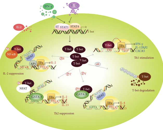
T-bet T-bet T-bet IL-12 IL-2 IL-2 TATA NFAT NFAT NFAT NFAT NFAT PS AP1 IL-2 suppression P P P P P T Y Y T S GATA-3 GATA-3 GATA GATA TFs TFs NFAT TATA TATA IL-2 IL-4 IL-5
IL-13 IL-4IL-5
IL-13 Th2 suppression TFs STAT1 T-bet T-bet T-bet T-bet T-bet degradation Th1 stimulation ub ub ub ub ub ubK T-bet T-bet T-bet T-bet IFN-𝛾 STAT1 STAT4 NF-𝜅B TBE TATA TFs IFN-𝛾 IL-12R𝛽2 CXCR3 NF-𝜅B
Figure 2: Multiple T-bet functions playing a role in Th cell differentiation. Induction of T-bet expression through activation of STAT1 and STAT4 directly stimulates the transcription of T-box-binding element-containing genes, such as IFNG, IL12RB2, and CXCR3, thereby enhancing Th1 cell development. T-Bet undergoes serine phosphorylation at S508 and then downregulates IL-2 production in Th1 cells by recruiting NF-𝜅B p65 from the IL2 promoter. Protein levels of T-bet in Th1 cells can be controlled by the ubiquitin-mediated proteasomal degradation pathway. Moreover, T-bet protein undergoes additional posttranslational modifications, for example, phosphorylation at T302 and Y525, which facilitates its suppression of the Th2 cytokine production that is induced by activation of NFAT and GATA-3.
Indeed, T302 is located in the DNA-binding T-box, as is
Y304 [28]. The T-box domain consists of several repeats of
𝛽-strands and 𝛼-helices and is involved in both dimerization
and DNA binding [16]. Muller and Herrmann predicted that
the𝛼-helices 𝛼H3 and 𝛼H4 in brachyury (T) are important
for the direct interaction of this protein with the minor and
major grooves of DNA [16]. However, T302 may not be
associated directly with the DNA grooves, regardless of its phosphorylation status. It would be interesting to know which upstream kinase and phosphatase regulates T302 phosphorylation and whether T302 phosphorylation affects other protein modifications of T-bet.
4.4. Ubiquitination of Lysine 313 in T-Bet. T-bet expression is critical for the transcriptional regulation of IFNG and for the development of Th1 cells, but the means of regulation of T-bet at the protein level is yet to be identified. Jang et al. have recently reported that T-bet undergoes ubiquitination-mediated proteasomal degradation during the later stages
of Th1 cell differentiation [29]. Of the 16 lysine residues
present in mouse T-bet protein, 11 are predominantly located
within the T-box domain, and the remaining 5 are located at the carboxyl-terminus (residues 326 through 530), while no lysine residues are present in the amino-terminus (residues 1 through 134). Interestingly, lysine residues within the T-box domain are preferentially ubiquitinated upon overexpression of ubiquitin. Further analysis has identified that mutation of lysine 313 (K313) decreases ubiquitination-mediated T-bet degradation and enhances the expression level of T-bet in the nucleus and the cytoplasm. Despite the increased levels of the K313 mutant, this mutation completely abrogated T-bet functions involving DNA binding, transcriptional acti-vation of IFNG, and suppression of IL-2 and Th2 cytokine
production. The crystal structure of the𝛼-helices of the
T-box domain bound to DNA strongly suggests that the amino group of K313 is associated with the phosphate of a DNA base via hydrogen-bond interaction. In addition, mutation of K313 also leads to failure to suppress IL-2 and Th2 cytokine production; however, the interaction with and suppression of GATA-3 and NF-𝜅B p65 are not altered by mutation of K313. Interestingly, NFAT interaction is abolished in K313-mutant T-bet, which is also strongly associated with an absence of
phosphorylation at T302. It is not clear yet whether and how K313 regulates T302 phosphorylation and vice versa.
5. T-Bet in Inflammatory and
Autoimmune Diseases
Since Mosmann et al. discovered Th1 and Th2 subsets that produce signature cytokines, IFN-𝛾, and 4, 5, and IL-13, respectively, and that modulate inflammatory and allergic
immune responses [30], further studies have identified novel
subsets of Th cells, such as Th17, TFH, and Treg cells [11,
21,23,24,31–34]. Extensive studies have also characterized
the cytokine signaling pathways and transcription factors involved in the regulation of immune responses to pathogens
[1, 35–54]. Importantly, T-bet plays a fundamental role in
controlling differentiation of several subsets of Th cells and
in modulating inflammatory and autoimmune diseases [10–
12].
T-Bet also functions as an antiasthmatic regulator. A deficiency in T-bet spontaneously leads to the development of asthmatic symptoms, which is characterized by increased eosinophil infiltration into the airway, mucus-secreting gob-let cell hyperplasia, and chronic airway remodeling with collagen accumulation and proliferative myofibroblasts; these
features are often also seen in asthmatic patients [55–57].
Restoration of T-bet expression shifts the immune balance to a Th1 response and prevents and attenuates pathologic lung
inflammation in vivo [58,59].
Moreover, T-bet is protected against intracellular path-ogenic infections. Abrogation of T-bet-induced IFN-𝛾 pro-duction resulted in higher susceptibility to intracellular path-ogens in vivo, including Mycobacterium tuberculosis,
Leish-mania major, and Salmonella typhimurium [18, 60, 61],
emphasizing the importance of IFN-𝛾 production by T-bet-expressing Th1 cells in the defense against bacterial infections. However, T-bet-deficient mice are resistant to infection by Listeria monocytogenes, because INF-𝛾 production by natural killer cells is both necessary and sufficient for the host defense
against L. monocytogenes [62].
Furthermore, T-bet can aggravate the development of inflammatory and autoimmune diseases, including inflam-matory bowel disease, experimental autoimmune encephalo-myelitis, inflammatory arthritis, and type I diabetes, as these inflammatory diseases are attenuated in the absence of
T-bet [6, 56, 63, 64]. These findings suggest that fine-tuning
of the immune response by modulation of T-bet expression could have beneficial effects for patients with chronic asthma, inflammatory bowel disease, arthritis, multiple sclerosis, and diabetes.
6. Conclusions and Perspectives
T-bet is a T-box domain-containing transcription factor that is typically involved in developmental regulation but exerts multiple functions in Th cell differentiation; transcriptional activation of IFN-𝛾-expressing Th1 cells, indirect suppression of Th2, Th17, and Treg cell development, and fine-modulation of IL-2 production in Th1 cells. This multitasking is not
surprising, as T-bet undergoes several posttranslational mod-ifications. Phosphorylation at Y525 plays a role in GATA-3 suppression during Th2 regulation, phosphorylation at S508 causes NF-𝜅B p65 suppression in the context of IL-2 regulation in Th1 cells, phosphorylation at T30IL-2 plays a role in fine-tuning IL-2 production, and ubiquitination at K313 plays a role in controlling T-bet protein stability. Thus, posttranslational modification of T-bet facilitates its func-tional diversity and the complexity of its modulation of
cytokine expression (Figure 2). It is not known whether the
various posttranslational modifications occur sequentially or simultaneously, whether one type of protein modification affects other modification, or whether changes in the post-translational modification of T-bet are related to the devel-opment of infectious and chronic inflammatory diseases. Further identification of novel protein modifications related to T-bet functions would provide valuable insights into the development of powerful therapeutic interventions for controlling chronic inflammatory and autoimmune diseases.
List of Abbreviations
BCL-6: B-cell lymphoma-6
CK: Casein kinase
GATA-3: GATA-binding protein 3
GSK-3: Glycogen synthase kinase-3
IFN: Interferon
IL: Interleukin
ITK: IL-2-inducible T cell kinase
NFAT: Nuclear factor of activated T cells
NF-𝜅B: Nuclear factor kappa B
ROR𝛾t: Retinoic acid-related orphan receptor gamma t
RUNX: Runt-related transcription factor
STAT: Signal transducer and activator of transcription
T-bet: T-Box protein expressed in T cells
Th: T helper
Treg: Regulatory T
TFH: Follicular T helper
Ub: Ubiquitin.
Conflict of Interests
No potential conflict of interests was disclosed.
Acknowledgment
This work was supported by Mid-Career Researcher Program through NRF Grant (2013R1A2A2A01068302).
References
[1] S. J. Szabo, S. T. Kim, G. L. Costa, X. Zhang, C. G. Fathman, and L. H. Glimcher, “A novel transcription factor, T-bet, directs Th1 lineage commitment,” Cell, vol. 100, no. 6, pp. 655–669, 2000. [2] A. A. Lighvani, D. M. Frucht, D. Jankovic et al., “T-bet is
rapidly induced by interferon-𝛾 in lymphoid and myeloid cells,” Proceedings of the National Academy of Sciences of the United States of America, vol. 98, no. 26, pp. 15137–15142, 2001.
[3] G. Lugo-Villarino, R. Maldonado-L´opez, R. Possemato, C. Pe˜naranda, and L. H. Glimcher, “T-bet is required for optimal production of IFN-𝛾 and antigen-specific T cell activation by dendritic cells,” Proceedings of the National Academy of Sciences of the United States of America, vol. 100, no. 13, pp. 7749–7754, 2003.
[4] M. J. Townsend, A. S. Weinmann, J. L. Matsuda et al., “T-bet regulates the terminal maturation and homeostasis of NK and V𝛼14i NKT cells,” Immunity, vol. 20, no. 4, pp. 477–494, 2004. [5] E. S. Hwang, S. J. Szabo, P. L. Schwartzberg, and L. H. Glimcher,
“T helper cell fate specified by kinase-mediated interaction of T-bet with GATA-3,” Science, vol. 307, no. 5708, pp. 430–433, 2005.
[6] J. Wang, J. W. Fathman, G. Lugo-Villarino et al., “Transcription factor T-bet regulates inflammatory arthritis through its func-tion in dendritic cells,” Journal of Clinical Investigafunc-tion, vol. 116, no. 2, pp. 414–421, 2006.
[7] W. S. Garrett, G. M. Lord, S. Punit et al., “Communicable ulcerative colitis induced by T-bet deficiency in the innate immune system,” Cell, vol. 131, no. 1, pp. 33–45, 2007.
[8] N. Powell, A. W. Walker, E. Stolarczyk et al., “The transcription factor T-bet regulates intestinal inflammation mediated by interleukin-7 receptor+ innate lymphoid cells,” Immunity, vol. 37, no. 4, pp. 674–684, 2012.
[9] C. S. Klose, E. A. Kiss, V. Schwierzeck et al., “A T-bet gradient controls the fate and function of CCR6-RORgammat+ innate lymphoid cells,” Nature, vol. 494, no. 7436, pp. 261–265, 2013. [10] V. Lazarevic and L. H. Glimcher, “T-bet in disease,” Nature
Immunology, vol. 12, no. 7, pp. 597–606, 2011.
[11] V. Lazarevic, L. H. Glimcher, and G. M. Lord, “T-bet: a bridge between innate and adaptive immunity,” Nature Reviews Immunology, vol. 13, no. 11, pp. 777–789, 2013.
[12] Y. Wang, J. Godec, K. Ben-Aissa et al., “The transcription factors T-bet and Runx are Reruired for the ontogeny of pathogenic interferon-gamma-producing T Helper 17 cells,” Immunity, vol. 40, no. 3, pp. 355–366, 2014.
[13] V. Wilson and F. L. Conlon, “The T-box family,” Genome Biology, vol. 3, no. 6, article 3008, 2002.
[14] V. E. Papaioannou and L. M. Silver, “The T-box gene family,” Bioessays, vol. 20, no. 1, pp. 9–19, 1998.
[15] F. L. Conlon, L. Fairclough, B. M. J. Price, E. S. Casey, and J. C. Smith, “Determinants of T box protein specificity,” Develop-ment, vol. 128, no. 19, pp. 3749–3758, 2001.
[16] C. W. Muller and B. G. Herrmann, “Crystallographic structure of the T domain-DNA complex of the Brachyury transcription factor,” Nature, vol. 389, no. 6653, pp. 884–888, 1997.
[17] S. Sinha, S. Abraham, R. M. Gronostajski, and C. E. Campbell, “Differential DNA binding and transcription modulation by three T-box proteins, T, TBX1 and TBX2,” Gene, vol. 258, no. 1-2, pp. 15–29, 2000.
[18] S. J. Szabo, B. M. Sullivan, C. Sternmann, A. R. Satoskar, B. P. Sleckman, and L. H. Glimcher, “Distinct effects of T-bet in Th1 lineage commitment and IFN-𝛾 production in CD4 and CD8 T cells,” Science, vol. 295, no. 5553, pp. 338–342, 2002.
[19] E. S. Hwang, J.-H. Hong, and L. H. Glimcher, “IL-2 production in developing Th1 cells is regulated by heterodimerization of RelA and T-bet and requires T-bet serine residue 508,” Journal of Experimental Medicine, vol. 202, no. 9, pp. 1289–1300, 2005. [20] A. N. Mathur, H.-C. Chang, D. G. Zisoulis et al., “T-bet is a
critical determinant in the instability of the IL-17-secreting T-helper phenotype,” Blood, vol. 108, no. 5, pp. 1595–1601, 2006.
[21] V. Lazarevic, X. Chen, J.-H. Shim et al., “T-bet represses TH 17 differentiation by preventing Runx1-mediated activation of the gene encoding ROR𝛾t,” Nature Immunology, vol. 12, no. 1, pp. 96–104, 2011.
[22] M. A. Koch, G. Tucker-Heard, N. R. Perdue, J. R. Killebrew, K. B. Urdahl, and D. J. Campbell, “The transcription factor T-bet controls regulatory T cell homeostasis and function during type 1 inflammation,” Nature Immunology, vol. 10, no. 6, pp. 595–602, 2009.
[23] S. Nakayamada, Y. Kanno, H. Takahashi et al., “Early Th1 cell differentiation is marked by a Tfh cell-like transition,” Immu-nity, vol. 35, no. 6, pp. 919–931, 2011.
[24] K. J. Oestreich, A. C. Huang, and A. S. Weinmann, “The lineage-defining factors T-bet and Bcl-6 collaborate to regulate Th1 gene expression patterns,” Journal of Experimental Medicine, vol. 208, no. 5, pp. 1001–1013, 2011.
[25] K. J. Oestreich, S. E. Mohn, and A. S. Weinmann, “Molecular mechanisms that control the expression and activity of Bcl-6 in TH1 cells to regulate flexibility with a TFH-like gene profile,” Nature Immunology, vol. 13, no. 4, pp. 405–411, 2012.
[26] E. L. Riddle, R. A. Schwartzman, M. Bond, and P. A. Insel, “Multi-tasking RGS proteins in the heart: the next therapeutic target?” Circulation Research, vol. 96, no. 4, pp. 401–411, 2005. [27] K. Moriwaki and F. K. Chan, “RIP3: a molecular switch for
necrosis and inflammation,” Genes and Development, vol. 27, no. 15, pp. 1640–1649, 2013.
[28] A. Chen, S.-M. Lee, B. Gao, S. Shannon, Z. Zhu, and D. Fang, “c-Abl-mediated tyrosine phosphorylation of the T-bet DNA-binding domain regulates CD4+ T-cell differentiation and allergic lung inflammation,” Molecular and Cellular Biology, vol. 31, no. 16, pp. 3445–3456, 2011.
[29] E. J. Jang, H. R. Park, J. H. Hong, and E. S. Hwang, “Lysine 313 of T-box is crucial for modulation of protein stability, DNA binding, and threonine phosphorylation of T-bet,” Journal of Immunology, vol. 190, no. 11, pp. 5764–5770, 2013.
[30] T. R. Mosmann, H. Cherwinski, and M. W. Bond, “Two types of murine helper T cell clone. I. Definition according to profiles of lymphokine activities and secreted proteins,” Journal of Immunology, vol. 136, no. 7, pp. 2348–2357, 1986.
[31] E. Bettelli, T. Korn, and V. K. Kuchroo, “Th17: the third member of the effector T cell trilogy,” Current Opinion in Immunology, vol. 19, no. 6, pp. 652–657, 2007.
[32] Y.-H. Wang and Y.-J. Liu, “The IL-17 cytokine family and their role in allergic inflammation,” Current Opinion in Immunology, vol. 20, no. 6, pp. 697–702, 2008.
[33] X. O. Yang, R. Nurieva, G. J. Martinez et al., “Molecular antag-onism and plasticity of regulatory and inflammatory T cell programs,” Immunity, vol. 29, no. 1, pp. 44–56, 2008.
[34] J. Zhu, H. Yamane, and W. E. Paul, “Differentiation of effector CD4+ T cell populations,” Annual Review of Immunology, vol. 28, pp. 445–489, 2010.
[35] S. Z. Ben-Sasson, G. Le Gros, D. H. Conrad, F. D. Finkelman, and W. E. Paul, “IL-4 production by T cells from naive donors. IL-2 is required for IL-4 production,” Journal of Immunology, vol. 145, no. 4, pp. 1127–1136, 1990.
[36] G. Le Gros, S. Z. Ben-Sasson, R. Seder, F. D. Finkelman, and W. E. Paul, “Generation of interleukin 4 (IL-4)-producing cells in vivo and in vitro: IL-2 and IL-4 are required for in vitro generation of IL-4-producing cells,” Journal of Experimental Medicine, vol. 172, no. 3, pp. 921–929, 1990.
[37] I. Iwamoto, S. Tomoe, H. Tomioka, K. Takatsu, and S. Yoshida, “Role of CD4+ T lymphocytes and interleukin-5 in antigen-induced eosinophil recruitment into the site of cutaneous late-phase reaction in mice,” Journal of Leukocyte Biology, vol. 52, no. 5, pp. 572–578, 1992.
[38] S. H. Gavett, D. J. O’Hearn, X. Li, S.-K. Huang, F. D. Finkelman, and M. Wills-Karp, “Interleukin 12 inhibits antigen-induced airway hyperresponsiveness, inflammation, and Th2 cytokine expression in mice,” Journal of Experimental Medicine, vol. 182, no. 5, pp. 1527–1536, 1995.
[39] F. Kontgen, R. J. Grumont, A. Strasser et al., “Mice lacking the c-rel proto-oncogene exhibit defects in lymphocyte proliferation, humoral immunity, and interleukin-2 expression,” Genes and Development, vol. 9, no. 16, pp. 1965–1977, 1995.
[40] A. K. Abbas, K. M. Murphy, and A. Sher, “Functional diversity of helper T lymphocytes,” Nature, vol. 383, no. 6603, pp. 787–793, 1996.
[41] J. Bliss, V. van Cleave, K. Murray et al., “IL-12, as an adjuvant, promotes a T helper 1 cell, but does not suppress a T helper 2 cell recall response,” Journal of Immunology, vol. 156, no. 3, pp. 887–894, 1996.
[42] I. C. Ho, M. R. Hodge, J. W. Rooney, and L. H. Glimcher, “The proto-oncogene c-maf is responsible for tissue-specific expression of interleukin-4,” Cell, vol. 85, no. 7, pp. 973–983, 1996.
[43] M. H. Kaplan, Y. L. Sun, T. Hoey, and M. J. Grusby, “Impaired IL-12 responses and enhanced development of Th2 cells in Stat4-deficient mice,” Nature, vol. 382, no. 6587, pp. 174–177, 1996. [44] J. A. Lederer, V. L. Perez, L. DesRoches et al., “Cytokine
tran-scriptional events during helper T cell subset differentiation,” Journal of Experimental Medicine, vol. 184, no. 2, pp. 397–406, 1996.
[45] A. Mori, M. Suko, O. Kaminuma et al., “IL-2-induced IL-5 synthesis, but not proliferation, of human CD4+ T cells is suppressed by FK506,” Journal of Immunology, vol. 158, no. 8, pp. 3659–3665, 1997.
[46] T. Nakamura, Y. Kamogawa, K. Bottomly, and R. A. Flavell, “Polarization of IL-4- and IFN-gamma-producing CD4+ T cells following activation of naive CD4+ T cells,” Journal of Immunology, vol. 158, no. 3, pp. 1085–1094, 1997.
[47] S. J. Szabo, A. S. Dighe, U. Gubler, and K. M. Murphy, “Reg-ulation of the interleukin (IL)-12R beta 2 subunit expression in developing T helper 1 (Th1) and Th2 cells,” Journal of Experimental Medicine, vol. 185, no. 5, pp. 817–824, 1997. [48] W. Zheng and R. A. Flavell, “The transcription factor GATA-3
is necessary and sufficient for Th2 cytokine gene expression in CD4 T cells,” Cell, vol. 89, no. 4, pp. 587–596, 1997.
[49] Y.-W. He, M. L. Deftos, E. W. Ojala, and M. J. Bevan, “ROR𝛾t, a novel isoform of an orphan receptor, negatively regulates Fas ligand expression and IL-2 production in T cells,” Immunity, vol. 9, no. 6, pp. 797–806, 1998.
[50] I.-C. Ho, D. Lo, and L. H. Glimcher, “c-maf Promotes T helper cell type 2 (Th2) and attenuates Th1 differentiation by both inter-leukin 4-dependent and -independent mechanisms,” Journal of Experimental Medicine, vol. 188, no. 10, pp. 1859–1866, 1998. [51] L. Rogge, D. D’Ambrosio, M. Biffi et al., “The role of Stat4 in
species-specific regulation of Th cell development by type I IFNs,” Journal of Immunology, vol. 161, no. 12, pp. 6567–6574, 1998.
[52] J. Zamorano, H. Y. Wang, R. Wang, Y. Shi, G. D. Longmore, and A. D. Keegan, “Regulation of cell growth by IL-2: role of STAT5
in protection from apoptosis but not in cell cycle progression,” Journal of Immunology, vol. 160, no. 7, pp. 3502–3512, 1998. [53] L. H. Glimcher and K. M. Murphy, “Lineage commitment in the
immune system: the T helper lymphocyte grows up,” Genes and Development, vol. 14, no. 14, pp. 1693–1711, 2000.
[54] W. Ouyang, M. L¨ohning, Z. Gao et al., “Stat6-independent GATA-3 autoactivation directs IL-4-independent Th2 develop-ment and commitdevelop-ment,” Immunity, vol. 12, no. 1, pp. 27–37, 2000.
[55] S. Finotto, M. Hausding, A. Doganci et al., “Asthmatic changes in mice lacking T-bet are mediated by IL-13,” International Immunology, vol. 17, no. 8, pp. 993–1007, 2005.
[56] M. F. Neurath, B. Weigmann, S. Finotto et al., “The transcription factor T-bet regulates mucosal T cell activation in experimental colitis and Crohn’s disease,” Journal of Experimental Medicine, vol. 195, no. 9, pp. 1129–1143, 2002.
[57] S. Finotto, M. F. Neurath, J. N. Glickman et al., “Development of spontaneous airway changes consistent with human asthma in mice lacking T-bet,” Science, vol. 295, no. 5553, pp. 336–338, 2002.
[58] T. Kiwamoto, Y. Ishii, Y. Morishima et al., “Transcription factors T-bet and GATA-3 regulate development of airway remodeling,” The American Journal of Respiratory and Critical Care Medicine, vol. 174, no. 2, pp. 142–151, 2006.
[59] J. W. Park, H. J. Min, J. H. Sohn et al., “Restoration of T-box-containing protein expressed in T cells protects against allergen-induced asthma,” Journal of Allergy and Clinical Immunology, vol. 123, no. 2, pp. 479–e6, 2009.
[60] B. M. Sullivan, O. Jobe, V. Lazarevic et al., “Increased suscep-tibility of mice lacking T-bet to infection with Mycobacterium tuberculosis correlates with increased IL-10 and decreased IFN-𝛾 production,” Journal of Immunology, vol. 175, no. 7, pp. 4593– 4602, 2005.
[61] R. Ravindran, J. Foley, T. Stoldasek, L. H. Glimcher, and S. J. McSorley, “Expression of T-bet by CD4 T cells is essential for resistance to Salmonella infection,” Journal of Immunology, vol. 175, no. 7, pp. 4603–4610, 2005.
[62] S. S. Way and C. B. Wilson, “Cutting edge: immunity and IFN-𝛾 production during Listeria monocytes infection in the absence of T-bet,” Journal of Immunology, vol. 173, no. 10, pp. 5918–5922, 2004.
[63] J. H. Esensten, M. R. Lee, L. H. Glimcher, and J. A. Bluestone, “T-bet-deficient NOD mice are protected from diabetes due to defects in both T cell and innate immune system function,” Journal of Immunology, vol. 183, no. 1, pp. 75–82, 2009. [64] E. Bettelli, B. Sullivan, S. J. Szabo, R. A. Sobel, L. H. Glimcher,
and V. K. Kuchroo, “Loss of T-bet, but not STAT1, prevents the development of experimental autoimmune encephalomyelitis,” Journal of Experimental Medicine, vol. 200, no. 1, pp. 79–87, 2004.
Submit your manuscripts at
http://www.hindawi.com
Stem Cells
International
Hindawi Publishing Corporation
http://www.hindawi.com Volume 2014
Hindawi Publishing Corporation
http://www.hindawi.com Volume 2014
INFLAMMATION
Hindawi Publishing Corporation
http://www.hindawi.com Volume 2014
Behavioural
Neurology
Endocrinology
International Journal of Hindawi Publishing Corporationhttp://www.hindawi.com Volume 2014 Hindawi Publishing Corporation
http://www.hindawi.com Volume 2014
Disease Markers
Hindawi Publishing Corporation
http://www.hindawi.com Volume 2014 BioMed
Research International
Oncology
Journal of Hindawi Publishing Corporationhttp://www.hindawi.com Volume 2014
Hindawi Publishing Corporation
http://www.hindawi.com Volume 2014
Oxidative Medicine and Cellular Longevity
Hindawi Publishing Corporation
http://www.hindawi.com Volume 2014
PPAR Research
The Scientific
World Journal
Hindawi Publishing Corporationhttp://www.hindawi.com Volume 2014
Immunology Research Hindawi Publishing Corporation
http://www.hindawi.com Volume 2014
Journal of
Obesity
Journal ofHindawi Publishing Corporation
http://www.hindawi.com Volume 2014
Hindawi Publishing Corporation
http://www.hindawi.com Volume 2014
Computational and Mathematical Methods in Medicine
Ophthalmology
Journal ofHindawi Publishing Corporation
http://www.hindawi.com Volume 2014
Diabetes Research
Journal of Hindawi Publishing Corporationhttp://www.hindawi.com Volume 2014
Hindawi Publishing Corporation
http://www.hindawi.com Volume 2014 Research and Treatment
AIDS
Hindawi Publishing Corporation
http://www.hindawi.com Volume 2014
Gastroenterology Research and Practice
Hindawi Publishing Corporation
http://www.hindawi.com Volume 2014