Metagenomic Analysis of Airborne Bacterial
Community and Diversity in Seoul, Korea,
during December 2014, Asian Dust Event
Seho Cha1, Sathiyaraj Srinivasan2, Jun Hyeong Jang1, Dongwook Lee1, Sora Lim1, Kyung Sang Kim2, Weonhwa Jheong3, Dong-Won Lee4, Eung-Roh Park5, Hyun-Mi Chung5, Joonho Choe6, Myung Kyum Kim2*, Taegun Seo1*
1 Department of Life Science, Dongguk University-Seoul, Goyang, South Korea, 2 Department of Bio &
Environmental Technology, Division of Environmental & Life Science, College of Natural Science, Seoul Women’s University, Seoul, South Korea, 3 Biosafety Research Team, Environmental Health Research Department, National Institute of Environmental Research, Incheon, South Korea, 4 Air Quality Research Division, Climate and Air Quality Research Department, National Institute of Environmental Research, Incheon, South Korea, 5 Water Supply and Sewerage Research Division, Environmental Infrastructure Research Department, National Institute of Environmental Research, Incheon, South Korea, 6 Department of Biological Sciences, Korea Advanced Institute of Science and Technology, Daejeon, South Korea
*[email protected](MKK);[email protected](TS)
Abstract
Asian dust or yellow sand events in East Asia are a major issue of environmental contamina-tion and human health, causing increasing concern. A high amount of dust particles, espe-cially called as particulate matter 10 (PM10), is transported by the wind from the arid and semi-arid tracks to the Korean peninsula, bringing a bacterial population that alters the ter-restrial and atmospheric microbial communities. In this study, we aimed to explore the bac-terial populations of Asian dust samples collected during November–December 2014. The dust samples were collected using the impinger method, and the hypervariable regions of the 16S rRNA gene were amplified using PCR followed by pyrosequencing. Analysis of the sequencing data were performed using Mothur software. The data showed that the number of operational taxonomic units and diversity index during Asian dust events were higher than those during non-Asian dust events. At the phylum level, the proportions of Proteobac-teria, ActinobacProteobac-teria, and Firmicutes were different between Asian dust and non-Asian dust samples. At the genus level, the proportions of the genus Bacillus (6.9%), Arthrobacter (3.6%), Blastocatella (2%), Planomicrobium (1.4%) were increased during Asian dust com-pared to those in non-Asian dust samples. This study showed that the significant relation-ship between bacterial populations of Asian dust samples and non-Asian dust samples in Korea, which could significantly affect the microbial population in the environment.
Introduction
Asian dust events in East Asia, including Korea and Japan, are a seasonal phenomenon, mostly in the early spring that influences the airborne environment and human health problems [1–
3]. Asian dust is known to originate from several arid regions of China, including the Gobi a1111111111 a1111111111 a1111111111 a1111111111 a1111111111 OPEN ACCESS
Citation: Cha S, Srinivasan S, Jang JH, Lee D, Lim
S, Kim KS, et al. (2017) Metagenomic Analysis of Airborne Bacterial Community and Diversity in Seoul, Korea, during December 2014, Asian Dust Event. PLoS ONE 12(1): e0170693. doi:10.1371/ journal.pone.0170693
Editor: Emmanuel Dias-Neto, AC Camargo Cancer
Hospital, BRAZIL
Received: June 8, 2016 Accepted: January 9, 2017 Published: January 25, 2017
Copyright:© 2017 Cha et al. This is an open access article distributed under the terms of theCreative Commons Attribution License, which permits unrestricted use, distribution, and reproduction in any medium, provided the original author and source are credited.
Data Availability Statement: All relevant data are
within the paper and its Supporting Information files.
Funding: This work was supported by the National
Institute of Environmental Research, Republic of Korea. The funder had no role in study design, data collection and analysis, decision to publish, or preparation of the manuscript.
Competing Interests: The authors have declared
desert and the Taklamakan desert, and the dust particles affect the surrounding countries, including China, Japan, and Korea [4–6], as well as Greenland and North America through the global transport of Asian dust particles [7–9]. Moreover, Asian dust events have increased from 77 days containing 8 days during winter (Dovember to February) in 1990s to 122 days containing 21 days during winter in 2000s (Korea meteorological administrate, KMA).
During Asian dust events, several hundred times the normal level of dust particles are pres-ent in the airborne environmpres-ent, and the dust particles contain various chemical componpres-ents as well as microorganisms [10–13]. Previous studies have shown that an increasing concentra-tion of the dust particles is associated with various human health problems and several chemi-cal components or heavy metals found in the dust particles are known to contribute various human diseases [14,15]. However, a report by Kimet al. showed that the airborne Hg
concen-tration was not different during Asian dust events [16], and Heet al. showed that heat-treated
Asian dust samples did not induce allergic lung inflammation compared to normal dust sam-ples, suggesting that unknown microbial materials could contribute some risk to human health [17]. In recent years, the chemical compositions of Asian dust particles have been identified comparatively, but the microbial community structure of the dust particles is not yet under-stood. Only limited studies have reported that the bacterial communities in an airborne envi-ronment were altered during Asian dust events, and exposure to some of the microorganisms could cause some human health problems [18–20]. These studies have provided a biological feature of Asian dust particles, and are useful researches to investigate environmental effects of Asian dust events. However, since an Asian dust event is a seasonal phenomenon and bacterial communities in Asian dust particles are influenced by a variety of conditions, including origin region, sampling method, sampling method, and period, an accumulation of bacterial infor-mation through a continuous monitoring is important to understand an alteration of bacterial community structure during Asian dust events.
In this study, we aimed to investigate an alteration of bacterial communities during Asian dust and non-Asian dust events in December 2014, South Korea, and focused to determine a detailed taxonomic feature. For the characterization of airborne bacterial structure, we per-formed a metagenomics analysis using the next-generation sequencing method to overcome the limitations of culture-based techniques, which can only analyze culturable bacteria in a specific kind of medium. We analyzed bacterial communities and diversity features from the Asian dust and non-Asian dust events, and statistically investigated different phyla and genera between the two events. These results will be used to understand the alteration of bacterial communities by transported dust particles during Asian dust events and to provide informa-tion on airborne bacteria during the period November to December, 2014 in South Korea.
Materials and Methods
Collection of air samples
Asian dust and non-Asian dust event samples were collected on the rooftop of Dongguk Uni-versity in Seoul, South Korea (GPS; 37.557 N, 127.002 E). No other commercial buildings sur-round the sampling building and it is free of the influence of other pollutant contamination. A liquid impinger connected with an aspirator was used to collect the dust samples on the roof-top (25 m height) of the building. The dust samples were collected in 300 ml of phosphate-buffered saline (PBS) for 10 to 24 hours (flow rate is 20 L/min). Asian dust event samples were collected from December 1 to December 2, 2014 and the non-Asian dust event samples were collected on November 26 and December 3, 2014 for comparison. The water-insoluble bioaer-osols were filtered using a polycarbonate filter membrane and used for the analysis [21].
Environmental information and backward trajectories of air during the
sampling period
Meteorological information, including humidity, temperature, and particulate matter 10 (PM10) concentration, was obtained from the Korea meteorological administration (KMA). For the backward trajectory analysis of transported dust particles, the hybrid single-particle Lagrangian integrated trajectory (HYSPLIT) model was used [22,23]. Global data assimilation system (GDAS) data provided by national centers for environmental prediction (NCEP) were used as meteorological prediction information for the trajectory analysis. The starting point of the backward trajectory analysis was the sampling location (GPS; 37.557 N, 127.002 E), and the starting time was 15 UTC (00 Korea standard time). To track the source of dust particles during the Asian dust and non-Asian dust events, 48 h backward trajectories were calculated at the height of 500 m.
DNA isolation
After filtering the collected bioaerosol through a polycarbonate filter membrane, half of the membrane was used to extract genomic DNA. The genomic DNA was extracted by a previ-ously described method [24]. In brief, half of the membrane was mixed with DNA extraction buffer (100 mM Tris-Cl, pH 8.0; 100 mM sodium-EDTA, pH 8.0; 100 mM sodium phosphate, pH 8.0; 1.5 M NaCl; 1% CTAB] and 1 mg of proteinase K, followed by incubation with shaking at 37˚C for 30 min. Then, the mixture was incubated with 2% SDS at 65˚C for 30 min. After centrifugation, the supernatants were mixed with an equal volume of chloroform:isoamylalco-hol (24:1, v/v). The aqueous layer was mixed with 70% isopropanol and the precipitated DNA pellet was washed with 70% ethanol. The genomic DNA pellet was dried and resuspended with distilled water.
Pyrosequencing and analysis
For amplification of the 16S rRNA gene region from the extracted DNA, a set of forward and reverse primers was constructed according to a previous report [25]. The forward primer sequence is 517F of the 16S rRNA gene region, GCCAGCAGCCGCGGTAAT, and the reverse primer sequence is 896R of the 16S rRNA gene region, CCGTACTCCCCAGGCGG. The region contains the fourth and fifth hypervariable regions of 16S rRNA gene sequences. For pyrose-quencing, proper adaptor and multiplex identifier tag sequences provided by the manufactur-er’s instructions (Macrogen, Korea) were conjugated with primer sequences. PCR was performed by following conditions: pre-denaturation at 95˚C for 3 min, 35 cycles of denatur-ation at 95˚C for 3 s, and annealing and extension at 64˚C for 15 s. After amplificdenatur-ation of the 16S rRNA gene region, equal amounts of each PCR product (30 ng) were collected in a new tube and pyrosequencing processes were performed by a GS FLX system (Roche, Switzerland). The sequences were submitted into the INSDC SRA (S1 File, BioProject and SRA accession numbers are PRJNA360153 and SRP096119, respectively). For analysis of the pyrosequencing results, we performed a standard procedure using Mothur software [26]. Briefly, trim.seq com-mand was used to remove short and low quality sequences. Since the length of amplified PCR product had more than 300 bp, a threshold length of sequence was determined as more than 200 bp. A threshold of quality score value was determined by a default value in Mothur soft-ware (qwindowaverage value in trim.seq command is 35). After trimming the unnecessary sequences, chimera.uchime command was used to remove potentially chimeric sequences. The total read numbers for November 26 and December 1 to December 3 were 7269, 7119, 7873, and 8483, respectively. Bacterial classification and statistical analysis were performed
using the SILVA database (release 119) [27] and Metastats software, respectively [28]. For cal-culating diversity indices, including OTU [29], Chao [30], and inversed simpson index [31], a taxonomic data-based phylotype analysis at species level was performed using Mothur soft-ware. Briefly, the bacterial classification data (taxonomy files in Mothur software) analyzed by the SILVA database were used to process phylotype command. We used a phylotype analysis instead of cutoff-based clustering, because several sequences were classified into a same taxo-nomic name even though the sequences had a different OTU at cutoff value of 0.03. In the present study, since we focused the respective taxonomic information for identifying a taxo-nomic characteristic between Asian dust and non-Asian dust events, the taxotaxo-nomic data-based phylotype analysis was used. After producing shared files at species level using phylotype command, the files were used to analyze diversity indices, including OTU, Chao, and inversed simpson index. Bacterial proportion results and statistical data were analyzed using shared files at phylum and genus level.
Results and Discussion
Environmental conditions during the sampling period
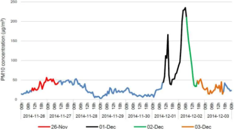
In order to determine any alterations to the bacterial community during Asian dust events, we collected airborne samples on November 26 and from December 1 to December 3. The relative humidity during the sampling period varied randomly around 57.6–79.3% and the tempera-ture was about -2.7–6˚C. No precipitation occurred during sampling period. The backward trajectory analysis showed that the air mass on November 26, 2014 originated from the East Sea. Meanwhile the air mass during December 1 to December 3 came from the desert area of Mongolia, passed over China, and arrived at the Korean peninsula for 2 days (HYSPLIT, data not shown). As shown inFig 1, the PM10 concentration increased from December 1, and was more than 200μg/m3
at 03:00–07:00 (Korea standard time, KST, UTC + 09) on December 2. Based on the hourly averaged PM10 concentration obtained from KMA and backward trajec-tory analysis, the collected airborne samples were classified into two group, Asian dust (Decem-ber 1 and Decem(Decem-ber 2) and non-Asian dust (Novem(Decem-ber 26 and Decem(Decem-ber 3). In this study, there are some different sampling durations between December 2 and other days. We, however, cautiously suggests that the different duration may not affect the following results since there are a similar biological characteristic between December 1 and 2. In addition, we performed SourceTracker software to confirm whether the samples were contaminated by other sources during sampling or following processes. Human microbiome project (HMP) data was used to
Fig 1. Concentration of PM10 during Asian dust and non-Asian dust events. Indicated date presents
sampling time and the time is Korea standard time, KST (UTC + 09).
compare a proportion of each sample under different source condition. In case of non-Asian dust as source, Asian dust samples had a relative high proportions, 0.8081 (December 1) and 0.7419 (December 2), but HMP data revealed clearly low proportion (0.1048). Moreover, the proportions of non-Asian dust samples (0.9007 in November 26) and 0.9564 in December 3) are clearly higher than HMP data (0.1876) in case of Asian dust as source.
Diversity analysis of the bacterial communities in the Asian dust and
non-Asian dust event samples
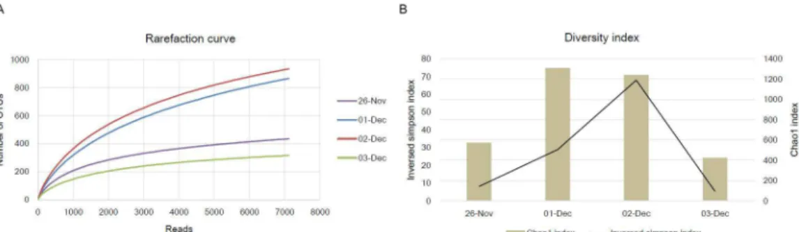
After pyrosequencing of amplified 16S rRNA gene products obtained from November 26 and December 1 to December 3, we analyzed the sequences using the standard operating proce-dure of Mothur software. For normalization of each sample, we randomly selected 7119 sequences presented the lowest sequences among each sample. To analyze an alteration of the diversity index caused by newly transported dust particles containing variable microorganisms during the Asian dust events, we first determined operational taxonomic units (OTUs) of each sample at the species level. As shown inFig 2A, the number of OTUs in the Asian dust sample were higher than those in the non-Asian dust sample (t-test, P = 0.02). In addition, the Chao1 index and the inversed Simpson index, which present a diversity index, were also increased in the Asian dust event samples (Fig 2B). Each diversity index was 2.55 times (t-test, P = 0.044) and 7.22 times (t-test, P = 0.13) higher than that for the non-Asian dust event samples. In a previous study, Jeonet al. also show that the diversity index, Chao1, of Asian dust event
sam-ples was 2.63 times higher than non-Asian dust event samsam-ples [32]. Indeed, while we expected that both richness (OTUs and Chao1) and evenness (Simpson) indices had a significantly dif-ference value between bacterial communities of Asian dust and non-Asian dust, only richness indices were significantly higher in the Asian dust samples. However, we observed a consistent result in the other period (in submission), suggesting that while various kind of microorgan-isms were transported by Asian dust particles, several taxonomic groups were presented as a relatively higher proportion in the dust particles. Beside the concerned issue, these results mean that newly transported microorganisms are present in the airborne environment during Asian dust events, leading to an alteration of the airborne microbial community compared to its normal status.
Characterization of bacterial community at the phylum level
Through the diversity analysis, we confirmed that the airborne microbial environment was affected by transported dust particles, which probably contain various microorganisms, during Asian dust events. We therefore classified the sequences obtained from pyrosequencing data according to the SILVA database. As a result, a total of 19 phyla were classified and several
Fig 2. Rarefaction curves and diversity index of the Asian dust and non-Asian dust event samples. doi:10.1371/journal.pone.0170693.g002
predominant phyla (more than 1% of total bacterial community) are presented inFig 3and
Table 1. From the results, we confirmed that the proportion of phylum Proteobacteria in the non-Asian dust samples was 1.6 times higher than the Asian dust samples. Meanwhile, the pro-portions of various phyla, including Actinobacteria, Acidobacteria, Firmicutes, and Gemmati-monadetes, were increased compared to those in the non-Asian dust event samples. To determine which phyla have a statistical difference between the two groups, we performed Metastats analysis using Mothur software. As shown inTable 2, the proportions of phyla Pro-teobacteria, Actinobacteria, and Firmicutes were significantly different between the two groups. In the Asian dust event samples, the phylum Proteobacteria was 0.62 times lower than in the non-Asian dust samples, and phyla Actinobacteria and Firmicutes were increased by 2.8 times and 5.6 times, respectively. These results mean that the proportion of the predominantly presented phylum, Proteobacteria, in the airborne environment was reduced with an increas-ing influx of newly transported microorganisms durincreas-ing Asian dust events. The influx of phyla Actinobacteria and Firmicutes during Asian dust events was also reported in previous work [32,33]. As similar with this results, the phylum Proteobacteria was predominantly presented
Fig 3. Bacterial communities (%) at the phylum level in the Asian dust and non-Asian dust event samples. Other phyla present lower than 1% of total bacteria community.
doi:10.1371/journal.pone.0170693.g003
Table 1. Bacterial communities (%) at the phylum level in the Asian dust and non-Asian dust event samples.
Phylum Bacterial communities (%)
26-Nov (non-Asian dust) 1-Dec (Asian dust) 2-Dec (Asian dust) 3-Dec (non-Asian dust)
Proteobacteria 85.29371 53.7716 51.83539 84.13297 Actinobacteria 9.739992 23.75334 17.50286 4.962867 Firmicutes 3.549319 15.69041 16.84237 2.298715 Deinococcus-Thermus Tr Tra 3.391337 6.695744 Acidobacteria Tr 3.258885 5.334688 Tr Cyanobacteria Tr Tr 1.079639 Tr Gemmatimonadetes Tr Tr 1.587705 Tr Verrucomicrobia Tr Tr 1.486092 Tr Others 1.416976 3.525776 0.939921 1.909702 a
Tr, trace, indicates a proportion lower than 1%.
during non-Asian dust events (53.1%), while Firmicutes presented as a relatively low propor-tion. The proportions of phyla Proteobacteria and Firmicues were changed into 15% and 53%, but the proportion of Actinobacteria was rarely altered during Asian dust events [32]. In addi-tion, Yamaguchiet al. showed that the phylum Actinobacteria was largely present in the Asian
dust event samples as well as in several source soils of Asian dust, including the Gobi desert and the Taklamakan desert [34]. Although there are several difference between these studies, Proteobacteria is one of predominant phylum during normal days, and proportions of Firmi-cutes and Actinobacteria were mainly altered during Asian dust events. However, since envi-ronmental conditions, sampling methods, and regions are different in each research, it is important to accumulate various database for understanding biological features of Asian dust particles.
Characterization of bacterial community at the genus level
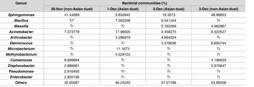
Next, we characterized the bacterial community at the genus level in the collected samples. For the analysis, we classified the pyrosequencing data at the genus level and confirmed that a total of 646 genera were present. Among them, 12 genera were found to make up more than 2% of the total proportion, as shown inFig 4andTable 3. During the non-Asian dust events, genus
Sphingomonas belong to phylum Proteobacteria was predominantly present at the proportion
of 45%, and generaAcinetobacter, Comamonas, Deinococcus, and Diaphorobacter were present
Table 2. Metastats analysis at the phylum level in the Asian dust and non-Asian dust event samples.
Phylum 1-Dec and 2-Dec (Asian dust) 26-Nov and 3-Dec P-valueb
Averagea Variance Std. error Averagea Variance Std. error
Proteobacteria 52.8035 1.87 0.9681 84.7133 0.67 0.5804 0.000167
Actinobacteria 20.6281 19.53 3.1252 7.3514 11.41 2.3886 0.036333
Firmicutes 16.2664 0.66 0.576 2.924 0.78 0.6253 0.017222
aAverage proportion of the phylum during indicated date.
bP<0.05 means that the proportion of indicated phylum has statistically difference.
doi:10.1371/journal.pone.0170693.t002
Fig 4. Bacterial communities (%) at the genus level in the Asian dust and non-Asian dust event samples. Other genera present lower than 2% of total bacteria community.
at proportions of 7.8%, 5.4%, 3.3%, and 2.9%, respectively. Although the genusSphingomonas
was still present predominantly in the Asian dust event samples, the proportion in the total genera was largely decreased to 14.6%. Instead of the decrement, generaBacillus, Arthrobacter, Microbacterium, and Methylobacterium were increased by 6.8, 4.8, 2.9, and 17.6 times,
respec-tively, in the Asian dust event samples. To determine the significantly different genera in their proportions between the two groups, we performed Metastats analysis. As a result, genera
Bacillus and Arthrobacter were found to be significantly increased by 6.8 and 4.8 times,
respec-tively (Table 4). In addition, among various genera that have proportion of lower than 2%, generaBlastocatella (1.99% of the Asian dust event samples) and Planomicrobium (1.43% of
the Asian dust event samples) were significantly increased by 16.2 and 6.9 times, respectively. These results reveal that generaBacillus and Planomicrobium belonging to the phylum
Firmi-cutes andArthrobacter belonging to the phylum Actinobacteria were major parts of the
bacte-rial community transported with dust particles in South Korea during the December 2014 Asian dust event. In particular, these genera have been known to find arid or semi-arid regions, suggesting that the feature of habitat may contribute a relatively high proportion of the genera in the Asian dust samples [35–37]. Among the identified genera,Bacillus has been
reported to induce severe diseases such as anthrax and food poisoning caused byBacillus anthracis and Bacillus cereus, respectively [38–41]. In our results, the most abundantBacillus
Table 3. Bacterial communities (%) at the genus level in the Asian dust and non-Asian dust event samples.
Genus Bacterial communities (%)
26-Nov (non-Asian dust) 1-Dec (Asian dust) 2-Dec (Asian dust) 3-Dec (non-Asian dust)
Sphingomonas 41.44999 9.832842 19.3573 48.96853 Bacillus Tra 7.262256 6.541344 Tr Massilia Tr Tr 5.182269 4.962867 Acinetobacter 7.373779 17.98005 4.458275 8.322527 Arthrobacter Tr 3.286979 4.064524 Tr Deinococcus Tr Tr 3.378636 6.695744 Microbacterium Tr 11.1673 Tr Tr Methylobacterium Tr 4.228122 Tr Tr Comamonas 6.699684 Tr Tr 4.196629 Diaphorobacter 2.888981 Tr Tr 2.970647 Pseudomonas 2.916495 Tr Tr Tr Enterobacter 2.820195 Tr Tr Tr Others 35.85087 46.24245 57.01766 23.88306 a
Tr, trace, indicates a proportion lower than 1%.
doi:10.1371/journal.pone.0170693.t003
Table 4. Metastats analysis at the genus level in the Asian dust and non-Asian dust event samples.
Genus 1-Dec and 2-Dec (Asian dust) 26-Nov and 3-Dec P-valueb
Averagea Variance Std. error Averagea Variance Std. error
Bacillus 6.9018 0.26 0.3605 1.0209 0.11 0.231 0.025584
Arthrobacter 3.6758 0.3 0.3888 0.7595 0.04 0.1347 0.039053
Blastocatella 1.9934 0.05 0.1532 0.1228 0.01 0.054 0.027008
Planomicrobium 1.4293 0.02 0.0949 0.2063 0.03 0.1238 0.036915
a
Average proportion of the genus during indicated date.
b
P<0.05 means that the proportion of indicated genus has statistically difference.
species isBacillus circulans (2.6%, data not shown). Although there are a few cases which reveal Bacillus circulans-associated diseases, a fatal sepsis case in an immunocompromised patient is
reported [42,43]. A relationship between disease and the other genera identified in this result is not well understood, but several reports have showed that the genusArthrobacter is found in
clinical samples [44,45]. An accumulation of bacterial information by continuous monitoring during Asian dust and non-Asian dust events should be necessary for establishing a relation-ship between human health and bacterial community during Asian dust events.
Conclusions
In this study, we characterized the airborne bacterial community during Asian dust and non-Asian dust events from November to December 2014. As a result, we found that the diversity of the airborne bacterial environment was increased by newly transported dust particles, which contain various microorganisms, during the Asian dust event, leading to a decrement of the proportion of the predominant bacteria in the airborne environment. During the Asian dust event on December 2014, the proportion of predominantly presented phylum Proteobac-teria was significantly reduced compared to the non-Asian dust event in November and December 2014. Meanwhile, phyla Actinobacteria and Firmicutes, especially generaBacillus, Arthrobacter, and Planomicrobium, were newly introduced with dust particles. In addition,
although the proportion was comparatively lower than the microorganisms mentioned above, various microorganisms were also transported during the Asian dust event. Indeed, the small proportion of newly transported microorganisms cannot be disregarded because the total amount (16S rRNA gene copy number) of microorganisms was dramatically increased (several hundreds to thousands) in the airborne environment during the Asian dust event [33]. In a further study, continuous monitoring of Asian dust and non-Asian dust event particles should be performed to determine the biological characterization of Asian dust particles. Moreover, various environmental effects, including soil environment, human health, and industrial prob-lems, affected by the precipitated microorganisms should also be evaluated.
Supporting Information
S1 File. Raw sequence file for bacterial 16S rRNA gene sequences during November 26, December 1, December 2, and December 3, 2014, South Korea. Files named by Nov 26 and Dec 3 are non-Asian events, and files named by Dec 1 and Dec 2 are Asian dust events. The files were submitted into the INSDC SRA (BioProject and SRA accession numbers are PRJNA360153 and SRP096119, respectively).
(ZIP)
Author Contributions
Conceptualization: SC MK TS. Data curation: SC SS JJ DL SL KK MK TS. Formal analysis: SC KK MK TS. Funding acquisition: TS. Investigation: SC MK TS. Methodology: SC SS TS. Project administration: MK TS.Resources: WJ DL EP JC HC MK TS. Software: SC KK.
Supervision: MK TS. Validation: TS. Visualization: SC TS. Writing – original draft: SC. Writing – review & editing: TS.
References
1. Kar A, Takeuchi K. Yellow dust: an overview of research and felt needs. J Arid Environ. 2004; 59 (1):167–87.
2. Kim YH, Kim KS, Kwak NJ, Lee KH, Kweon SA, Lim Y. Cytotoxicity of yellow sand in lung epithelial cells. J Biosciences. 2003; 28(1):77–81.
3. Sun JM, Liu TS, Lei ZF. Sources of heavy dust fall in Beijing, China on April 16, 1998. Geophys Res Lett. 2000; 27(14):2105–8.
4. Kwon HJ, Cho SH, Chun YS. The effects of the Asian dust events on daily mortality in Seoul, Korea. Epidemiology. 2000; 11(4):S88–S.
5. Kim KH, Kim MY. The effects of Asian Dust on particulate matter fractionation in Seoul, Korea during spring 2001. Chemosphere. 2003; 51(8):707–21. doi:10.1016/S0045-6535(03)00036-5PMID: 12668030
6. Mori I, Nishikawa M, Tanimura T, Quan H. Change in size distribution and chemical composition of kosa (Asian dust) aerosol during long-range transport. Atmos Environ. 2003; 37(30):4253–63.
7. Bory AJM, Biscaye PE, Grousset FE. Two distinct seasonal Asian source regions for mineral dust deposited in Greenland (NorthGRIP). Geophys Res Lett. 2003; 30(4).
8. Duce RA, Unni CK, Ray BJ, Prospero JM, Merrill JT. Long-range atmospheric transport of soil dust from Asia to the tropical north pacific: temporal variability. Science. 1980; 209(4464):1522–4. doi:10.1126/ science.209.4464.1522PMID:17745962
9. Kellogg CA, Griffin DW. Aerobiology and the global transport of desert dust. Trends Ecol Evol. 2006; 21 (11):638–44. doi:10.1016/j.tree.2006.07.004PMID:16843565
10. Kim KH, Choi GH, Kang CH, Lee JH, Kim JY, Youn YH, et al. The chemical composition of fine and coarse particles in relation with the Asian Dust events. Atmos Environ. 2003; 37(6):753–65.
11. Nakano T, Yokoo Y, Nishikawa M, Koyanagi H. Regional Sr-Nd isotopic ratios of soil minerals in north-ern China as Asian dust fingerprints. Atmos Environ. 2004; 38(19):3061–7.
12. Cheng TT, Lu DR, Wang GC, Xu YF. Chemical characteristics of Asian dust aerosol from Hunshan Dake sandland in Northern China. Atmos Environ. 2005; 39(16):2903–11.
13. Hua NP, Kobayashi F, Iwasaka Y, Shi GY, Naganuma T. Detailed identification of desert-originated bacteria carried by Asian dust storms to Japan. Aerobiologia. 2007; 23(4):291–8.
14. Reichhardt T. Weighing the health risks of airborne particulates. Environ Sci Technol. 1995; 29 (8):360A–4A. doi:10.1021/es00008a744PMID:22676121
15. Pritchard RJ, Ghio AJ, Lehmann JR, Winsett DW, Tepper JS, Park P, et al. Oxidant generation and lung injury after particulate air pollutant exposure increase with the concentrations of associated metals. Inhal Toxicol. 1996; 8(5):457–77.
16. Kim MY, Shin JY, Cho SC, Kim J, Lee GW, Kim KH. Environmental Mobilization Characteristics of Total Gaseous Mercury in the Western Coast of Korea During the Yellow Sand Period, 2001. Journal of Korean Earth Science Society. 2001; 22(6):480–90.
17. He M, Ichinose T, Yoshida S, Nishikawa M, Mori I, Yanagisawa R, et al. Airborne Asian sand dust enhances murine lung eosinophilia. Inhal Toxicol. 2010; 22(12):1012–25. doi:10.3109/08958378.2010. 510151PMID:20849355
18. Choi DS, Park YK, Oh SK, Yoon HJ, Kim JC, Seo WJ, et al. Distribution of airborne microorganisms in yellow sands of Korea. J Microbiol. 1997; 35(1):1–9.
19. Lee S, Choi B, Yi SM, Ko G. Characterization of microbial community during Asian dust events in Korea. Science of the Total Environment. 2009; 407(20):5308–14. doi:10.1016/j.scitotenv.2009.06.052 PMID:19631361
20. Maki T, Puspitasari F, Hara K, Yamada M, Kobayashi F, Hasegawa H, et al. Variations in the structure of airborne bacterial communities in a downwind area during an Asian dust (Kosa) event. Science of the Total Environment. 2014; 488:75–84. doi:10.1016/j.scitotenv.2014.04.044PMID:24815557
21. Whon TW, Kim MS, Roh SW, Shin NR, Lee HW, Bae JW. Metagenomic characterization of airborne viral DNA diversity in the near-surface atmosphere. J Virol. 2012; 86(15):8221–31. doi:10.1128/JVI. 00293-12PMID:22623790
22. Draxier RR, Hess GD. An overview of the HYSPLIT_4 modelling system for trajectories, dispersion and deposition. Aust Meteorol Mag. 1998; 47(4):295–308.
23. Draxler RR. The use of global and mesoscale meteorological model data to predict the transport and dispersion of tracer plumes over Washington, D. C. Weather Forecast. 2006; 21(3):383–94.
24. Islam MR, Sultana T, Joe MM, Cho JC, Sa TM. Comparisons of direct extraction methods of microbial DNA from different paddy soils. Saudi J Biol Sci. 2012; 19(3).
25. Vasileiadis S, Puglisi E, Arena M, Cappa F, Cocconcelli PS, Trevisan M. Soil Bacterial Diversity Screen-ing UsScreen-ing SScreen-ingle 16S rRNA Gene V Regions Coupled with Multi-Million Read GeneratScreen-ing SequencScreen-ing Technologies. Plos One. 2012; 7(8).
26. Schloss PD, Westcott SL, Ryabin T, Hall JR, Hartmann M, Hollister EB, et al. Introducing mothur: Open-Source, Platform-Independent, Community-Supported Software for Describing and Comparing Microbial Communities. Appl Environ Microb. 2009; 75(23):7537–41.
27. Quast C, Pruesse E, Yilmaz P, Gerken J, Schweer T, Yarza P, et al. The SILVA ribosomal RNA gene database project: improved data processing and web-based tools. Nucleic Acids Res. 2013; 41(D1): D590–D6.
28. White JR, Nagarajan N, Pop M. Statistical Methods for Detecting Differentially Abundant Features in Clinical Metagenomic Samples. Plos Comput Biol. 2009; 5(4).
29. Sneath RR, Sokal PHA. Numerical Taxonomy, San Francisco: W.H. Freeman. 1973
30. Chao A. Non-parametric estimation of the number of classes in a population. Scand J Statist. 1984; 11:265–270
31. Simpson EH. Measurement of diversity. Nature. 1949; 163:688–688
32. Jeon EM, Kim HJ, Jung K, Kim JH, Kim MY, Kim YP, et al. Impact of Asian dust events on airborne bac-terial community assessed by molecular analyses. Atmos Environ. 2011; 45(25):4313–21.
33. Yamaguchi N, Park J, Kodama M, Ichijo T, Baba T, Nasu M. Changes in the Airborne Bacterial Commu-nity in Outdoor Environments following Asian Dust Events. Microbes Environ. 2014; 29(1):82–8. doi: 10.1264/jsme2.ME13080PMID:24553107
34. Yamaguchi N, Ichijo T, Sakotani A, Baba T, Nasu M. Global dispersion of bacterial cells on Asian dust. Sci Rep-Uk. 2012; 2.
35. Foesel BU, Rohde M, Overmann J. Blastocatella fastidiosa gen. nov., sp. nov., isolated from semiarid savanna soil—The first described species of Acidobacteria subdivision 4. Syst Appl Microbiol. 2013; 36:82–89. doi:10.1016/j.syapm.2012.11.002PMID:23266188
36. Kellogg CA, Griffin DW, Garrison VH, Peak K, Royall N, Smith RR, Shinn EA. Characterization of aero-solized bacteria and fungi from desert dust events in Mali, West Africa. Aerobiologia. 2004; 20(2):99– 110.
37. Kwaasi AA, Parhar RS, al-Mohanna FA, Harfi HA, Collison KS, al-Sedairy ST. Aeroallergens and viable microbes in sandstorm dust. Potential triggers of allergic and nonallergic respiratory ailments. Allergy. 1998; 53(3):255–65. PMID:9542605
38. Dixon TC, Meselson M, Guillemin J, Hanna PC. Medical progress: anthrax. N. Engl. J. Med. 1999; 341:815–826. doi:10.1056/NEJM199909093411107PMID:10477781
39. Dixon TC, Fahd A, Koehler T, Swanson J, Hanna PC. Early events in anthrax pathogenesis: intracellu-lar survival of B. anthracis and its escape from RAW264.7 macrophages. Cell Microbiol. 2000; 2:453– 463. PMID:11207600
40. Ehling-Schulz M, Fricker M, Scherer S. Bacillus cereus, the causative agent of an emetic type of food-borne illness. Mol Nutr Food Res. 2004; 48(7):479–487. doi:10.1002/mnfr.200400055PMID: 15538709
41. Guinebretière MH, Broussolle V, Nguyen-The C. Enterotoxigenic Profiles of poisoning and Food-borne Bacillus cereus Strains. J Clin Microbiol. 2002; 40(8):3053–3056. doi: 10.1128/JCM.40.8.3053-3056.2002PMID:12149378
42. Alebouyeh M, Orimi P, Azimi-rad M, Tajbakhsh M, Tajeddin E, Sherafat S, Nazemalhosseini mojarad E, Zali MR. Fatal sepsis by Bacillus circulans in an immunocompromised patient. Iran J Microbiol. 2011; 3 (3):156–158. PMID:22347600
43. Logan NA, Old DC, Dick HM. Isolation of Bacillus circulans from a wound infection. J Clin Pathol. 1985; 38(7):838–839. PMID:4019805
44. Funke G, Hutson RA, Bernard KA, Pfyffer GE, Wauters G, Collins MD. Isolation of Arthrobacter spp. from clinical specimens and description of Arthrobacter cumminsii sp. nov. and Arthrobacter woluwen-sis sp. nov. J Clin Microbiol. 1996; 34:2356–2363. PMID:8880479
45. Bernasconi E, Valsangiacomo C, Peduzzi R, Carota A, Moccetti T, Funke G. Arthrobacter woluwensis subacute infective endocarditis: case report and review of the literature. Clin Infect Dis. 2004; 38(4): e27–31. doi:10.1086/381436PMID:14765360