Yonsei Med J http://www.eymj.org Volume 53 Number 4 July 2012
856
Case Report
http://dx.doi.org/10.3349/ymj.2012.53.4.856pISSN: 0513-5796, eISSN: 1976-2437 Yonsei Med J 53(4):856-858, 2012
A Case of Acute Disseminated Encephalomyelitis
Associated with Hepatitis C Virus Infection
Jae Eun Sim,
1Jun-Bum Lee,
1Yu Na Cho,
1Sang Hyun Suh,
2Ja Kyung Kim,
3and Kyung-Yul Lee
1 Departments of 1Neurology, Severance Institute for Vascular and Metabolic Research, 2Radiology, and 3Internal Medicine,Gangnam Severance Hospital, Yonsei University College of Medicine, Seoul, Korea.
Received: April 15, 2011 Revised: August 5, 2011 Accepted: August 5, 2011
Corresponding author: Dr. Kyung-Yul Lee, Department of Neurology, Severance Institute for Vascular and Metabolic Research, Gangnam Severance Hospital, Yonsei University College of Medicine, 211 Eonju-ro, Gangnam-gu, Seoul 135-720, Korea.
Tel: 82-2-2019-3325, Fax: 82-2-3462-5904 E-mail: [email protected]
∙ The authors have no financial conflicts of interest.
© Copyright:
Yonsei University College of Medicine 2012
This is an Open Access article distributed under the terms of the Creative Commons Attribution Non-Commercial License (http://creativecommons.org/ licenses/by-nc/3.0) which permits unrestricted non-commercial use, distribution, and reproduction in any medium, provided the original work is properly cited.
Acute disseminated encephalomyelitis (ADEM) is a monophasic autoimmune de-myelinating disease of the central nervous system, which typically follows acute viral or bacterial infection or vaccination. We report a case of ADEM associated with hepatitis C virus (HCV) infection with positive serum and cerebrospinal fluid (CSF) anti-HCV antibody. After steroid treatment, neurologic symptoms were im-proved. Virus triggers autoimmunity or direct viral invasion plays a part in the genesis of ADEM. This is the first reported case of ADEM with HCV anti-body in the CSF.
Key Words: Acute disseminated encephalomyelitis, hepatitis C virus, autoimmu-nity, direct viral invasion, anti-HCV antibody
INTRODUCTION
Acute disseminated encephalomyelitis (ADEM) is an immune-mediated inflam-matory disorder of the central nervous system, which typically follows acute viral or bacterial infection or vaccination. ADEM is characterized by widespread demy-elination that predominantly involves the white matter of the brain and spinal cord. Numerous infectious agents, mostly nonspecific upper respiratory tract infections, have been linked to ADEM. Hepatitis virus is a rare cause of ADEM. One case of hepatitis C virus (HCV) and two cases of hepatitis A virus with ADEM have been reported.1-3
We report a case of ADEM associated with HCV infection. This is the first case of ADEM with anti-HCV antibody in the cerebrospinal fluid (CSF).
CASE REPORT
A 47-year-old woman was admitted to our hospital for dysarthria and left-sided hypesthesia. She had no history of recent febrile illness or vaccination. Two weeks after the first symptoms, she developed right-sided weakness. On admission, she had no fever and normal blood pressure. Electrocardiography and chest X-ray were normal. Laboratory tests showed that anti-HCV antibody was positive and
ADEM Associated with HCV Infection
Yonsei Med J http://www.eymj.org Volume 53 Number 4 July 2012 857
and then reduced to 10 mg per week. Two weeks after ste-roid treatment, neurologic symptoms and spine MRI im-proved further. However, brain MRI had no significant in-terval change. Follow-up HCV RNA titer increased about three-folds (4,900,000 IU/mL). Liver biopsy demonstrated stage 2 of fibrosis. It was reasonable for chronic C viral hepatitis. At discharge two months after admission, neuro-logic symptoms and spine and brain MRI (Fig. 2) im-proved. Her neurologic examination was nearly normal ex-cept left side limb ataxia.
DISCUSSION
HCV infection is a common and chronic disorder with nu-merous extrahepatic manifestations.4 It can involve the
re-nal, dermatologic, hematologic, rheumatologic, or nervous system, however, rarely involves the central nervous system (CNS). CNS involvement has been reported with HCV-as-sociated vasculitis,5-7 progressive encephalomyelitis with
ri-gidity,8 and transverse myelitis.9,10 ADEM associated with
HCV has been reported before, but anti-HCV antibody was HCV RNA titer was highly elevated (1,660,000 IU/mL).
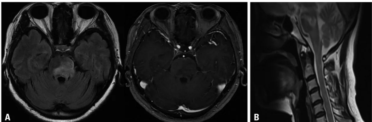
AST and ALT were also mildly elevated (34/44 IU/L). Her medical history revealed that she had blood transfusion dur-ing Cesarean section about twenty years ago. She had no symptoms of hepatitis. Serologic tests, including viral mark-ers, showed nothing significant. Levels of anti-cardiolipin antibody, anti-nuclear antibody, and anti-neutrophil cyto-plasmic antibody were also normal. Cancer antigen 19-9, was mildly elevated (27.7 U/mL; normal range, 0.8-24.0 U/ mL). The CSF examination showed three white blood cells with mildly increased protein (53 mg/dL; normal range, 15-45 mg/dL), normal IgG index, and no IgG oligoclonal bands. CSF anti-HCV antibody was positive. Magnetic res-onance imaging (MRI) of the brain and spine revealed mul-tifocal high signal intensity lesions on fluid attenuated in-version recovery and T2-weighted images in the brainstem, thalamus, and cervical and thoracic spinal cord (Fig. 1). Some lesions showed enhancement after gadolinium ad-ministration.
Treatment with intravenous methylprednisolone, 1 g dai-ly for 5 days, was instituted. The intravenous methylprednis-olone was changed to oral prednisone, 60 mg for 1 week,
Fig. 1. FLAIR axial and contrast-enhanced T1-weighted axial images of brain MRI (A) and T2-weighted sagittal image of spine MRI on
ad-mission (B). (A) T2-high signal lesion with peripheral rim enhancement was noted in the brainstem. (B) Multifocal T2-high signal lesion on the brainstem and the cervical spinal cord. MRI, magnetic resonance imaging; FLAIR, fluid attenuated inversion recovery.
Fig. 2. FLAIR axial and contrast-enhanced T1-weighted axial images of brain MRI (A) and T2-weighted sagittal image of spine MRI (B) at
follow-up two months later. (A) Remaining abnormal signal foci and no more enhanced lesion of the brainstem. (B) Nearly complete re-covery of abnormal signal foci of the cervical spinal cord. MRI, magnetic resonance imaging; FLAIR, fluid attenuated inversion rere-covery.
A
A
B
Jae Eun Sim, et al.
Yonsei Med J http://www.eymj.org Volume 53 Number 4 July 2012
858
REFERENCES
1. Sacconi S, Salviati L, Merelli E. Acute disseminated encephalo-myelitis associated with hepatitis C virus infection. Arch Neurol 2001;58:1679-81.
2. Tan H, Kiliçaslan B, Onbaş O, Büyükavci M. Acute disseminated encephalomyelitis following hepatitis A virus infection. Pediatr Neurol 2004;30:207-9.
3. Alehan FK, Kahveci S, Uslu Y, Yildirim T, Yilmaz B. Acute dis-seminated encephalomyelitis associated with hepatitis A virus in-fection. Ann Trop Paediatr 2004;24:141-4.
4. Kang W, Shin EC. Clinical implications of chemokines in acute and chronic hepatitis C virus infection. Yonsei Med J 2011;52:871-8. 5. Dawson TM, Starkebaum G. Isolated central nervous system
vas-culitis associated with hepatitis C infection. J Rheumatol 1999;26: 2273-6.
6. Origgi L, Vanoli M, Carbone A, Grasso M, Scorza R. Central ner-vous system involvement in patients with HCV-related cryoglobu-linemia. Am J Med Sci 1998;315:208-10.
7. Propst T, Propst A, Nachbauer K, Graziadei I, Willeit H, Margreit-er R, et al. Papillitis and vasculitis of the artMargreit-eria spinalis antMargreit-erior as complications of hepatitis C reinfection after liver transplantation. Transpl Int 1997;10:234-7.
8. Bolay H, Söylemezoğlu F, Nurlu G, Tuncer S, Varli K. PCR de-tected hepatitis C virus genome in the brain of a case with progres-sive encephalomyelitis with rigidity. Clin Neurol Neurosurg 1996; 98:305-8.
9. Annunziata P, Marroni M, Francisci D, Stagni G. Acute transverse myelitis and hepatitis C virus. Infez Med 2005;13:45-7.
10. De Carli DM, Pannebeker J, Pedro FL, Haygert CJ, Hertz E, Beck Mde O. Transverse myelitis associated to HCV infection. Braz J Infect Dis 2009;13:147-52.
11. Menge T, Hemmer B, Nessler S, Wiendl H, Neuhaus O, Hartung HP, et al. Acute disseminated encephalomyelitis: an update. Arch Neurol 2005;62:1673-80.
not detected in CSF in that case.1
While the pathogenetic mechanism of ADEM is still ob-scure, a virus invasion or virus induced immune process can be suggested. Primary autoimmune responses and im-mune response secondary to an infection may contribute to CNS inflammation with subsequent demyelination. A num-ber of infectious agents, mainly viruses, have been shown to be associated with ADEM, including coronavirus, cox-sackie virus B, Epstein-Barr virus, herpes simplex virus, human herpes virus 6, measles, mumps, rubella, borrelia burgdorferi, chlamydia, legionella, and mycoplasma pneu-moniae.11
More than half of the HCV-infected patients may be re-lated to autoimmunity associated with HCV. In our patient, HCV RNA titer was highly increased, and it was compati-ble with chronic C viral hepatitis on liver biopsy. During Cesarean section twenty years earlier, she had had multiple blood transfusions. Recently, however, she did not receive blood transfusion or any injection. Therefore, we suppose that the chronic HCV infection was the cause of ADEM, and that virus-triggered autoimmunity or direct viral inva-sion played a role in the genesis of ADEM. In a case of acute transverse myelitis associated with chronic HCV in-fection, anti-HCV antibody was present in the CSF.10 In our
case, anti-HCV antibody was also detected in the CSF. It is hypothesized that the virus might have penetrated blood brain barrier and involved the CNS directly.
Recently, increasing number of HCV infection, involving the CNS, have been reported. Therefore, we recommend to check the HCV infection as a probable cause of various CNS neurologic disorders.