ABSTRACT
Background: Gender-related factors might play an important role in the development of reflux esophagitis (RE) and symptomatic gastro-esophageal reflux disease (GERD). We aimed to evaluate the prevalence and risk factors for RE and symptomatic GERD and determine whether gender specific differences exist.
Methods: This study was conducted on a health cohort consisting of 10,158 participants who underwent comprehensive health screening. Lifestyles and gastrointestinal symptoms were investigated using a self-reported structured questionnaire. Questionnaires about menstrual status were added for the women.
Results: The prevalence of RE in men was significantly higher than that in women (10.6% vs. 2.0%, P < 0.001); however, symptomatic GERD showed predominance in women
(6.2% vs. 2.5%, P < 0.001). Although the prevalence of RE gradually increased with the
duration of menopause stratified by decade (P = 0.007), that of symptomatic GERD rapidly
increased across the menopausal transit in women. Apart from common risk factors of obesity and current smoking for RE, over 70 years of age in women and hiatal hernia and hypertriglyceridemia in men were significant risk factors. In symptomatic GERD, high somatization was a common risk factor. Excessive alcohol drinking was a significant risk factor in men, but not in women.
Conclusion: This study showed a predominance of RE in men, but a predominance of symptomatic GERD in women. In women, dynamic increase in the prevalence of GERD is closely related to the menopause conditions and its duration. There are specific risk factors for RE and symptomatic GERD according to gender differences.
Keywords: Gastro-Esophageal Reflux Disease; Gender Distribution; Menopause; Prevalence; Risk Factors
INTRODUCTION
Gastro-esophageal reflux disease (GERD), defined as the abnormal reflux of gastric contents into the esophagus, is a chronic disease that results in reflux symptoms and complications, such as reflux esophagitis (RE) or Barrett's esopahgus.1,2 In recent decades, there has been
a significant rise in the prevalence of GERD worldwide including Korea.3 The prevalence of
Original Article
Received: Jan 22, 2019 Accepted: May 10, 2019 Address for Correspondence: Hye-Kyung Jung, MD
Department of Internal Medicine, Ewha Womans University School of Medicine, 1071 Anyangcheon-ro, Yangcheon-gu, Seoul 07985, Korea.
E-mail: [email protected]
© 2019 The Korean Academy of Medical Sciences.
This is an Open Access article distributed under the terms of the Creative Commons Attribution Non-Commercial License (https:// creativecommons.org/licenses/by-nc/4.0/) which permits unrestricted non-commercial use, distribution, and reproduction in any medium, provided the original work is properly cited.
ORCID iDs Sang Yoon Kim
https://orcid.org/0000-0002-1155-1495 Hye-Kyung Jung https://orcid.org/0000-0002-6653-5214 Jiyoung Lim https://orcid.org/0000-0002-3252-9035 Tae Oh Kim https://orcid.org/0000-0002-6369-5618 A Reum Choe https://orcid.org/0000-0002-2552-7066 Chung Hyun Tae
https://orcid.org/0000-0002-0764-7793 Ki-Nam Shim https://orcid.org/0000-0003-4004-6292 Chang Mo Moon https://orcid.org/0000-0003-2550-913X Seong-Eun Kim https://orcid.org/0000-0002-6310-5366
Sang Yoon Kim , Hye-Kyung Jung , Jiyoung Lim , Tae Oh Kim , A Reum Choe , Chung Hyun Tae , Ki-Nam Shim , Chang Mo Moon , Seong-Eun Kim , and Sung-Ae Jung
Department of Internal Medicine, Ewha Womans University School of Medicine, Seoul, Korea
Gender Specific Differences in
Prevalence and Risk Factors for
Gastro-Esophageal Reflux Disease
Gastroenterology &
Sung-Ae Jung
https://orcid.org/0000-0001-7224-2867 Disclosure
The authors have no potential conflicts of interest of disclose.
Author Contributions
Conceptualization: Jung HK. Data curation: Jung HK, Kim SY, Lim J, Kim TO, Choe AR, Tae CH. Formal analysis: Jung HK, Kim SY. Investigation: Jung HK, Kim SY. Methodology: Jung HK, Kim SY. Writing - original draft: Kim SY. Writing - review & editing: Jung HK, Shim KN, Moon CM, Kim SE, Jung SA.
RE was 3.4% in 1997,3,4 but increased to 6.2% in 2008,5 while that of symptomatic GERD
was 3.5% in 2001,6 but increased to 7.1% in 2007 in Korea.7 Consequently, the clinical
significance and risk factors for RE and symptomatic GERD are gaining importance. Several studies reported that the common risk factors for RE and GERD are age,5 obesity,8,9
hiatal hernia,10 alcohol drinking,11 and smoking.11,12 In addition, gender-related factors
might contribute in the pathogenesis and development of RE and symptomatic GERD.13,14
The studies of risk factors for RE and symptomatic GERD according to gender are still rare. Investigation of gender-related differences in RE and symptomatic GERD is important for providing better understanding of these pathogenesis, prevention and treatment for both men and women patients.13,14 The study aimed to evaluate the gender-related differences in
the prevalence and risk factors for RE and symptomatic GERD.
METHODS
Study participants
This study was retrospectively conducted in a large cohort of apparently healthy Korean men and women who underwent a comprehensive annual or biennial health examination at Ewha Womans University Mokdong Hospital from January 2015 to December 2016. In Korea, the Industrial Safety and Health Act stipulates that medical checkups are provided free of charge to all employees annually or every two years. In the case of having health examination twice during this study period, the former examination was considered here. Two-third of the participants from the health promotion center were employees of several companies and their spouses. The rest of them took a health screening at their own expense.
Among a total of 14,104 individuals, 10,680 participants (75.7%), who responded to the questionnaire and underwent upper endoscopy screening, were enrolled. Of them, 10,158 participants were finally eligible after exclusion of those with prior major abdominal surgery (n = 33), gastric cancer (n = 8), and active peptic ulcer disease (n = 481). We measured the levels of high density lipoprotein-cholesterol (HDL-C), hypertriglyceride, and fasting venous glucose with the Hitachi 7600 analyzer (Hitachi Ltd., Tokyo, Japan). According to the National Cholesterol Education Program Adult Treatment Panel III criteria, low HDL-C levels were defined as less than 40 mg/dL in men and less than 50 mg/dL in women, and hypertriglyceridemia was defined as triglyceride levels greater than 150 mg/dL. Weight (to the nearest 0.1 kg), height (to the nearest 0.1 cm), waist circumference (WC) (to the nearest 0.1 cm), and blood pressure were measured by well-trained nurses. Based on the modified criteria of Asian-Pacific guidelines of the World Health Organization, the participants were categorized according to the body mass index (BMI) as follows: normal (< 23 kg/m2),
overweight (23–24.9 kg/m2) and obese (≥ 25 kg/m2). The WC was measured at the midpoint
between the lower border of rib cage and iliac crest. Central obesity was identified as a WC greater than 90 cm in men and that WC greater than 85 cm in women, according to the clinical practice guidelines for overweight and obesity in Korean Society for the Study of Obesity.15 Diabetes mellitus (DM) was determined based on the diagnostic criteria of the
American Diabetes Association or self-reported history of DM. Hypertension (HTN) was defined as a mean systolic blood pressure greater than 140 mmHg, a mean diastolic blood pressure greater than 90 mmHg, or a self-reported history of HTN.
Endoscopic examinations and definition of RE
Upper endoscopy was performed by experienced certified gastroenterologists. The presence and severity of RE were classified by the Los Angeles (LA) classification as grade A–D (LA-A to D) and were based on the longest length of a mucosal break and the confluence of erosions.16 Hiatal hernia was defined as a circular extension of the gastric mucosa 2 cm or
more above the diaphragmatic indentation from the endoscopic findings.
Questionnaires and definition of symptomatic GERD
Symptomatic GERD was defined as heartburn or acid regurgitation at least twice per week for the past 6 months or interference with daily life within the past year.1,17 Korean Rome
III questionnaires, which reveled good reliability and satisfactory construct validity, was used to evaluate symptomatic GERD.18 The Cronbach's alpha for heartburn was 0.82 and
that for acid regurgitation was 0.84.18,19 Extra-esophageal symptoms such as dysphagia,
chronic cough, chest pain, and hoarseness were not considered as symptomatic GERD because of limited evidence of causal relationship with reflux.1 The participants were also
instructed to complete structured questionnaires for somatization symptom checklist (SSC) scores.20 These SSC scores include 17 non-gastrointestinal (GI) symptoms or illnesses.
The participants were asked to indicate how troublesome each symptom was (intensity on a scale of 0 to 4, indicating no problem to extremely troublesome) and how frequent each symptom occurred (frequency on a scale of 0 to 4, indicating no problem to daily occurrence) during the past year using the five-Likert scale. These scores indicate the degree of overall psychosomatic distress.20,21 The minimum score possible was 0 and the maximum was 272.
In measuring health-related quality of life (HRQoL), the Korean version of the EuroQol-5 Dimension (EQ-5D) was used.22 The EQ-5D records the degree of self-reported problems
on five dimensions as follows; mobility, self-care, usual activities, pain/discomfort, anxiety/ depression. Previous study has demonstrated its cross-cultural adaptation and validation on upper GI disorders and HRQoL.21
Questionnaires and lifestyle habits
Each participant completed a structured questionnaire which included demographic factors, medical history, lifestyle habits (smoking and alcohol drinking), and status of menopause (pre- vs. post-menopause). Smoking was categorized as non-smoking, past smoking and current smoking, with the latter defined as having regularly smoked more than 1.5 pack-years since the past year. Alcohol drinking was classified into none and excessive alcohol drinking, with the latter defined as more than 14 standard drinks per week in men and more than 7 standard drinks per week in women during the previous 6 months. A standard drink was defined as a cup of alcohol containing 12 grams, regardless of types of alcohol.
Statistical analysis
Differences in variables between men and women were analyzed by using the Student's t-test or Mann-Whitney U test and χ2 test. Student's t-test was used to compare continuous
variables between two groups, and the χ2 test to compare categorical variables. Because
HDL-C, triglyceride, and WC were skewed data, median (interquartile range) and non-parametric tests (Mann-Whitney U test) were used to compare between two groups. Multiple logistic regression was performed for assessing predictor variables selected from the univariate analyses. The odds ratios (OR) and 95% confidence intervals (CI) were computed from the estimated coefficients in the regression. The statistical analyses were carried out using SPSS version 21.0 for Windows software (SPSS Inc., Chicago, IL, USA). A two tailed
Ethics statement
This study was approved by the Institutional Review Board (IRB) of Ewha University Mokdong Hospital (IRB No. 2018-08-026-002). The requirement for written informed consent was waved because we were using anonymous data and questionnaires collected regularly during the health checkup process. This study followed the principles of the Declaration of Helsinki.
RESULTS
Participant characteristics
A total of 10,158 participants were enrolled in this study (Table 1). Their mean age was 47.1 ± 10.2 years and 50.9% of them were men. The overall prevalence of RE was 6.4% (651/10,158); 4.5% (457/10,158) in LA-A, 1.8% (182/10,158) in LA-B, and 0.1% (12/10,158) in LA-C. Compared to women, the incidence of obesity, DM, HTN, and hypertriglyceridemia were more common in men. Also, the lifestyle of smoking and excessive alcohol drinking was more frequent in men (Table 1). Among women participants, 1,709 of them (34.2%) had the menopause status, excluding 319 (6.4%) due to missing data. The incidence of obesity, DM, HTN, and hypertriglyceridemia in women participants with menopause were significantly higher in those with pre-menopause. Frequency of smoking and excessive alcohol drinking in pre-menopause women was also more common than those with menopause (Supplementary Table 1).
Prevalence of RE
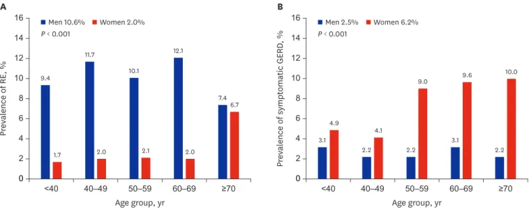
The prevalence of RE in men was significantly higher than that in women (10.6% vs. 2.0%, P < 0.001) (Table 1). In women, its prevalence increased with age, but there were no
significant differences with age in men. Below the age of 70, the prevalence of RE in men was significantly higher than that in women; however, above the age of 70, this difference disappeared and the prevalence became similar (Fig. 1A).
Prevalence of symptomatic GERD
The prevalence of symptomatic GERD in women was significantly higher compared to that in men in all age groups (2.5% vs. 6.2%, P < 0.001) (Table 1). In women, its prevalence
dramatically increased above the age of 50, and high prevalence persisted since then (Fig. 1B).
Clinical presentation of RE and symptomatic GERD according to menopause
In women, the spectrum in GERD varied depending on the duration of menopause. The prevalence of RE and symptomatic GERD increased with age in women (Fig. 1A and B), particularly after menopause. During the menopausal period, the prevalence of RE was significantly increased as the duration of menopause stratified by decade became longer (P = 0.007) (Fig. 2). According to BMI adjusted logistic analysis, over 30 years of menopausewas a significant risk factor in the occurrence of RE (OR, 5.29; 95% CI, 1.52–18.48; P = 0.009)
(Table 2). In contrast, the prevalence of symptomatic GERD increased during the first 20 years after menopause, but tended to decrease thereafter (P < 0.001) (Fig. 2).
Comparison of risk factors for RE between men and women participants
According to the univariate analysis in men, being overweight, obesity, HTN,hypertriglyceridemia, central obesity (over 90 cm of WC), past smoking, current smoking, excessive alcohol consumption, and hiatal hernia were significant risk factors for RE (Table 3); however, age was not significant. Multivariate analysis in men revealed that being overweight (OR, 1.57; 95% CI, 1.18–2.09; P = 0.002), obesity (OR, 1.81; 95% CI, 1.34–2.44; P < 0.001),
hypertriglyceridemia (OR, 1.28; 95% CI, 1.06–1.55; P = 0.012), current smoking (OR, 1.67;
95% CI, 1.30–2.16; P < 0.001), and hiatal hernia (OR, 2.54; 95% CI, 1.81–3.62; P < 0.001) were
all independent risk factors for RE (Table 3). By univariate analysis in women, over 70 years of age, obesity, hypertriglyceride, central obesity (over 85 cm of WC), current smoking, and hiatal hernia were significant risk factors for RE (Table 3). Multivariate analysis in women revealed that obesity and smoking were significantly independent risk factors and their ORs were about twice as high as that of men (OR, 3.17; 95% CI, 1.79–5.61; P < 0.001; OR, 3.47; 95%
CI, 1.61–7.48; P = 0.001, respectively). Unlike the men participants, over 70 years of age was the
moderate risk factor for RE in women (OR, 2.90; 95% CI, 1.19–7.04; P = 0.019) (Table 3).
Table 1. Clinical characteristics of participants
Characteristics Total (n = 10,158) Men (n = 5,166) Women (n = 4,992) P value
Age, yr 47.1 ± 10.2 47.7 ± 10.0 46.5 ± 10.4 < 0.001 Age group, yr < 0.001 < 40 2,323 (22.9) 1,034 (20.0) 1,289 (25.8) 40–49 3,791 (37.3) 1,924 (37.2) 1,867 (37.4) 50–59 2,968 (29.2) 1,660 (32.1) 1,308 (26.2) 60–69 821 (8.1) 413 (8.0) 408 (8.2) ≥ 70 255 (2.5) 135 (2.6) 120 (2.4) BMI, kg/m2 23.6 ± 3.2 24.9 ± 2.9 22.4 ± 3.0 < 0.001 BMI group, kg/m2 < 0.001 Normal, < 23.0 4,488 (44.2) 1,319 (25.5) 3,169 (63.5) Overweight, 23–24.9 2,497 (24.6) 1,510 (29.2) 987 (19.8) Obese, ≥ 25.0 3,173 (31.2) 2,337 (45.2) 836 (16.7) Diabetes mellitus 730 (7.2) 547 (10.6) 183 (3.7) < 0.001 Hypertension 3,309 (32.6) 2,107 (40.8) 1,202 (24.1) < 0.001 HDL-C, mg/dL Median (IQRs) 55.0 (47.0–65.0) 50.0 (43.0–58.0) 61.0 (52.0–70.0) < 0.001 < 40 (M), < 50 (W) 1,534 (15.1) 630 (12.2) 904 (18.1) < 0.001 Triglyceride, mg/dL Median (IQRs) 98.0 (68.0–146.0) 124.0 (86.0–179.5) 79.0 (57.0–110.0) < 0.001 ≥ 150 2,436 (24.0) 1,869 (36.2) 567 (11.4) < 0.001 Waist circumference, cm Median (IQRs) 81.0 (73.0–88.0) 86.0 (82.0–91.0) 73.0 (68.0–79.0) < 0.001 ≥ 90 (M), ≥ 85 (W) 2,199 (21.6) 1,665 (32.2) 534 (10.7) < 0.001 Smoking status < 0.001 Never 5,803 (59.0) 1,362 (26.7) 4,441 (93.8) Past 2,250 (22.9) 2,085 (40.9) 165 (3.5) Current 1,781 (18.1) 1,654 (32.4) 127 (2.7)
Excessive alcohol drinking 2,913 (28.7) 1,614 (31.2) 1,299 (26.0) < 0.001
Hiatal hernia, No < 0.001 No 9,915 (97.6) 4,962 (96.1) 4,953 (99.2) Yes 243 (2.4) 204 (3.9) 39 (0.8) SSC scores 14.4 ± 18.2 11.3 ± 14.8 17.6 ± 20.7 < 0.001 LA classification < 0.001 None 9,507 (93.6) 4,617 (89.4) 4,890 (98.0) A 457 (4.5) 388 (7.5) 69 (1.4) B 182 (1.8) 151 (2.9) 31 (0.6) C 12 (0.1) 10 (0.2) 2 (0.0) Symptomatic GERD 435 (4.3) 127 (2.5) 308 (6.2) < 0.001 Presence of menopause -No - NA 2,964 (59.4) Yes - NA 1,709 (34.2) Missing data - NA 319 (6.4)
Data are shown as number (%), mean ± standard deviation or median (IQRs).
BMI = body mass index, HDL-C = high density lipoprotein-cholesterol, IQR = interquartile range, M = men, W = women, SSC = somatization symptom checklist, LA = Los Angeles, GERD = gastro-esophageal reflux disease, NA = not available.
Comparison of risk factors for symptomatic GERD between men and women
The univariate analysis revealed that current smoking, excessive alcohol drinking, and SSC scores were related to symptomatic GERD in men. By multivariate analysis, SSC scores were0 ≥70 60–69 50–59 40–49 <40 9.4 1.7 11.7 2.0 10.1 2.1 12.1 2.0 7.4 6.7 3.1 4.9 2.2 4.1 2.2 9.0 3.1 9.6 2.2 10.0 14 12 10 8 4 2 Pr eval ence of RE , % Age group, yr A B 16 16 6 0 ≥70 60–69 50–59 40–49 <40 14 12 10 8 4 2 Pr eval ence of sympt omatic GERD , % Age group, yr P < 0.001 P < 0.001 6
Men 10.6% Women 2.0% Men 2.5% Women 6.2%
Fig. 1. Prevalence of RE and symptomatic GERD by gender and age. (A) Prevalence of RE. (B) Prevalence of symptomatic GERD. Blue bars indicate men and red bars indicate women participants.
RE = reflux esophagitis, GERD = gastro-esophageal reflux disease.
0 30–39 20–29 10–19 0–9 Premenopause 1.8 21.9 4.0 1.9 22.8 10.2 3.1 12.0 23.5 4.5 24.3 9.8 11.5 7.7 24.8 14 12 10 8 4 2 Pr eval ence, % Duration of menopause, yr 20 28 27 26 25 24 23 22 21 20 18 16 6 symptomatic GERD RE BMI P = 0.007 P < 0.001
Fig. 2. Prevalence of RE and symptomatic GERD with BMI according to duration of menopause. Blue line indicates symptomatic GERD, red line indicates RE, and dotted green line indicates BMI.
RE = reflux esophagitis, GERD = gastro-esophageal reflux disease, BMI = body mass index. Table 2. Prevalence of reflux esophagitis according to duration of menopause and adjustment for BMI
Duration of menopause Unadjusted OR Unadjusted 95% CI P value BMI-adjusted OR BMI-adjusted 95% CI P value
RE, yr, ref. premenopause 1
0–9 1.09 0.66–1.79 0.748 0.99 0.60–1.64 0.971
10–19 1.74 0.88–3.46 0.112 1.48 0.74–2.96 0.267
20–29 2.57 1.01–6.55 0.049 2.01 0.78–5.18 0.149
30–39 7.16 2.09–24.59 0.002 5.29 1.52–18.48 0.009
revealed as the scores which increased by one point, OR increased by 1.03 (95% CI, 1.02–1.04;
P < 0.001). Excessive alcohol drinking (OR, 1.84; 95% CI, 1.26–2.70; P = 0.002) was also a
significant risk factor for symptomatic GERD in men (Table 4). However, unlike RE, obesity, or other metabolic components were not associated with symptomatic GERD in men. Table 3. Comparison of risk factors for reflux esophagitis between men and women
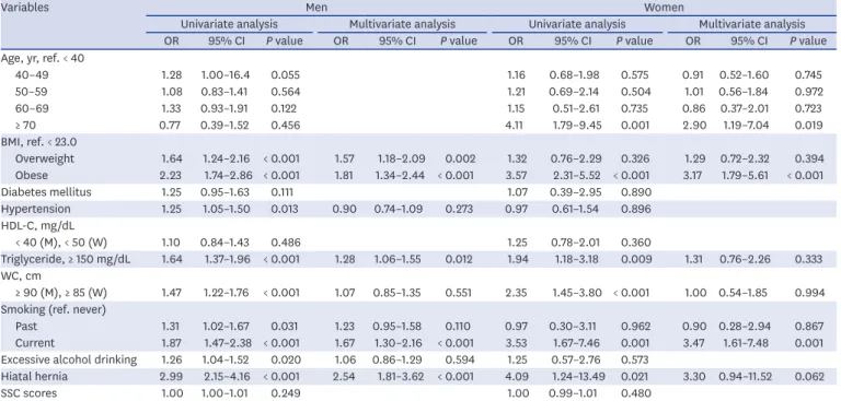
Variables Men Women
Univariate analysis Multivariate analysis Univariate analysis Multivariate analysis OR 95% CI P value OR 95% CI P value OR 95% CI P value OR 95% CI P value
Age, yr, ref. < 40
40–49 1.28 1.00–16.4 0.055 1.16 0.68–1.98 0.575 0.91 0.52–1.60 0.745 50–59 1.08 0.83–1.41 0.564 1.21 0.69–2.14 0.504 1.01 0.56–1.84 0.972 60–69 1.33 0.93–1.91 0.122 1.15 0.51–2.61 0.735 0.86 0.37–2.01 0.723 ≥ 70 0.77 0.39–1.52 0.456 4.11 1.79–9.45 0.001 2.90 1.19–7.04 0.019 BMI, ref. < 23.0 Overweight 1.64 1.24–2.16 < 0.001 1.57 1.18–2.09 0.002 1.32 0.76–2.29 0.326 1.29 0.72–2.32 0.394 Obese 2.23 1.74–2.86 < 0.001 1.81 1.34–2.44 < 0.001 3.57 2.31–5.52 < 0.001 3.17 1.79–5.61 < 0.001 Diabetes mellitus 1.25 0.95–1.63 0.111 1.07 0.39–2.95 0.890 Hypertension 1.25 1.05–1.50 0.013 0.90 0.74–1.09 0.273 0.97 0.61–1.54 0.896 HDL-C, mg/dL < 40 (M), < 50 (W) 1.10 0.84–1.43 0.486 1.25 0.78–2.01 0.360 Triglyceride, ≥ 150 mg/dL 1.64 1.37–1.96 < 0.001 1.28 1.06–1.55 0.012 1.94 1.18–3.18 0.009 1.31 0.76–2.26 0.333 WC, cm ≥ 90 (M), ≥ 85 (W) 1.47 1.22–1.76 < 0.001 1.07 0.85–1.35 0.551 2.35 1.45–3.80 < 0.001 1.00 0.54–1.85 0.994 Smoking (ref. never)
Past 1.31 1.02–1.67 0.031 1.23 0.95–1.58 0.110 0.97 0.30–3.11 0.962 0.90 0.28–2.94 0.867 Current 1.87 1.47–2.38 < 0.001 1.67 1.30–2.16 < 0.001 3.53 1.67–7.46 0.001 3.47 1.61–7.48 0.001 Excessive alcohol drinking 1.26 1.04–1.52 0.020 1.06 0.86–1.29 0.594 1.25 0.57–2.76 0.573
Hiatal hernia 2.99 2.15–4.16 < 0.001 2.54 1.81–3.62 < 0.001 4.09 1.24–13.49 0.021 3.30 0.94–11.52 0.062
SSC scores 1.00 1.00–1.01 0.249 1.00 0.99–1.01 0.480
OR = odds ratio, CI = confidence interval, BMI = body mass index, HDL-C = high density lipoprotein-cholesterol, M = men, W = women, WC = waist circumference, SSC = somatization symptom checklist.
Table 4. Comparison of risk factors for symptomatic gastro-esophageal reflux disease between men and women
Variables Men Women
Univariate analysis Multivariate analysis Univariate analysis Multivariate analysis OR 95% CI P value OR 95% CI P value OR 95% CI P value OR 95% CI P value
Age, yr, ref. < 40
40–49 0.70 0.44–1.11 0.132 0.83 0.59–1.16 0.272 0.86 0.59–1.25 0.429 50–59 0.71 0.44–1.15 0.168 1.93 1.41–2.65 < 0.001 1.79 1.24–2.60 0.002 60–69 1.02 0.53–1.96 0.958 2.06 1.36–3.12 0.001 2.16 1.29–3.63 0.004 ≥ 70 0.71 0.22–2.36 0.578 2.16 1.13–4.13 0.020 1.18 0.68–1.31 0.728 BMI, ref. < 23.0 Overweight 0.74 0.45–1.22 0.239 0.97 0.72–1.31 0.848 Obese 1.00 0.66–1.52 0.999 0.96 0.69–1.32 0.776 Diabetes mellitus 0.72 0.37–1.38 0.316 0.88 0.46–1.67 0.686 Hypertension 0.77 0.53–1.11 0.155 1.30 1.01–1.68 0.042 0.94 0.68–1.31 0.723 HDL-C, mg/dL < 40 (M), < 50 (F) 0.89 0.51–1.56 0.683 0.94 0.69–1.27 0.672 Triglyceride, ≥ 150 mg/dL 1.19 0.83–1.70 0.345 0.93 0.64–1.35 0.713 WC, cm ≥ 90 (M), ≥ 85 (F) 0.97 0.66–1.41 0.858 1.11 0.78–1.59 0.561
Smoking, ref. never
Past 1.49 0.91–2.44 0.115 1.44 0.84–2.46 0.190 1.59 0.92–2.74 0.096 1.14 0.59–2.22 0.692 Current 1.85 1.13–3.05 0.015 1.48 0.85–2.57 0.163 1.97 1.11–3.48 0.020 1.35 0.68–2.67 0.391 Excessive alcohol drinking 2.22 1.54–3.20 < 0.001 1.84 1.26–2.70 0.002 1.53 1.01–2.33 0.045 1.31 0.82–2.09 0.261
Hiatal hernia 0.58 0.18–1.85 0.358 0.00 0.00–0.00 0.998
SSC scores 1.03 1.02–1.04 < 0.001 1.03 1.02–1.04 < 0.001 1.03 1.02–1.03 < 0.001 1.02 1.02–1.03 < 0.001 OR = odds ratio, CI = confidence interval, BMI = body mass index, HDL-C = high density lipoprotein-cholesterol, M = men, W = women, WC = waist circumference, SSC = somatization symptom checklist.
In univariate analysis, age, HTN, current smoking, excessive alcohol drinking, and SSC scores were significant risk factors for symptomatic GERD in women. Multivariate analysis showed that 50-59 and 60-69 years of age (OR, 1.79; 95% CI, 1.24–2.60; P = 0.002 and OR, 2.16; 95% CI, 1.29–3.63; P = 0.004, respectively) and SSC scores (OR, 1.02; 95% CI, 1.02–1.03; P < 0.001) were independent
risk factors for symptomatic GERD in women (Table 4). Meanwhile, obesity and current smoking were not independent risk factors for symptomatic GERD, compared to that of RE.
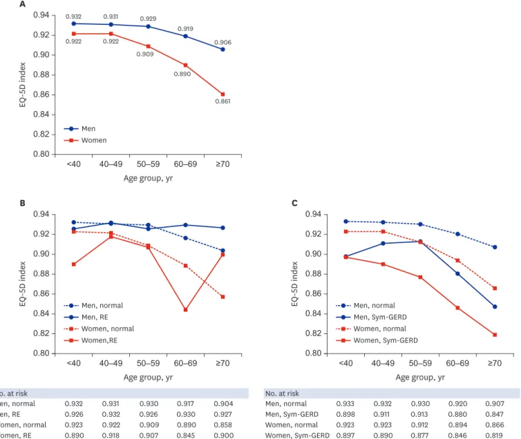
Health related quality of life of GERD according to gender
The general HRQoL in woman participants was significantly lower than that in man participants. EQ5D index was 0.93 ± 0.04 in men and 0.92 ± 0.06 in woman participants, respectively (P < 0.001). EQ-5D index decreased with age for both man and woman participants
(Fig. 3A). There was no significant difference of EQ-5D index according to the presence of RE
0.80 ≥70 60–69 50–59 40–49 <40 0.932 0.931 0.929 0.919 0.906 0.922 0.922 0.909 0.890 0.861 0.94 0.92 0.90 0.88 0.84 0.82 EQ -5D inde x Age group, yr A 0.86 Men Women No. at risk Men, RE Men, normal 0.927 0.926 0.904 0.932 0.932 0.931 0.926 0.930 0.930 0.917 Women, RE Women, normal 0.900 0.890 0.858 0.923 0.918 0.922 0.907 0.909 0.845 0.890 No. at risk Men, Sym-GERD Men, normal 0.847 0.898 0.907 0.933 0.9110.932 0.9130.930 0.8800.920 Women, Sym-GERD Women, normal 0.819 0.897 0.866 0.923 0.890 0.923 0.877 0.912 0.846 0.894 0.80 ≥70 60–69 50–59 40–49 <40 0.94 0.92 0.90 0.88 0.84 0.82 EQ -5D inde x Age group, yr B 0.86 0.80 ≥70 60–69 50–59 40–49 <40 0.94 0.92 0.90 0.88 0.84 0.82 EQ -5D inde x Age group, yr C 0.86 Men, normal Women, normal Men, RE Women,RE Men, normal Women, normal Men, Sym-GERD Women, Sym-GERD
Fig. 3. Presentation of Korean version of the EQ-5D by gender and age. (A) Total participants. (B) Participants with RE. (C) Participants with Sym-GERD. Blue line indicates man and red line indicates woman participants.
in both man and woman participants (Fig. 3B). However, EQ-5D index in symptomatic GERD participants of both genders was lower than that in normal participants. EQ-5D index showed a sharp decline after 40 years of age in woman participants with symptomatic GERD, while it declined sharply after 50 years of age in man participants with symptomatic GERD (Fig. 3C).
DISCUSSION
The present study shows dynamic clinical manifestations and different risk factors for GERD according to gender. RE shows a predominance in men of about 5:1 ratio between men and women, but this difference disappeared above the age of 70. Meanwhile, symptomatic GERD exhibits a woman dominance and this difference becomes more apparent at the peri-menopausal period. GERD is the spectrum of various manifestations caused by gastro-esophageal reflux, and RE does not necessarily match the symptom as an indication of damage to the esophageal mucosa caused by backflow.1 In this study, symptomatic GERD
among RE and RE among symptomatic GERD were only 5.2% and 7.8%, respectively, and the association between the two diseases was low.
Aging, especially over 70 years of age, was a woman-specific risk factor for RE. Commonly, women gain more weight after menopause because hormonal change substantially contributes to central fat deposition and obesity.23 Deleterious changes in adipokines occur
with estrogen decrease at menopause, which could influence on the fatty acid metabolism and increase visceral adiposity.24 The pathophysiology of central fat deposition was also
supported by previous studies in animal models. Ovariectomized mice showed adipocyte hypertrophy, adipose tissue inflammation and the development of hepatic steatosis with decreased energy expenditure.25 In fact, BMI is a well-known risk factor for RE.8,10 Increased
BMI during menopause might affect the rise of the prevalence of RE; hence, we conducted BMI adjusted analysis in this study. As a result, longer duration of menopause still revealed stronger association with the development of RE, despite adjustment for BMI. Thus, a long duration of menopause which is related to marked reduction of estrogen level, might contribute to the pathogenesis of RE later in life.
Recent studies in experimental animal models have manifested that 17β-estradiol, one form of estrogen, may play a protective role in increasing esophageal mucosal resistance by expression of tight junction proteins, such as occludin.26 Furthermore, 17β-estradiol
inhibits the transcription factor p65/relA which promotes the expression of
pro-inflammatory mediators and inflammation.27 This anti-inflammatory activity of estrogen
might contribute to mucosal resistance to acid. Therefore, we suggest that the development of RE in women aged over 70 years could be caused by the long duration of low estrogen level. Similarly, previous studies revealed that the roles of estrogen in animal models of GI tracts were associated with colonic epithelial permeability and mucosal immunity, affecting inflammatory bowel disease.28 Unfortunately, research on how long the low level of estrogen
contribute to increase the development of RE has not been well established to date. The present study shows that the protective effects of estrogen on esophageal mucosa might be considerably decreased when the decline in estrogen level lasts for over 30 years during menopause, not at the beginning or transition of menopause.
Previous meta-analysis of 28 studies for RE showed a pooled men/women prevalence ratio of 1.57/1 (95% CI, 1.40; 1.76/1).29 Most studies included in this meta-analysis had estimated
and compared the men/women ratio of RE in patients with symptomatic GERD. In the present study, however, the majority was free of symptoms and examined for regular health examination. Under the similar background of study participants who underwent regular medical check-up, several recent studies reported that the man predominance in RE has been evident, ranging from 3.1/1–4.1/1, consistent with our results.30,31 They suggest that there is
a gender related difference in the influence of eating habits and lifestyle factors such as high caloric intake, alcohol drinking or current smoking on the prevalence of RE.
Symptomatic GERD, which is mainly diagnosed on the basis of the presence of heartburn or acid regurgitation, presents different clinical features and risk factors from RE.6,7 Several
previous studies presented that difference in gender distribution exists in symptomatic GERD.32,33 Although there is not a significantly big difference between both genders, women
have a relatively high prevalence of symptomatic GERD. However, we revealed that prevalence of symptomatic GERD in women was significantly higher compared to men in all age groups. We suggest the possibility for this discrepancy. Symptomatic GERD is diagnosed using different questionnaire methodology for each study, for instance methods of survey sampling (self-paid medical checkup, stomach cancer screening program, or random sampling), symptom definition (once to twice per week reflux symptoms or composite scores), and data acquisition (interview or self-report questionnaires).34 Therefore, the definition of GERD
based on different methodology remains a limitation for most observational studies, which may affect determination of the prevalence of GERD.21,34
With regard to the association between BMI and symptomatic GERD, previous studies have been controversial; some studies have noted that higher BMI is associated with symptomatic GERD,9,10 whereas other studies have shown negative relations between BMI
and symptomatic GERD.35 The present study shows no association between obesity and
symptomatic GERD.
SSC scores, which indicate the degree of psychosomatic complaints,20 is a significantly
independent predictor for symptomatic GERD in both genders. Psychosomatic complaints induce each individual to lower their threshold values and increase the perception of GERD symptoms with the change of biological responses.36 Similarly, EQ-5D index tends
to decrease with age in both genders. However, women aged over 50 years show sharper decline of EQ-5D index than men in symptomatic GERD. This can be interpreted as resulting primarily from the profound decrease in estrogen around menopause, based on gender difference. The median age of menopause is 51 and decline in estrogen level is maintained thereafter. The hormonal transition around menopause, especially estrogen, substantially contribute to increased menopausal symptoms including hot flushes, sleep disturbance, weight gain, mood symptoms, and somatic complaints.23 Although acid reflux is a major
factor contributing to GERD symptoms, the increase in acid perception is not associated with the presence or absence of esophageal mucosal damage. These findings suggest various factors including non-acid reflux component (such as bile, pepsin or gas), increased mucosal permeability, central sensitization of spinal sensory neurons, and up-regulation of peripheral pain receptors (such as nociceptor transient receptor potential vanilloid type 1 [TRPV1]) could contribute greater to symptom perception in patients with non-erosive reflux disease.37
Regardless of pathologic acid reflux, multiple predisposing factors are associated with triggering reflux symptoms. Further studies toward cellular, molecular, and environmental factors should be required to understand the mechanisms of reflux perception in patients with non-erosive reflux disease.
On the other hand, the prevalence of symptomatic GERD tends to dramatically increase within 20 years of menopause, but that tends to decrease after 20 years of menopause. A recent report showed that menopausal symptoms could persist for an average of 10 years or longer.38 This might explain that menopause-induced symptoms are closely related to a high
prevalence of symptomatic GERD within 20 years of menopause. In case of cardiovascular disease (CVD), there were similar results to this study.39,40 The transition of menopause
induces the vasomotor hot flushes. Particularly, severe night-time hot flushes in recently menopausal women are associated with an increase of systolic blood pressure and heart rate.39 Meanwhile, the long duration of menopause gradually induces the adverse changes in
body fat distribution and vascular remodeling that can increase the prevalence of CVD later in life.40 Taken together, we propose that the duration of menopause acts on the development
of RE, while the transition of menopause has an influence on symptomatic GERD. Further studies are needed to determine the role of estrogen in the pathogenesis of RE and symptomatic GERD, which might lead to effective prevention and treatment.
We have believed that excessive alcohol drinking and current smoking are closely related to gender difference. In relation to gender-role norms and stereotypes, previous studies described that alcohol drinking and smoking were generally presented as a man behavior.41
With regard to RE, current smoking is a common risk factor in both genders. However, OR of current smoking in women is about twice as high as those in men. As for symptomatic GERD, excessive alcohol drinking was the most important risk factor in men, but not in women. Overall, the present study suggests that there is a gender-related difference in the influence of alcohol drinking and smoking on the occurrence of RE and symptomatic GERD. We identified a considerable number of silent RE patients in a large health-screening cohort. When we applied the strict definition of GERD symptoms to heartburn or acid regurgitation at least twice per week for the past 6 months or interfere with daily life within the past year, the prevalence of symptomatic GERD was 4.3% in this study. According to LA classification defined as RE grade A to D, that of RE was observed as 6.4%. Among RE patients, 94.8% had no typical GERD symptoms, which indicates silent RE. Previous study from Italy showed the silent RE in 77.9% of patients with RE (122/1,033, 11.8%).42 However, that study is based
on participants who visited a hospital for dyspeptic symptom evaluation. They may not be a representative subgroup of the general population and thus, the percentage of silent RE may be relatively less common than our regular health examination study. On the other hand, several classification systems for endoscopic assessment of RE currently exist, which could have caused some confusion with diagnosis of RE.16,43,44 Minimal change esophagitis is
widely accepted as part of the spectrum of RE in Japan, using a modified LA classification system with additional grades M and N.44 In a nationwide multi-center prospective study of
healthy Korea population, minimal change lesions of the esophagus, which is considered as early endoscopic findings of RE, was found in 11.9% and RE was observed in 7.91%.45 We
thought that differences in the percentage of silent RE from earlier reported studies are likely to be associated with variation in endoscopic classification system of RE.
The present study has several strengths. First, this study enrolled a large sample of 10,158 ethnically homogenous participants who underwent comprehensive medical checkups. Second, the present study was conducted with a well-validated methodology. We used structured questionnaires for the diagnosis of symptomatic GERD, and assessment of SSC. RE was evaluated with upper endoscopy, the gold standard for the diagnosis of esophagitis, by experienced gastroenterologists.
We acknowledge some limitations. First, this study was conducted at only one center, which may limit the generalization. Second, two-thirds of the participants from the health promotion center were offered a medical examination from the workplace as a reward and only one-third paid for the examination themselves. Therefore, their socioeconomic status was probably better than the general population. These might lead to a potential selection bias, not identical to population-based studies. Third, there was no information about the current medication history, including the use of acid secretion inhibitory agents or nonsteroidal anti-inflammatory drugs. The use of these drugs might be a confounding factor for the analysis of RE and symptomatic GERD. Fourth, hormonal level related to menopause such as estradiol and follicle stimulating hormone were not undergone in this study. Instead of laboratory tests, questionnaire was used to identified menopause. Finally, sufficient participants were not available for analysis because only 21 women were taking the exogenous sex hormone. Therefore, we were not able to evaluate the influence of hormone replacement therapy during menopause.
In conclusion, this study is a large cross-sectional study demonstrating an association between diverse clinical manifestations of GERD and gender differences. Especially, the effect of menopause may play a critical role in the pathogenesis of chronic inflammatory condition of esophagus in women. Similar to the expression of vasomotor symptoms, the presentation of GERD symptoms is rapidly increasing during perimenopausal period. These findings are particularly relevant given the current issue in life style modification to manage GERD such as dietary controls or physical activity after menopause in women. Future research requires priori studies of the pathogenesis of sex hormones on chronic inflammation of the esophagus, in addition to the obesity that progresses with longer menopause.
SUPPLEMENTARY MATERIAL
Supplementary Table 1
Clinical characteristics of women according to menopausal status Click here to view
REFERENCES
1. Vakil N, van Zanten SV, Kahrilas P, Dent J, Jones R; Global Consensus Group. The Montreal definition and classification of gastroesophageal reflux disease: a global evidence-based consensus. Am J Gastroenterol
2006;101(8):1900-20.
PUBMED
2. Dodds WJ, Hogan WJ, Helm JF, Dent J. Pathogenesis of reflux esophagitis. Gastroenterology 1981;81(2):376-94.
PUBMED
3. Lee SJ, Song CW, Jeen YT, Chun HJ, Lee HS, Um SH, et al. Prevalence of endoscopic reflux esophagitis among Koreans. J Gastroenterol Hepatol 2001;16(4):373-6.
PUBMED | CROSSREF
4. Kim JI, Kim SG, Kim N, Kim JG, Shin SJ, Kim SW, et al. Changing prevalence of upper gastrointestinal disease in 28 893 Koreans from 1995 to 2005. Eur J Gastroenterol Hepatol 2009;21(7):787-93.
PUBMED | CROSSREF
5. Park CH, Kim KO, Baek IH, Choi MH, Jang HJ, Kae SH, et al. Differences in the risk factors of reflux esophagitis according to age in Korea. Dis Esophagus 2014;27(2):116-21.
6. Cho YS, Choi MG, Jeong JJ, Chung WC, Lee IS, Kim SW, et al. Prevalence and clinical spectrum of gastroesophageal reflux: a population-based study in Asan-si, Korea. Am J Gastroenterol 2005;100(4):747-53.
PUBMED | CROSSREF
7. Yang SY, Lee OY, Bak YT, Jun DW, Lee SP, Lee SH, et al. Prevalence of gastroesophageal reflux disease symptoms and uninvestigated dyspepsia in Korea: a population-based study. Dig Dis Sci 2008;53(1):188-93.
PUBMED | CROSSREF
8. Chung SJ, Kim D, Park MJ, Kim YS, Kim JS, Jung HC, et al. Metabolic syndrome and visceral obesity as risk factors for reflux oesophagitis: a cross-sectional case-control study of 7078 Koreans undergoing health check-ups. Gut 2008;57(10):1360-5
PUBMED | CROSSREF
9. El-Serag HB, Graham DY, Satia JA, Rabeneck L. Obesity is an independent risk factor for GERD symptoms and erosive esophagitis. Am J Gastroenterol 2005;100(6):1243-50.
PUBMED | CROSSREF
10. de Vries DR, van Herwaarden MA, Smout AJ, Samsom M. Gastroesophageal pressure gradients in gastroesophageal reflux disease: relations with hiatal hernia, body mass index, and esophageal acid exposure. Am J Gastroenterol 2008;103(6):1349-54.
PUBMED | CROSSREF
11. Labenz J, Jaspersen D, Kulig M, Leodolter A, Lind T, Meyer-Sabellek W, et al. Risk factors for erosive esophagitis: a multivariate analysis based on the ProGERD study initiative. Am J Gastroenterol
2004;99(9):1652-6.
PUBMED | CROSSREF
12. Matsuzaki J, Suzuki H, Kobayakawa M, Inadomi JM, Takayama M, Makino K, et al. Association of visceral fat area, smoking, and alcohol consumption with reflux esophagitis and Barrett's esophagus in Japan.
PLoS One 2015;10(7):e0133865.
PUBMED | CROSSREF
13. Asanuma K, Iijima K, Shimosegawa T. Gender difference in gastro-esophageal reflux diseases. World J Gastroenterol 2016;22(5):1800-10.
PUBMED | CROSSREF
14. Kim YS, Kim N, Kim GH. Sex and gender differences in gastroesophageal reflux disease. J Neurogastroenterol Motil 2016;22(4):575-88.
PUBMED | CROSSREF
15. Lee SY, Park HS, Kim DJ, Han JH, Kim SM, Cho GJ, et al. Appropriate waist circumference cutoff points for central obesity in Korean adults. Diabetes Res Clin Pract 2007;75(1):72-80.
PUBMED | CROSSREF
16. Kusano M, Ino K, Yamada T, Kawamura O, Toki M, Ohwada T, et al. Interobserver and intraobserver variation in endoscopic assessment of GERD using the “Los Angeles” classification. Gastrointest Endosc
1999;49(6):700-4.
PUBMED | CROSSREF
17. Shaw MJ, Talley NJ, Beebe TJ, Rockwood T, Carlsson R, Adlis S, et al. Initial validation of a diagnostic questionnaire for gastroesophageal reflux disease. Am J Gastroenterol 2001;96(1):52-7.
PUBMED | CROSSREF
18. Song KH, Jung HK, Min BH, Youn YH, Choi KD, Keum BR, et al. Development and validation of the Korean Rome III questionnaire for diagnosis of functional gastrointestinal disorders. J Neurogastroenterol Motil 2013;19(4):509-15.
PUBMED | CROSSREF
19. Cronbach LJ. Coefficient alpha and the internal structure of tests. Psychometrika 1951;16(3):297-334.
20. Attanasio V, Andrasik F, Blanchard EB, Arena JG. Psychometric properties of the SUNYA revision of the Psychosomatic Symptom Checklist. J Behav Med 1984;7(2):247-57.
PUBMED | CROSSREF
21. Choi JY, Jung HK, Song EM, Shim KN, Jung SA. Determinants of symptoms in gastroesophageal reflux disease: nonerosive reflux disease, symptomatic, and silent erosive reflux disease. Eur J Gastroenterol Hepatol
2013;25(7):764-71.
PUBMED | CROSSREF
22. Seong SS, Choi CB, Sung YK, Park YW, Lee HS, Uhm WS, et al. Health-related quality of life using EQ-5D in Koreans. J Korean Rheum Assoc 2004;11(3):254-62.
23. Thurston RC, Sowers MR, Sternfeld B, Gold EB, Bromberger J, Chang Y, et al. Gains in body fat and vasomotor symptom reporting over the menopausal transition: the study of women's health across the nation. Am J Epidemiol 2009;170(6):766-74.
24. Lee CG, Carr MC, Murdoch SJ, Mitchell E, Woods NF, Wener MH, et al. Adipokines, inflammation, and visceral adiposity across the menopausal transition: a prospective study. J Clin Endocrinol Metab
2009;94(4):1104-10.
PUBMED | CROSSREF
25. Rogers NH, Perfield JW 2nd, Strissel KJ, Obin MS, Greenberg AS. Reduced energy expenditure and increased inflammation are early events in the development of ovariectomy-induced obesity. Endocrinology
2009;150(5):2161-8.
PUBMED | CROSSREF
26. Honda J, Iijima K, Asanuma K, Ara N, Shiroki T, Kondo Y, et al. Estrogen enhances esophageal barrier function by potentiating occludin expression. Dig Dis Sci 2016;61(4):1028-38.
PUBMED | CROSSREF
27. Ghisletti S, Meda C, Maggi A, Vegeto E. 17beta-estradiol inhibits inflammatory gene expression by controlling NF-kappaB intracellular localization. Mol Cell Biol 2005;25(8):2957-68.
PUBMED | CROSSREF
28. Verdú EF, Deng Y, Bercik P, Collins SM. Modulatory effects of estrogen in two murine models of experimental colitis. Am J Physiol Gastrointest Liver Physiol 2002;283(1):G27-36.
PUBMED | CROSSREF
29. Cook MB, Wild CP, Forman D. A systematic review and meta-analysis of the sex ratio for Barrett's esophagus, erosive reflux disease, and nonerosive reflux disease. Am J Epidemiol 2005;162(11):1050-61.
PUBMED | CROSSREF
30. Mizuta A, Adachi K, Furuta K, Ohara S, Morita T, Koshino K, et al. Different sex-related influences of eating habits on the prevalence of reflux esophagitis in Japanese. J Gastroenterol Hepatol 2011;26(6):1060-4.
PUBMED | CROSSREF
31. Song HJ, Shim KN, Yoon SJ, Kim SE, Oh HJ, Ryu KH, et al. The prevalence and clinical characteristics of reflux esophagitis in Koreans and its possible relation to metabolic syndrome. J Korean Med Sci
2009;24(2):197-202.
PUBMED | CROSSREF
32. Lin M, Gerson LB, Lascar R, Davila M, Triadafilopoulos G. Features of gastroesophageal reflux disease in women. Am J Gastroenterol 2004;99(8):1442-7.
PUBMED | CROSSREF
33. Nam SY, Park BJ, Cho YA, Ryu KH. Gender-specific effect of micronutrient on non-erosive reflux disease and erosive esophagitis. J Neurogastroenterol Motil 2019;25(1):82-90.
PUBMED | CROSSREF
34. Wu JC. Gastroesophageal reflux disease: an Asian perspective. J Gastroenterol Hepatol 2008;23(12):1785-93.
PUBMED | CROSSREF
35. Kim N, Lee SW, Cho SI, Park CG, Yang CH, Kim HS, et al. The prevalence of and risk factors for erosive oesophagitis and non-erosive reflux disease: a nationwide multicentre prospective study in Korea. Aliment Pharmacol Ther 2008;27(2):173-85.
PUBMED | CROSSREF
36. Bradley LA, Richter JE, Pulliam TJ, Haile JM, Scarinci IC, Schan CA, et al. The relationship between stress and symptoms of gastroesophageal reflux: the influence of psychological factors. Am J Gastroenterol
1993;88(1):11-9.
PUBMED
37. Bredenoord AJ. Mechanisms of reflux perception in gastroesophageal reflux disease: a review. Am J Gastroenterol 2012;107(1):8-15.
PUBMED | CROSSREF
38. Avis NE, Crawford SL, Greendale G, Bromberger JT, Everson-Rose SA, Gold EB, et al. Duration of menopausal vasomotor symptoms over the menopause transition. JAMA Intern Med 2015;175(4):531-9.
PUBMED | CROSSREF
39. Tuomikoski P, Haapalahti P, Ylikorkala O, Mikkola TS. Vasomotor hot flushes and 24-hour ambulatory blood pressure in recently post-menopausal women. Ann Med 2010;42(3):216-22.
PUBMED | CROSSREF
40. El Khoudary SR, Thurston RC. Cardiovascular implications of the menopause transition: endogenous sex hormones and vasomotor symptoms. Obstet Gynecol Clin North Am 2018;45(4):641-61.
PUBMED | CROSSREF
41. Erol A, Karpyak VM. Sex and gender-related differences in alcohol use and its consequences: contemporary knowledge and future research considerations. Drug Alcohol Depend 2015;156:1-13.
42. Zagari RM, Law GR, Fuccio L, Pozzato P, Forman D, Bazzoli F. Dyspeptic symptoms and endoscopic findings in the community: the Loiano-Monghidoro study. Am J Gastroenterol 2010;105(3):565-71.
PUBMED | CROSSREF
43. Genta RM, Spechler SJ, Kielhorn AF. The Los Angeles and Savary-Miller systems for grading esophagitis: utilization and correlation with histology. Dis Esophagus 2011;24(1):10-7.
PUBMED | CROSSREF
44. Hoshihara Y. Endoscopic findings of GERD. Nihon Rinsho 2004;62(8):1459-64.
PUBMED
45. Lee JH, Kim N, Chung IK, Jo YJ, Seo GS, Kim SW, et al. Clinical significance of minimal change lesions of the esophagus in a healthy Korean population: a nationwide multi-center prospective study. J Gastroenterol Hepatol 2008;23(7 Pt 1):1153-7.