Circulation Journal
Official Journal of the Japanese Circulation Society http://www.j-circ.or.jp
ethnic disparity in CAD risk remain to be elucidated.5–8
In the recent past, coronary artery calcification (CAC) has been identified as a novel and robust marker of atherosclerotic progression,9–11 and has been shown to correlate well with
total coronary plaque burden.12,13 CAC score can be utilized as an
accurate and reliable tool for detecting subclinical CAD, and has pidemiologic evidence indicates that the prevalence
and severity of coronary artery disease (CAD) vary depending on ethnicity.1 Notably, Asian populations
have a lower burden of CAD compared with Western subjects, in spite of the similar conventional risk factors.2–4 Thus, to
date, the precise mechanisms responsible for the observed
E
Received July 31, 2016; revised manuscript received August 17, 2016; accepted August 24, 2016; released online September 21, 2016 Time for primary review: 11 days
Division of Cardiology, Yonsei Cardiovascular Center, Yonsei University College of Medicine, Seoul (D.H., J.H.L., H.-J.C.), Korea; Dalio Institute of Cardiovascular Imaging, New York-Presbyterian Hospital and the Weill Cornell Medical College, New York, NY (D.H., B.ó.H., J.H.L., F.Y.L., J.K.M.); Department of Imaging, Cedars Sinai Medical Center, Los Angeles, CA (H.G.), USA; Department of Internal Medicine, Seoul National University Hospital Healthcare System Gangnam Center, Seoul National University College of Medicine, Seoul (S.-Y.C.); Department of Radiology, Seoul National University Bundang Hospital, Seoul (E.J.C.); Division of Cardiol-ogy, Department of Medicine, Sungkyunkwan University School of Medicine, Heart Stroke and Vascular Institute, Samsung Medical Center, Seoul (J.S.); Department of Internal Medicine, Gangnam Heartscan Seoul Clinic, Seoul (H.-W.H.); Department of Radiology, Gangnam Heartscan Clinic, Seoul (S.H.P.), Korea; and Tennessee Heart and Vascular Institute, Hendersonville, Nashville, TN (T.C.), USA
The first two authors contributed equally to this work (D.H., B.ó.H.).
Mailing address: Hyuk-Jae Chang, MD, PhD, Division of Cardiology, Yonsei Cardiovascular Center, Yonsei University College of Medicine, 50 Yonsei-ro, Seodaemun-gu, Seoul 03722, Korea. E-mail: hjchang@yuhs.ac
ISSN-1346-9843 doi: 10.1253/circj.CJ-16-0762
All rights are reserved to the Japanese Circulation Society. For permissions, please e-mail: cj@j-circ.or.jp
Prevalence and Distribution of Coronary Artery
Calcification in Asymptomatic United
States and Korean Adults
– Cross-Sectional Propensity-Matched Analysis –
Donghee Han, MD; Bríain ó Hartaigh, PhD; Heidi Gransar, BSc; Ji Hyun Lee, MD;
Su-Yeon Choi, MD, PhD; Eun Ju Chun, MD, PhD; Jidong Sung, MD, PhD;
Hae-Won Han, MD, PhD; Sung Hak Park, MD, PhD; Tracy Callister, MD;
Fay Y. Lin, MD; James K. Min, MD; Hyuk-Jae Chang, MD, PhD
Background: The incidence of coronary artery disease (CAD) varies depending on ethnicity, but the precise differ-ences remain to be firmly established. This study therefore evaluated the disparity in coronary artery calcification (CAC), as a marker of CAD, in asymptomatic US and Korean adults.
Methods and Results: CAC score was compared between asymptomatic Korean (n=15,128) and US (n=7,533) adults. Propensity score matching was performed according to age, gender, hypertension, diabetes, dyslipidemia, and current smoking, which generated 2 cohorts of 5,427 matched pairs. Both cohorts were categorized according to age group: 45–54, 55–64, and 65–74 years. Overall, the prevalence of CAC score >0, >100, and >400 in Korean adults was lower than in US adults (P<0.001, all). According to increasing age groups, the likelihood of CAC was most often lower in Korean adults, especially in Korean women. The odds of having CAC >400 in Korean adults aged 65–74 years was 0.66 (95% CI: 0.48–0.91) overall, 0.78 (95% CI: 0.52–1.19) in men, and 0.50 (95% CI: 0.29–0.86) in women, compared with US counterparts.
Conclusions: Korean adults have a lower prevalence and severity of atherosclerotic burden as assessed on CAC, compared with US adults, but the disparity in CAC according to ethnicity may decline with older age. (Circ J 2016; 80: 2349 – 2355)
Key Words: Atherosclerosis; Computed tomography; Coronary artery calcium; Ethnicity
ORIGINAL ARTICLE
Clinical Risk Factors
The following risk factors were collected in both cohorts: age, gender, hypertension, diabetes mellitus, dyslipidemia, and current smoking status. Hypertension was defined as self-reported history of high blood pressure or use of anti-hypertensive medica-tions. Diabetes was defined as baseline use of anti-diabetic medication, history of elevated blood glucose >126 mg/dl or Hba1c >6.5%. Dyslipidemia was considered present for any individual reporting a history of high total cholesterol, high low-density lipoprotein cholesterol, low high-density lipoprotein cholesterol, high triglycerides, or current use of lipid-lowering therapy. Cigarette smoking was defined as active smoker at the time of scanning. All measurements were similarly defined in both study cohorts.
CAC Image Acquisition
Individuals in the US cohort underwent CAC testing using either a C-100 or C-150 Ultrafast electron beam computed tomography (EBCT) scanner (Imatron, South San Francisco, CA, USA). Images were obtained using a 100-ms scanning time and were reconstructed with a 3-mm slice thickness. In the Korean cohort, CAC scanning was performed using a >16-slice multi-detector computed tomography (MDCT) scanner in all centers. Specific CT types used in each center included the Philips Brilliance 256 iCT, Philips Brilliance 40 channel MDCT, Siemens 16-slice Sensation, and GE 64-slice Lightspeed. All 3 Korean sites performed scans using a standard prospec-tive or retrospecprospec-tive method with a 225–400-ms gantry rota-tion time. Image data were reconstructed with a 2.5–3-mm slice thickness. In both cohorts, CAC score was calculated according to Agatston et al.19
Statistical Analysis
Propensity score matching was used to match the Korean subjects to the US subjects according to the following conven-proven useful for prediction of numerous adverse health
out-comes.14 The question, however, of whether changing trends in
the presence and severity of CAC are responsible for the ethnic disparity in CAD risk remain to be firmly established.15–17
We therefore investigated ethnic disparity, if any, regarding subclinical coronary atherosclerosis as measured with CAC using data from 2 large observational cohort studies. Specifically, we compared the presence and severity of CAC in asymptomatic US and Korean adults after accounting for conventional CAD risk factors based on a propensity score matching approach.
Methods
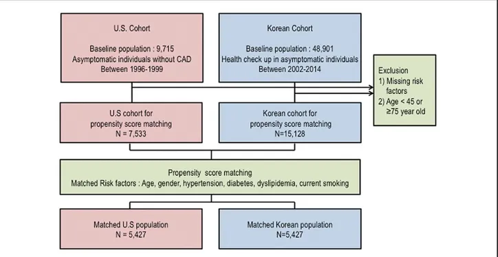
SubjectsThe current investigation utilized data for 2 individual study cohorts of asymptomatic US and Korean adults (Figure 1). The US study sample enrolled 9,715 consecutive asymptom-atic individuals without known CAD, who were referred by their general internist to undergo CAC testing at a single site between January 1996 and December 1999 (Tennessee Heart and Vascular Institute, Hendersonville, TN, USA). Information regarding the Korean subjects was derived from the KOrea Initiatives on Coronary Artery calcification (KOICA) registry.18
The KOICA registry included 48,901 single-ethnicity asymp-tomatic individuals who underwent a health examination at 3 health-care centers between December 2002 and July 2014 (Severance Check-up Health Care Center, Seoul National University Healthcare System Gangnam Center, Samsung Medical Center, Seoul, South Korea). The appropriate institu-tional review board committees approved the study protocol across each site. Given that the study was designed retrospec-tively using medical records, informed consent was waived by the board.
2351 CAC in US and Korean Subjects
matched patient groups were compared using paired t-test for continuous variables and the McNemar test for categorical variables. We calculated the 50th, 75th, 90th, and 95th percentile of CAC and used an exponential model to estimate each per-centile of CAC according to age group as well as gender. On multivariable logistic regression analysis, odds ratios (OR) and 95% confidence intervals (CI) were used to estimate the likelihood of any, moderate, or severe CAC according to each clinical risk factor in both study cohorts. In addition, a separate conditional logistic regression analysis was carried out for the case-control matching to determine the odds of Korean adults having any, moderate, or severe CAC with reference (eg, binary reference=0) to the US adults. Two-tailed P<0.05 was considered significant, and all statistical analysis was performed using STATA version 13.1 (StataCorp LP, College Station, TX, USA).
Results
Clinical CharacteristicsClinical characteristics before and after propensity matching are listed in Table 1. Prior to matching, the prevalence of tional risk factors: age, gender, hypertension, diabetes mellitus,
dyslipidemia, and current smoking. Propensity scores were calculated using a non-parsimonious multiple logistic regression model separately per gender to ensure that the balancing property of the covariates was satisfied. Subsequently, the propensity scores were used to match the 2 cohorts using the Mahalanobis nearest-neighbor matching algorithm with a caliper of 0.001.20
Subsequently, a post-match cohort of 5,427 pairs was generated. The propensity-matched population was further categorized according to age group (eg, 45–54, 55–64, and 65–74 years) to determine whether a graded association existed between CAD and age on the background of ethnicity. For the purpose of this study, CAC was used to detect the presence of sub-clinical atherosclerosis, and subjects were categorized according to the severity of CAC using the following scores: >0, presence of any CAC; >100, moderate CAC; and >400, severe CAC. Continuous variables are expressed as mean ± SD, and cate-gorical variables are reported as counts with proportions. Comparison of baseline characteristics between both cohorts was done using Student’s t-test for continuous variables and Pearson’s chi-squared test for categorical parameters. The
Table 1. Baseline Clinical Characteristics
Before matching After matching
US adults
(n=7,533) Korean adults (n=15,128) P-value US adults (n=5,427) Korean adults (n=5,427) P-value
Age (years) 56.2±7.7 56.2±6.9 0.599 56.1±7.6 56.1±7.4 0.613 45–54 3,608 (47.9) 6,919 (45.7) 2,592 (47.8) 2,573 (47.4) 55–64 2,601 (34.5) 6,169 (40.8) 1,955 (36.0) 2,000 (36.9) 65–74 1,324 (17.6) 2,040 (13.5) 880 (16.2) 854 (15.7) Male 4,298 (57.1) 10,398 (68.7) <0.001 3,351 (61.6) 3,351 (61.6) 1.000 Hypertension 3,331 (44.2) 7,106 (47.0) <0.001 2,511 (46.3) 2,543 (46.9) 0.218 Diabetes 652 (8.7) 2,744 (18.1) <0.001 587 (10.8) 574 (10.6) 0.373 Dyslipidemia 4,817 (64.0) 3,396 (22.5) <0.001 2,743 (50.5) 2,740 (50.5) 0.791 Current smoking 2,974 (39.5) 3,230 (21.4) <0.001 1,644 (30.3) 1,644 (30.3) 1.000 CAC score 137.55±398.3 55.9±2,228.0 <0.001 134.3±403.6 49.4±174.1 <0.001 Log(CAC+1) 2.3±2.5 1.4±2.1 <0.001 2.2±2.5 1.4±2.0 <0.001 CAC categories >0 3,998 (53.1) 5,688 (37.6) <0.001 2,835 (52.2) 2,005 (36.9) <0.001 >100 1,784 (23.7) 1,748 (11.6) <0.001 1,234 (22.7) 615 (11.3) <0.001 >400 711 (9.44) 517 (3.4) <0.001 495 (9.1) 169 (3.1) <0.001
Data given as mean ± SD or n (%). CAC, coronary artery calcification.
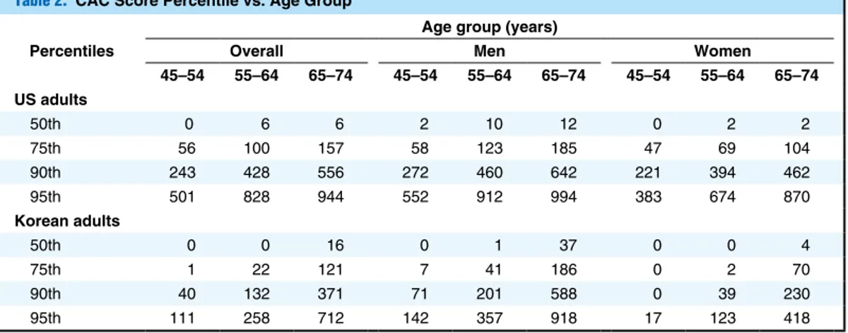
Table 2. CAC Score Percentile vs. Age Group Percentiles
Age group (years)
Overall Men Women
45–54 55–64 65–74 45–54 55–64 65–74 45–54 55–64 65–74 US adults 50th 0 6 6 2 10 12 0 2 2 75th 56 100 157 58 123 185 47 69 104 90th 243 428 556 272 460 642 221 394 462 95th 501 828 944 552 912 994 383 674 870 Korean adults 50th 0 0 16 0 1 37 0 0 4 75th 1 22 121 7 41 186 0 2 70 90th 40 132 371 71 201 588 0 39 230 95th 111 258 712 142 357 918 17 123 418
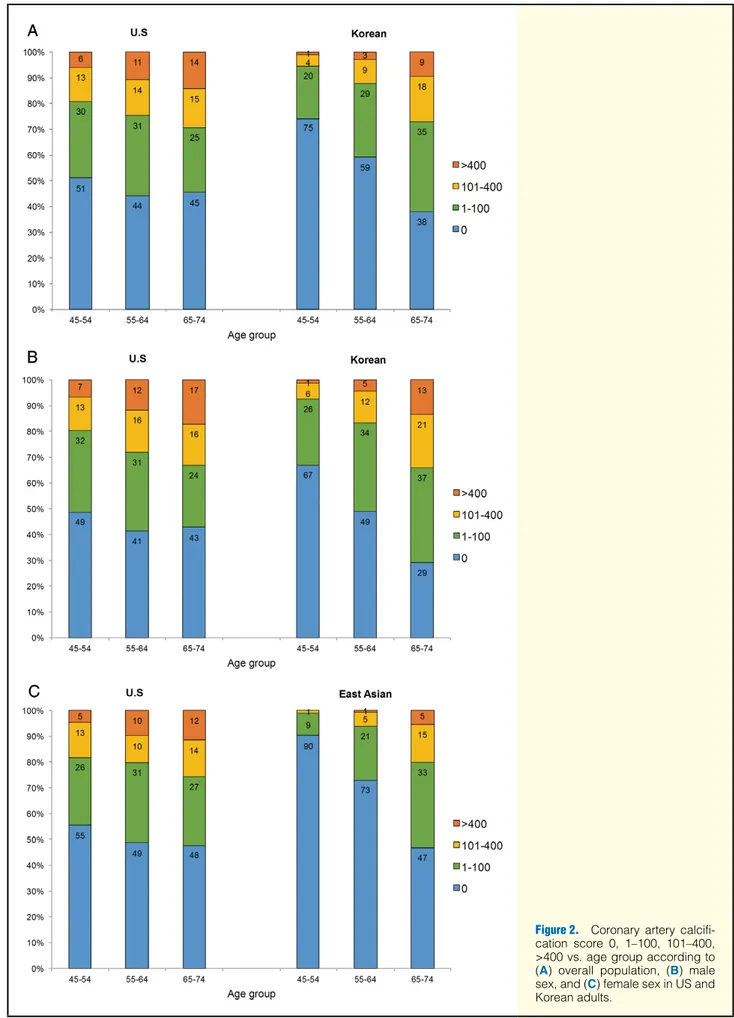
Figure 2. Coronary artery calcifi-cation score 0, 1–100, 101–400, >400 vs. age group according to (A) overall population, (B) male sex, and (C) female sex in US and Korean adults.
2353 CAC in US and Korean Subjects
That is, the prevalence and degree of CAC severity was higher in US adults compared with Korean adults. These findings did not differ materially when stratified by gender.
Clinical Risk Factors and CAC
The relationship between clinical risk factors and presence of any, moderate, or severe CAC within each cohort is given in
Table 3. On multivariable logistic regression analysis, all risk factors were associated with any, moderate, or severe CAC in the US cohort, with the exception of dyslipidemia, which was unrelated to severe CAC. Conversely, current smoking was not associated with any, moderate, or severe CAC in Korean adults. In addition, dyslipidemia also had a non-significant relationship with severe CAC in the Korean cohort. When comparing OR for clinical risk factors between cohorts, Korean adults had higher OR for age and gender compared with US adults, although, US adults had higher OR for hyper-tension, diabetes mellitus, dyslipidemia, and current smoking compared with Korean adults.
Ethnic Disparity and Likelihood of CAC
On logistic regression analysis the odds for any, moderate, or clinical risk factors significantly differed between cohorts.
Prevalent hypertension and diabetes mellitus was higher among Korean subjects. Conversely, prevalent dyslipidemia and current smoking was higher in US compared with Korean adults. As expected, following propensity score matching, all baseline characteristics were matched and neither risk factor differed significantly between study cohorts. Overall, CAC score differed between cohorts irrespective of propensity score matching. Both mean and log CAC differed significantly between study cohorts, with an increase in the severity of CAC categories becoming more prominent in the US vs. Korean adults (Table 1).
CAC vs. Age and Gender
Table 2 lists CAC score for estimated percentiles according to age and gender between study cohorts. Overall, U.S adults had higher CAC score percentile compared with Korean adults. Similar trends were found across age groups when CAC score was categorized according to gender. Figure 2
reports the prevalence and severity of CAC according to age between US and Korean adults. On visual inspection, the prevalence and severity of CAC appeared to differ by ethnicity.
Table 3. Multivariate Indicators of CAC
CAC score >0 CAC score >100 CAC score >400
OR 95% CI P-value OR 95% CI P-value OR 95% CI P-value
US adults
Age per 10 years 1.15 1.07–1.24 <0.001 1.31 1.21–1.43 <0.001 1.59 1.41–1.79 <0.001
Male 1.28 1.14–1.43 <0.001 1.33 1.15–1.53 <0.001 1.39 1.13–1.70 0.002 Hypertension 1.64 1.47–1.84 <0.001 1.66 1.46–1.90 <0.001 2.08 1.70–2.55 <0.001 Diabetes 1.82 1.51–2.20 <0.001 2.25 1.87–2.71 <0.001 2.67 2.11–3.37 <0.001 Dyslipidemia 1.39 1.24–1.56 <0.001 1.18 1.03–1.35 0.020 1.13 0.92–1.37 0.236 Smoking 1.73 1.53–1.96 <0.001 1.80 1.57–2.07 <0.001 1.71 1.40–2.08 <0.001 Korean adults
Age per 10 years 2.69 2.46–2.93 <0.001 3.06 2.71–3.44 <0.001 3.80 3.07–4.69 <0.001
Male 3.28 2.87–3.75 <0.001 3.16 2.56–3.89 <0.001 4.14 2.76–6.22 <0.001
Hypertension 1.40 1.24–1.58 <0.001 1.56 1.29–1.86 <0.001 1.75 1.25–2.44 0.001
Diabetes 1.47 1.21–1.78 <0.001 1.48 1.15–1.90 0.003 1.49 0.97–2.29 0.069
Dyslipidemia 1.25 1.11–1.42 <0.001 1.26 1.03–1.49 0.017 1.21 0.87–1.68 0.262
Smoking 1.02 0.89–1.16 0.810 1.04 0.86–1.27 0.702 1.25 0.89–1.75 0.204
CAC, coronary artery calcification; CI, confidential interval; OR, odds ratio.
Table 4. Likelihood of Having CAC in Korean as Reference US Adults by Age and Gender
Age categories CAC score >0 CAC score >100 CAC score >400
OR 95% CI P-value OR 95% CI P-value OR 95% CI P-value
Overall 45–54 years 0.36 0.32–0.42 <0.001 0.23 0.18–0.29 <0.001 0.16 0.10–0.25 <0.001 55–64 years 0.57 0.50–0.66 <0.001 0.45 0.38–0.55 <0.001 0.28 0.20–0.38 <0.001 65–74 years 1.41 1.13–1.77 0.003 0.94 0.74–1.19 0.583 0.66 0.48–0.91 0.011 Korean men 45–54 years 0.48 0.41–0.55 <0.001 0.30 0.24–0.39 <0.001 0.20 0.12–0.32 <0.001 55–64 years 0.75 0.63–0.90 0.002 0.55 0.44–0.69 <0.001 0.37 0.26–0.53 <0.001 65–74 years 1.87 1.36–2.59 <0.001 1.08 0.79–1.49 0.627 0.78 0.52–1.19 0.248 Korean women 45–54 years 0.13 0.09–0.19 <0.001 0.04 0.02–0.11 <0.001 0.03 0.01–0.24 <0.001 55–64 years 0.36 0.28–0.47 <0.001 0.27 0.18–0.39 <0.001 0.11 0.05–0.24 <0.001 65–74 years 1.05 0.77–1.45 0.744 0.77 0.53–1.11 0.165 0.50 0.29–0.86 0.009 Abbreviations as in Table 3.
CAC in the presence of hypertension and diabetes, whereas Korean adults had higher odds with advancing age and male gender. In the light of these findings, although known cardiac risk factors are considered to be well-established indicators of CAD, the effect on the development of coronary atherosclerosis could vary depending on ethnicity and geographic region.
Initially, in this study, we found that the effect of each clinical risk factor on the presence and severity of CAC dif-fered between Korean and US adults. Hence, we attempted to correct for multiple clinical risk factors that might have impacted on ethnicity and risk of CAD by using propensity score matching. Despite this, a substantial difference in the presence and severity of CAC persisted between Korean and U.S. adults. Unmeasured differences in diet and physical activity may account for the differential impact of risk factors and gender on CAC.23–25 Life course effects may also mediate
differences between age cohorts and geographic regions over time.26 Undoubtedly, additional studies are needed to further
assess the interplay between CAD risk factors and CAC, and whether the relationship differs depending on ethnicity.
In this study, when comparing subclinical CAD in both cohorts, we used CAC as a marker of coronary atherosclerosis. CAC score is a reliable and accurate marker for prediction of subclinical atherosclerosis. An additional strength, however, is that screening for CAC is perhaps easily reproducible when utilizing different types of scanners, such as EBCT or MDCT. Indeed, a previous study reported that estimated percentiles of CAC scores were similar when using either EBCT or MDCT.17
Another study observed that CAC score did not differ materially according to different protocols and/or workstations, thus, highlighting the ease of access in obtaining CAC measurements irrespective of the type of scanner used.27,28
This study is not without limitations. Although the Korean and US cohorts were large in sample size, both may not be truly representative of general populations in South Korea and the USA, respectively. In particular, subjects belonging to the Korean cohort were self-referred, while the US cohort was derived from a single enrollment center in Tennessee referred by physicians. Thus, differential referral bias at the 2 centers cannot be excluded. There was no specific ethnic information available for the US cohort, although by way of context, Caucasian subjects represented approximately 79.7% of the population residing in the State of Tennessee in 2014. Only limited clinical measures were available in the US cohort, and therefore, the potential mechanisms that perhaps explain the observed ethnic disparities in the distribution and severity of CAD were unable to be thoroughly examined in the current analysis, and clearly warrant further investigation. Although CAC is a reliable and reproducible measure of atherosclerosis, the finding of zero CAC does not guarantee absence of CAD because the non-calcified plaque could not be evaluated on CAC scanning. Hence, caution is needed when interpreting these findings given the possibility of underestimation of the prevalence and severity of CAD. The cross-sectional nature of this study limits the long-term comparison between CAC and the potential ethnic disparity. The period in which CAC was obtained differed substantially between the US (eg, 1996–1999) and Korean cohorts (eg, 2002–2014). Therefore, we cannot discount the possibility that any differences in the timing of CAC measurements may have influenced the present findings. More recently, there has been some decline in the incidence of CAD within Western societies,29 whereas, by contrast, the
health and socioeconomic burden of CAD continues to rise exponentially across certain Asian nations, particularly in South Korea.30 Hence, it should be anticipated that the ethnic
severe CAC score were lower in Korean adults compared with US adults (reference group; Table 4). The lowest OR were found for severe CAC (eg, CAC >400), and this trend appeared most prominent among Korean women than Korean men. CAC score tended to increase with advancing age among Korean men and women, although most OR remained lower compared with US adults, with the exception of Korean men aged 65–74 years, who had an 87% (95% CI: 36–159%, P<0.001) increased likelihood of having any CAC compared with US men of a similar age.
Discussion
In this study, we examined the prevalence of coronary athero-sclerosis on CAC score between 2 countries that varied in ethnicity. Based on propensity score matching, the main findings were: (1) significant difference in the prevalence and severity of CAC between US and Korean adults; (2) variation between US and Korean adults in the relationship between clinical risk factors and presence of any, moderate, or severe CAC; (3) lower burden of coronary atherosclerosis in Korean adults compared with US adults, which was more prominent in Korean women; and (4) attenuation of the disparity in CAC between US and Korean adults with advancing age. To our knowledge, this is the largest asymptomatic population-based study to evaluate the ethnic differences in subclinical coronary atherosclerosis between Asian and Western subjects.
Few prior epidemiologic studies have indicated that East Asians, including Chinese, Korean, and Japanese subjects, have lower CAD burden measured on CAC compared with Western subjects.4,16,17 In 1 study on the presence and severity of CAC
in 16,560 individuals within 4 ethnic groups, Asian subjects had a lower CAC burden compared with Caucasian subjects, even after adjustment for coronary risk factors.15 The
Multi-Ethnic Study of Atherosclerosis (MESA) also reported similar observations,17 whereby Chinese adults had a lower
preva-lence of CAC compared with Caucasian subjects, especially among women. More recently, Fujiyoshi et al compared CAC score between Japanese men and US Caucasian men, based on age group.4 That study showed that Japanese men had a lower
burden of coronary atherosclerosis than US Caucasian men. Taken together, these findings underline the lower prevalence and severity of CAC in Asian populations compared with Western Caucasian populations. A potential drawback of the extant literature, however, is the relatively small sample sizes of the Asian subjects in these investigations. The current study supports as well as extends upon these studies given the large sample size representative of Asian individuals, even after propensity score matching (n=5,427).
To date, the precise mechanisms that might influence ethnic disparity in the distribution and severity of CAD are not well understood. Previous studies have noted that the influence of certain CAD risk factors on the development and progression of atherosclerosis might differ as a function of ethnicity. For instance, the Seven Countries Study indicated that although higher cholesterol was indicative of increased absolute CAD mortality rate, the absolute level of CAD mortality appeared to differ significantly on the background of ethnicity.21 In
another study on the association between obesity and coronary atherosclerosis, increase in body mass index was independently associated with CAC in Japanese, Korean, and Caucasian subjects, but not among Japanese-American subjects.22 The
results also suggest that the likelihood of the presence and severity of CAC differed between the Asian and Western populations. Specifically, US adults had higher odds for having
2355 CAC in US and Korean Subjects
project. J Am Coll Cardiol 2005; 46: 807 – 814.
11. Detrano R, Guerci AD, Carr JJ, Bild DE, Burke G, Folsom AR, et al. Coronary calcium as a predictor of coronary events in four racial or ethnic groups. N Engl J Med 2008; 358: 1336 – 1345.
12. Rumberger JA, Simons DB, Fitzpatrick LA, Sheedy PF, Schwartz RS. Coronary artery calcium area by electron-beam computed tomography and coronary atherosclerotic plaque area: A histopathologic correlative study. Circulation 1995; 92: 2157 – 2162.
13. Sangiorgi G, Rumberger JA, Severson A, Edwards WD, Gregoire J, Fitzpatrick LA, et al. Arterial calcification and not lumen stenosis is highly correlated with atherosclerotic plaque burden in humans: A histologic study of 723 coronary artery segments using nondecalcifying methodology. J Am Coll Cardiol 1998; 31: 126 – 133.
14. Han D, ó Hartaigh B, Gransar H, Yoon JH, Kim KJ, Kim MK, et al. Incremental benefit of coronary artery calcium score above tradi-tional risk factors for all-cause mortality in asymptomatic Korean adults. Circ J 2015; 79: 2445 – 2451.
15. Bild DE, Detrano R, Peterson D, Guerci A, Liu K, Shahar E, et al. Ethnic differences in coronary calcification: The Multi-Ethnic Study of Atherosclerosis (MESA). Circulation 2005; 111: 1313 – 1320. 16. Budoff MJ, Nasir K, Mao S, Tseng PH, Chau A, Liu ST, et al. Ethnic
differences of the presence and severity of coronary atherosclerosis.
Atherosclerosis 2006; 187: 343 – 350.
17. McClelland RL, Chung H, Detrano R, Post W, Kronmal RA. Distribution of coronary artery calcium by race, gender, and age: Results from the Multi-Ethnic Study of Atherosclerosis (MESA).
Circulation 2006; 113: 30 – 37.
18. Lee JH, ó Hartaigh B, Han D, Park HE, Choi SY, Sung J, et al. Reas-sessing the usefulness of coronary artery calcium score among vary-ing racial and ethnic groups by geographic locations: Relevance of the Korea Initiatives on Coronary Artery Calcification Registry. J
Cardiovasc Ultrasound 2015; 23: 195 – 203.
19. Agatston AS, Janowitz WR, Hildner FJ, Zusmer NR, Viamonte M Jr, Detrano R. Quantification of coronary artery calcium using ultrafast computed tomography. J Am Coll Cardiol 1990; 15: 827 – 832. 20. Imbens GW. The role of the propensity score in estimating
dose-response functions. Biometrika 2000; 87: 706 – 710.
21. Verschuren WM, Jacobs DR, Bloemberg BP, Kromhout D, Menotti A, Aravanis C, et al. Serum total cholesterol and long-term coronary heart disease mortality in different cultures. Twenty-five-year follow-up of the seven countries study. JAMA 1995; 274: 131 – 136. 22. Fujiyoshi A, Sekikawa A, Shin C, Masaki K, David Curb J, Ohkubo T,
et al. A cross-sectional association of obesity with coronary calcium among Japanese, Koreans, Japanese Americans, and U.S. whites.
Eur Heart J Cardiovasc Imaging 2013; 14: 921 – 927.
23. Miedema MD, Petrone A, Shikany JM, Greenland P, Lewis CE, Pletcher MJ, et al. Association of fruit and vegetable consumption during early adulthood with the prevalence of coronary artery calcium after 20 years of follow-up: The Coronary Artery Risk Development in Young Adults (CARDIA) Study. Circulation 2015; 132: 1990 – 1998.
24. Bertoni AG, Whitt-Glover MC, Chung H, Le KY, Barr RG, Mahesh M, et al. The association between physical activity and subclinical ath-erosclerosis: The Multi-Ethnic Study of Atherosclerosis. Am J
Epi-demiol 2009; 169: 444 – 454.
25. Mori K, Ishida T, Yasuda T, Hasokawa M, Monguchi T, Sasaki M, et al. Serum trans-fatty acid concentration is elevated in young patients with coronary artery disease in Japan. Circ J 2015; 79: 2017 – 2025.
26. Kim D, Diez Roux AV, Kiefe CI, Kawachi I, Liu K. Do neighbor-hood socioeconomic deprivation and low social cohesion predict coronary calcification? The CARDIA study. Am J Epidemiol 2010; 172: 288 – 298.
27. Fujiyoshi A, Kadowaki T, Kadowaki S, Sekikawa A, Ohkubo T, Miura K, et al. Comparability in coronary artery calcium scores on CT scan between two community-based cohort studies. Int J Cardiol 2011; 149: 244 – 245.
28. Weininger M, Ritz KS, Schoepf UJ, Flohr TG, Vliegenthart R, Costello P, et al. Interplatform reproducibility of CT coronary calcium scoring software. Radiology 2012; 265: 70 – 77.
29. Wijeysundera HC, Machado M, Farahati F, Wang X, Witteman W, van der Velde G, et al. Association of temporal trends in risk factors and treatment uptake with coronary heart disease mortality, 1994 – 2005. JAMA 2010; 303: 1841 – 1847.
30. Chang HS, Kim HJ, Nam CM, Lim SJ, Jang YH, Kim S, et al. The socioeconomic burden of coronary heart disease in Korea. J Prev
Med Public Health 2012; 45: 291 – 300. disparity in CAC observed in the current study might differ
compared with that in forthcoming studies.
Conclusions
The prevalence and severity of subclinical coronary atheroscle-rosis measured using CAC appeared lower in Korean adults compared with US adults. These findings remained robust after controlling for multiple CAD risk factors on propensity score matching. Notably, the reduction in the presence of CAC was more prominent among Korean women, although the ethnic disparity in CAC tended to diminish with advancing age. Further studies are now needed to disentangle the mecha-nisms responsible for the disparity observed in the prevalence and severity of CAC between Asian and Western populations.
Acknowledgments
This work was supported by Leading Foreign Research Institute Recruitment Program through the National Research Foundation of Korea (NRF) funded by the Ministry of Science, ICT and Future Planning (MSIP; 2012027176) and funded in part by a generous gift from the Dalio Institute of Cardiovascular Imaging and the Michael Wolk Foundation.
Disclosures
J.K.M. serves as a consultant to HeartFlow. He is also on the scientific advisory board for Arineta, has a research agreement with GE Healthcare, and has ownership in MDDX. All other authors declare no conflicts of interest.
References
1. Yusuf S, Reddy S, Ounpuu S, Anand S. Global burden of cardiovas-cular diseases: Part I: General considerations, the epidemiologic transition, risk factors, and impact of urbanization. Circulation 2001; 104: 2746 – 2753.
2. Liu J, Hong Y, D’Agostino RB Sr, Wu Z, Wang W, Sun J, et al. Predictive value for the Chinese population of the Framingham CHD risk assessment tool compared with the Chinese Multi-Provincial Cohort Study. JAMA 2004; 291: 2591 – 2599.
3. Ueshima H, Sekikawa A, Miura K, Turin TC, Takashima N, Kita Y, et al. Cardiovascular disease and risk factors in Asia: A selected review. Circulation 2008; 118: 2702 – 2709.
4. Fujiyoshi A, Miura K, Ohkubo T, Kadowaki T, Kadowaki S, Zaid M, et al. Cross-sectional comparison of coronary artery calcium scores between Caucasian men in the United States and Japanese men in Japan: The multi-ethnic study of atherosclerosis and the Shiga epi-demiological study of subclinical atherosclerosis. Am J Epidemiol 2014; 180: 590 – 598.
5. Anand SS, Yusuf S, Vuksan V, Devanesen S, Teo KK, Montague PA, et al. Differences in risk factors, atherosclerosis, and cardiovascular disease between ethnic groups in Canada: The Study of Health Assessment and Risk in Ethnic groups (SHARE). Lancet 2000; 356: 279 – 284.
6. Meadows TA, Bhatt DL, Cannon CP, Gersh BJ, Rother J, Goto S, et al. Ethnic differences in cardiovascular risks and mortality in atherothrombotic disease: Insights from the Reduction of Athero-thrombosis for Continued Health (REACH) registry. Mayo Clin
Proc 2011; 86: 960 – 967.
7. Tillin T, Hughes AD, Mayet J, Whincup P, Sattar N, Forouhi NG, et al. The relationship between metabolic risk factors and incident cardiovascular disease in Europeans, South Asians, and African Caribbeans: SABRE (Southall and Brent Revisited) – a prospective population-based study. J Am Coll Cardiol 2013; 61: 1777 – 1786. 8. Yusuf S, Reddy S, Ounpuu S, Anand S. Global burden of
cardiovas-cular diseases: Part II: Variations in cardiovascardiovas-cular disease by specific ethnic groups and geographic regions and prevention strategies.
Circulation 2001; 104: 2855 – 2864.
9. Greenland P, LaBree L, Azen SP, Doherty TM, Detrano RC. Coronary artery calcium score combined with Framingham score for risk prediction in asymptomatic individuals. JAMA 2004; 291: 210 – 215. 10. Taylor AJ, Bindeman J, Feuerstein I, Cao F, Brazaitis M, O’Malley PG.
Coronary calcium independently predicts incident premature coronary heart disease over measured cardiovascular risk factors: Mean three-year outcomes in the Prospective Army Coronary Calcium (PACC)