Oriental Pharmacy and Experimental Medicine 2010 10(4), 288-293
Lindera erythrocarpa Makino extract reduces obesity induced by high-fat
diet in rats
Meejung Ahn
1, Wonjun Yang
2, Sohi Kang
2, Min-Chul Kang
3, Ryeo Kyeong Ko
3, Gi-Ok Kim
3,#and
Taekyun Shin
2,*
1
Department of Anatomy, College of Medicine, Jeju National University, Jeju 690-756, South Korea; 2Department of Veterinary Anatomy, College of Veterinary Medicine and Veterinary Medical Research Institute, Jeju National University, Jeju 690-756, South Korea; 3Bio-industry Development center, Jeju Technopark, Jeju 690-121, South Korea
Received for publication September 15, 2010; accepted December 6, 2010
SUMMARY
Lindera erythrocarpa Makino (LE) is widely distributed on Jeju Island, where it has been used for various traditional therapies. Effects of a crude extract of LE were examined in rats with obesity induced by a high-fat diet (HFD). Anti-obesity effects were followed in rats receiving orally administered vehicle, 100mg/kg extract, or 250 mg/kg LE extract, for 56 days. LE extract (250 mg/kg) suppressed increases in body weight and epididymal fat, with amelioration of fatty changes in the liver. Additionally, serum levels of alanine aminotransferase, aspartate aminotransferase, and total cholesterol were significantly decreased compared with those of vehicle-treated groups (p < 0.05). These results suggest that oral administration of LE extract reduced rat obesity induced by HFD, possibly through the reduction of fat accumulation.
Key words: Lindera erythrocarpa Makino; Obesity; Rat; Cholesterol
INTRODUCTION
Obesity is considered to be a disorder of energy balance, occurring when energy expenditure is no longer in equilibrium with daily energy intake, affecting body-weight homeostasis (Van et al., 2008). Obesity can be induced by the intake of high dietary fat, relative to internal fat metabolism. Obesity also refers to the status of an organism that
has accumulated body fat due to various reasons; the primary cause is a high-fat diet. Obesity is reaching epidemic proportions worldwide, and is correlated with various co-morbidities, among which the most relevant are dyslipidemia (Fried et al., 2007), diabetes mellitus type 2 (Pagotto et al., 2008), fatty liver (Maroviæ, 2008), and cardiovascular diseases such as heart failure and coronary heart disease (Artham et al., 2008).
There have been many studies exploring the potential of various plant extracts for the development of anti-obesity drugs (Birari and Bhutani, 2007). Simple medicinal preparations, from sources including grape-seed (Terra et al., 2010), Aesculus hippocastanum (Avci et al., 2010), Crocus sativus (Gout et al., 2010), and Alpinia officinarum (Xia et al., 2010), often mediate beneficial responses due to
*Correspondence: Taekyun Shin, Department of Veterinary Anatomy, College of Veterinary Medicine and Veterinary Medical Research Institute, Jeju National University, Jeju 690-756, South Korea. Tel: +82-64 754 3363; Fax: +82- 64 756-3354; E-mail: [email protected] #Co-correspondence: Gi-Ok Kim, Bio-industry Deve-lopment center, Jeju Technopark, Jeju 690-121, South Korea. Tel: +82-64 720 2333; Fax: +82- 64 720 2331; E-mail: [email protected]
their active chemical constituents.
Lindera erythrocarpa Makino (LE), a widely distributed species, is found on Jeju Island, where it has been used in various traditional therapies. Lucidone, which has been isolated from LE, has been shown to have physiological activity, including inflammatory (Senthil et al., 2010) and anti-cancer effects (Wang et al., 2008; Oh et al., 2005).
The aim of the present study was to evaluate the anti-obesity activity of a LE extract in rats, examining body weight, food intake, lipid profiles, and hepatic function markers in blood and in liver tissue.
MATERIALS AND METHODS
Material preparation
All solvents used in this experiment were of analytical grade. Whole LE plants were collected from Jeju Island in August 2004. A voucher specimen (06 - 105) was deposited at the Jeju Bio-Industry Development Center, Hi-Tech Industry Development Institute, Jeju, Korea. The fresh stem bark of LE was washed and dried in a hot air stream at 40°C for three days. The dried material (560 g) was pulverized in a mill, and extracted with stirring in 70% aqueous ethanol at room temperature for three days. The extract was filtered to remove insoluble materials, and the filtrate concentrated to yield 68.2 g of a gummy residue.
Animal experiments
Sprague-Dawley rats (6-week-old male rats weighing 169.3 ± 1.25 g) were purchased from the animal center of the Korea Research Institute of Bioscience and Biotechnology (Seoul, Korea). All were fed on a normal diet for three days to stabilize their metabolic status. All experiments were carried out in accordance with the Jeju National University guidelines for the care and use of laboratory animals. Diet and drug administration
A total of 16 rats were divided into four groups: vehicle-treated low-fat diet control (LFD, n = 4),
vehicle-treated high-fat diet control (HFD, n = 4), 100mg/kg LE extract-treated high-fat diet (LE100, n = 4), and 250 mg/kg LE extract-treated high-fat diet (LE250, n = 4 ).
The low-fat diet provided 10% of total Kcal as fat (D12450B, Korea Research Institute of Bioscience and Biotechnology, Seoul, Korea) and the high-fat diet provided 60% of total Kcal as fat (D12492). Each rat was fed an equal amount of one diet, ranging from 17.2 to 19.8 g/d during the feeding period. LE crude extract was dissolved in corn oil and administered orally every day, according to the method described by Oka et al. (2008). Handling of animals was the same for all groups, and did not affect weight gain. The vehicle-treated groups were fed a lab diet and received oral corn oil daily for 56 days, using a stomach tube during the entire study. Body weight and food consumption were measured once per week.
Blood sampling for serological analysis
At the end of the study period, animals were sacrificed, and heart blood samples were collected. These were allowed to coagulate at room temperature, and were centrifuged at 3500 rpm for 15 min at room temperature to collect the serum fraction. The serum levels of alanine aminotransferase (ALT) and aspartate aminotransferase (AST) were measured as an assay of liver function, and total cholesterol (T-CHO), triglyceride (TG), low-density lipoprotein (LDL), and high-density lipoprotein (HDL) were assayed by Chemon Co., Ltd. (Suwon, Korea) to determine the lipid profile.
Sampling and tissue preparation
To study the effect of LE extract on obesity induced by a high-fat diet, we sacrificed rats by CO2 inhalation at the end of the study. For histology, livers were embedded in paraffin after fixation in 4% parafor-maldehyde in phosphate-buffered saline (PBS) at pH 7.4. Paraffin tissue sections (5 µm thickness) were cut on a rotary microtome (Leica, Nussloch, Germany) and stained with hematoxylin and eosin.
Epididymal white adipose tissue (WAT) was taken from the region around the testis and epididymis (Rokling-Andersen et al., 2009) and weighed. Statistical analysis
The data represent means ± S.E. The data were analyzed using one-way analysis of variance (ANOVA), followed by a Tukey-Kramer post hoc test for multiple comparisons. In all cases, p < 0.05 was considered significant.
RESULTS
Effect of diet treatments on body weight and adipose tissue
The rate of weight gain was shown to be significantly increased in rats in the vehicle-treated HFD group, as compared to that of the vehicle-treated LFD group (p < 0.05). The weight gain in the LE250 group, but not the LE100 group, was significantly decreased relative to that of the HFD group (p < 0.01) (Fig. 1). Food consumption was not significantly different among the four groups (Fig. 2).
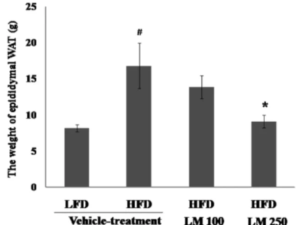
The weight of epididymal WAT was significantly increased in the vehicle-treated HFD group compared with those of vehicle-treated LFD group; the weight of epididymal WAT in the LE250 group, but not
the LE100 group, was significantly reduced compared with that of HFD controls (p < 0.05) (Fig. 3). Histological analysis of liver tissue
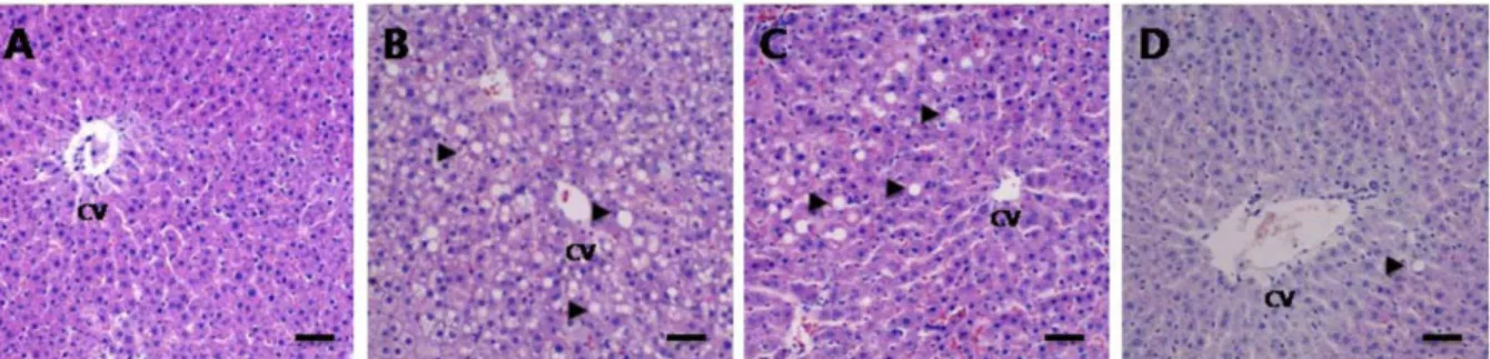
Histological examination showed no fatty changes or hemorrhage in the livers of vehicle-treated LFD rats (Fig. 4A). In those of vehicle-treated HFD rats, histological evidence of fatty change, hemorrhage, and edema were noted (Fig. 4B). In LE100 rats, fatty changes were found, though less than those Fig. 1. The rate of weight gain in rats fed LFD, HFD,
or HFD with LE extract. Values are mean±S.E. ##p < 0.01 compared with vehicle-treated LFD rat; *p < 0.05 compared with vehicle-treated HFD rat.
Fig. 2. Effect of LE extract on food consumption during 56 days in gram/day/rat. Food consumption was steady over this period except at the 2nd and 3rd weeks.
Fig. 3. The weight of epididymal WAT in rats fed LFD, HFD, or HFD with LE extract. Values are mean ± S.E. #p < 0.05 compared with vehicle-treated LFD control rat; *p < 0.05 compared with vehicle-treated HFD control rat.
observed in the livers of vehicle-treated HFD rats (Fig. 4C). Pathological lesions in LE250 rats, however, including fatty changes, hemorrhages, and edema were reduced relative to those seen in vehicle-treated HFD rats (Fig. 4D).
Effect of LE extract on hepatic enzymes and lipid profile
Serum ALT concentration was significantly increased
in the HFD group (295.1 ± 17.1) (p < 0.05) compared with the LFD group (167.4 ± 17.9), while that in the LE250 group was significantly decreased (160.2 ± 25.6) (p < 0.05) relative to those of the LE100 and HFD groups (Table 1). Serum AST concentration was not significantly changed in the LE100 group. Serum T-CHO, LDL, and HDL levels were significantly higher in the HFD group compared to the LFD group (p < 0.05), respectively. In the LE250 Fig. 4. Effect of LE extract on fatty change in rat liver tissue in obesity induced by high-fat diet. Representative images showing the fatty change in liver sections stained with hematoxylin and eosin, from vehicle-treated LFD control rat (A), vehicle-treated HFD control rat (B), LE100-treated HFD rat (C), LE250-treated HFD rat (D). Scale bars represent 100 µm. CV: central vein.
Table 1. Effect of Lindera erythrocarpa Makino crude extract on liver enzyme activity in obese rats
LFD HFD LE100 LE250
AST(IU/l) 167.4 ± 17.9a 280.0 ± 9.9# 261.9 ± 51.0 187.7 ± 19.9*
ALT(IU/l) 22.9 ± 1.1 42.9 ± 3.6## 58.1 ± 16.0 41.9 ± 4.0
a
Values are mean ± S.E.
LFD: low fat diet; HFD: high fat diet; LE100: daily oral administration of Lindera erythrocarpa Makino extract, 100 mg/kg body weight; LE250: daily oral administration of Lindera erythrocarpa Makino extract, 250 mg/kg body weight; AST, aspartate aminotransferase; ALT, alanine aminotransferase. #Significant difference at the p < 0.05 or ##
p < 0.01 level relative to vehicle-treated LFD group or vehicle-treated HFD group, respectively. *Significant difference at the p < 0.05 level relative to vehicle-treated HFD group and LE100-treated HFD groups.
Table 2. Effect of Lindera erythrocarpa Makino crude extract on plasma lipid profile in obese rats
LFD HFD LE100 LE250
T-CHO(mg/dl) 84.0 ± 4.7 127.7 ± 7.5# 93.3 ± 13.0 91.3 ± 7.9*
TG(mg/dl) 155.3 ± 22.6 125.7 ± 12.3 152.3 ± 29.7 126.0 ± 32.6
LDL(mg/dl) 8.8 ± 0.5 11.3 ± 1.0# 11.3 ± 1.9 13.0 ± 1.2
HDL(mg/dl) 49.4 ± 2.9 64.2 ± 1.5# 50.6 ± 7.9* 51.7 ± 5.4*
aValues are mean ± S.E. LFD: low fat diet; HFD: high fat diet; LE100: daily oral administration of Lindera erythrocarpa Makino extract, 100 mg/kg body weight; LE250: daily oral administration of Lindera erythrocarpa Makino extract, 250 mg/kg body weight; T-CHO, total cholesterol; TG, triglyceride; LDL, low-density lipoprotein; HDL, high-density lipoprotein. #Significant difference at the p < 0.05 level compared with the vehicle-treated LFD group and vehicle-vehicle-treated HFD group. *Significant difference at the p < 0.05 level compared with the vehicle-treated HFD group and LE100-treated HFD groups.
group, serum T-CHO and HDL levels, but not TG, were significantly lower than those in the HFD group (p < 0.05) (Table 2). Biochemical markers in LE100 group were not significantly altered.
DISCUSSION
We have confirmed that oral administration of LE extract at a dose of 250 mg/kg body weight reduces body weight gain induced by a high-fat diet in rats. This inhibition of weight gain and of fat accumulation was shown to be dose dependent in response to LE extract.
Consumption of the high-fat diet led to obesity because it facilitated the development of a positive energy balance leading to an increase in visceral fat deposition. This led to abdominal obesity in particular. Moreover, it is reported that HFD feeding is accompanied by molecular adaptations that favor fat storage in muscle rather than its oxidation (Schrauwen-Hinderling et al., 2005).
In addition to the high-fat diet, obesity has been known to be associated with increased appetite. LE extract may induce decreased appetite in rats fed a high-fat diet. This assumption is supported by a report that several isoflavan constituents of licorice are unique phytoestrogens, inhibiting serotonin re-uptake and thereby increasing the levels of serotonin in synaptic clefts. This enhances satiety and resembles the action of sibutramine, a synthetic drug used to suppress appetite in the treatment of obesity (Ofir et al., 2003).
Fatty changes in the liver are known to be associated with obesity (James et al., 1999). In the present study, HFD rat livers were enlarged and yellowish, indicating liver steatosis, whereas liver color of the LE250 rats on HFD was less changed relative to normal controls, suggesting that LE250 may reduce the accumulation of fat in the liver.
Serum AST and ALT levels are clinically and toxicologically important indicators, rising as a result of tissue damage caused by toxicants or disease conditions. Biochemical studies showed
significant elevation of ALT, AST, triglycerides, cholesterol, and albumin in the livers of rats treated with a methanol extract of Mitragyna speciosa Korth (Harizal et al., 2010). In the present study, the activities of serum AST, but not ALT, were significantly reduced in the LE250 group compared with those of control HFD rats. However, the LE100 group did not show changes in the levels of AST or ALT. Taken together, these results indicate that daily oral administration of high-dose LE (250 mg/kg) protects the liver from fatty change induced by a high fat diet, partly through a decrease in AST. Effects on both high-density and low-density lipoprotein levels differed in this study from those seen in a previous study (Nammi et al., 2009). We found that LE extract administration produced significant decreases in serum total cholesterol and high-density lipoprotein, but not in triglyceride or low-density lipoprotein. In the earlier obesity study, a high-fat diet induced the increase of total cholesterol, triglyceride, and low-density lipoprotein, and decreased the level of high-density lipoprotein (Nammi et al., 2009). The mechanisms responsible for the above results remain to be studied.
Taken together, our results demonstrate that LE extract has hepatoprotective, hypolipidemic, and anti-obesity effects in HFD-induced rat obesity.
ACKNOWLEDGEMENTS
This research was supported by Jeju bio-industry Development center, Jeju Technopark.
REFERENCES
Amin KA, Nagy MA. (2009) Effect of carnitine and herbal mixture extract on obesity induced by high fat diet in rats. Diabetol. Metab. Syndr. 16, 1-14. Artham SM, Lavie CJ, Milani RV, Ventura HO. (2008)
The obesity paradox: impact of obesity on the prevalence and prognosis of cardiovascular diseases. Postgrad. Med. 120, 34-41.
Avci G, Küçükkurt I, Küpeli Akkol E, Ye ilada E. (2010) Effects of escin mixture from the seeds of
s
Aesculus hippocastanum on obesity in mice fed a high fat diet. Pharm. Biol. 48, 247-252.
Birari RB, Bhutani KK. (2007) Pancreatic lipase inhibitors from natural sources: unexplored potential. Drug. Discov. Today. 12, 879-889.
Fried M, Hainer V, Basdevant A, Buchwald H, Deitel M, Finer N, Greve JW, Horber F, Mathus-Vliegen E, Scopinaro N, Steffen R, Tsigos C, Weiner R, Widhalm K; Bariatric Scientific Collaborative Group Expert Panel. (2007) Interdisciplinary European guidelines for surgery for severe (morbid) obesity. Obes. Surg. 17, 260-270.
Gout B, Bourges C, Paineau-Dubreuil S. (2010) Satiereal, a Crocus sativus L extract, reduces snacking and increases satiety in a randomized placebo-controlled study of mildly overweight, healthy women. Nutr. Res. 30, 305-313.
Harizal SN, Mansor SM, Hasnan J, Tharakan JK, Abdullah J. (2010) Acute toxicity study of the standardized methanolic extract of Mitragyna speciosa Korth in rodent. J. Ethnopharmacol. 131, 404-409.
James O, Day C. (1999) Non-alcoholic steatohepatitis: another disease of affluence. Lancet, J Ethnopharmacol. 353, 1634–1636.
Maroviæ D. (2008) Elevated body mass index and fatty liver. Srp. Arh. Celok Lek. 136, 122-125. Nammi S, Sreemantula S, Roufogalis BD. (2009)
Protective effects of ethanolic extract of Zingiber officinale rhizome on the development of metabolic syndrome in high-fat diet-fed rats. Basic Clin. Pharmacol. Toxicol. 104, 366-373.
Ofir R, Tamir S, Khatib S, Vaya J. (2003) Inhibition of serotonin reuptake by licorice constituents. J. Mol. Neurosci. 20, 135-140.
Oh HM, Choi SK, Lee JM, Lee SK, Kim HY, Han DC, Kim HM, Son KH, Kwon BM. (2005) Cyclopen-tenediones, inhibitors of farnesyl protein transferase and anti-tumor compounds, isolated from the fruit of Lindera erythrocarpa Makino. Bioorg. Med. Chem. 13, 6182-6187.
Pagotto U, Vanuzzo D, Vicennati V, Pasquali R. (2008) Pharmacological therapy of obesity. Ital Cardiol (Rome). 9, 83S-93S.
Rokling-Andersen MH, Rustan AC, Wensaas AJ, Kaalhus O, Wergedahl H, Røst TH, Jensen J, Graff BA, Caesar R, Drevon CA. (2009) Marine n-3 fatty acids promote size reduction of visceral adipose depots, without altering body weight and composition, in male Wistar rats fed a high-fat diet. Br. J. Nutr. 102, 995-1006.
Schrauwen-Hinderling VB, Kooi ME, Hesselink MK, Moonen-Kornips E, Schaart G, Mustard KJ, Hardie DG, Saris WH, Nicolay K, Schrauwen P. (2005) Intramyocellular lipid content and molecular adaptations in response to a 1-week high-fat diet. Obes. Res. 13, 2088-2094.
Senthil Kumar KJ, Hsieh HW, Wang SY. (2010) Anti-inflammatory effect of lucidone in mice via inhibition of NF-kappaB/MAP kinase pathway. Int. Immun-opharmacol. 10, 385-392.
Terra X, Pallarés V, Ardèvol A, Bladé C, Fernández-Larrea J, Pujadas G, Salvadó J, Arola L, Blay M. (2010) Modulatory effect of grape-seed procyanidins on local and systemic inflammation in diet-induced obesity rats. J. Nutr. Biochem. doi:10.1016/j.jnutbio. 2010.03.006.
Van Herpen NA, Schrauwen-Hinderling VB. (2008) Lipid accumulation in non-adipose tissue lipotoxicity. Physiol. Behav. 23, 231-241.
Wang SY, Lan XY, Xiao JH, Yang JC, Kao YT, Chang ST. (2008) Antiinflammatory activity of Lindera erythrocarpa fruits. Phytother. Res. 22, 213-216. Woo MN, Bok SH, Lee MK, Kim HJ, Jeon SM, Do
GM, Shin SK, Ha TY, Choi MS. (2008) Anti-obesity and hypolipidemic effects of a proprietary herb fiber combination (S&S PWH) in rats fed high-fat diets. J. Med. Food 11, 169-178.
Xia DZ, Yu XF, Wang HM, Ren QY, Chen BM. (2010) Anti-obesity and hypolipidemic effects of ethanolic extract from Alpinia officinarum Hance (Zingiberaceae) in rats fed high-fat diet. J. Med. Food 13, 785-791.