Relation between carotid artery
FDG uptake and cardiovascular risk in
asymptomatic adults
by
Dong Hyun Lee
Major in Medicine
Department of Medical Sciences
The Graduate School, Ajou University
Relation between carotid artery
FDG uptake and cardiovascular risk in
asymptomatic adults
by
Dong Hyun Lee
A Dissertation Submitted to The Graduate School of Ajou University
in Partial Fulfillment of the Requirements for the Degree of
Master of Medicine
Supervised by
Su Jin Lee, M.D., Ph.D.
Major in Medicine
Department of Medical Sciences
The Graduate School, Ajou University
This certifies that the dissertation
of Dong Hyun Lee is approved.
SUPERVISORY COMMITTEE
(Sign)
Su Jin Lee
(Sign)
Joon-Kee Yoon
(Sign)
Young-Sil An
The Graduate School, Ajou University
December, 18th, 2014
ABSTRACT
-Rel
at
i
on bet
ween car
ot
i
dar
t
er
y FDG upt
akeand
car
di
ovascul
arr
i
sk i
n asympt
omat
i
cadul
t
s
We investigated the relation between carotid artery FDG uptake and cardiovascularriskbasedontheFramingham riskscore(FRS),andevaluated the possible role of FDG uptake in terms of risk stratification of asymptomaticadults.Weevaluated 290adultswho underwentFDG PET/CT aspartofgeneralhealth screens.Wecalculated target-to-background ratios, correctedforpre-scanbloodglucoselevels,andobtained“TBRglu”valuesfor both common carotid arteries. The FRS and the presence/absence of metabolic syndrome were recorded for each subject.Relationships among TBRglu values,metabolic syndrome status,and clinical parameters were assessed.Carotidartery FDG uptakewassignificantlyassociatedwithclinical risk factors.Stepwise multiple regression analysis revealed thattriglyceride levels,diabetes,and metabolic syndrome were independentdeterminants of high TBRglu.Of subjects with metabolic syndrome,those exhibiting high carotid artery FDG uptake had significantly higherlevels ofhigh sensitivity C-reactive protein (hsCRP). In subjects who did not have metabolic syndrome,FRS were significantly elevated in those exhibiting high carotid artery FDG uptakecompared tothosewith low uptake(13.1± 7.0vs.8.2± 7.4),aswasalsotrueofsubjectswith thesyndrome(21.8± 16.0vs.13.5± 11.9).High carotid FDG uptake is significantly associated with clinicalrisk factorsand agreaterFRS.Ofsubjectswith metabolicsyndrome,thosewith high carotid uptakehad significantly higherhsCRP concentrationsand FRSs. Therefore,carotid artery FDG activity may serve as a possible biomarker
allowingcardiovascularriskstratificationofasymptomaticpopulations. ___________________________________________________________________
Key words: atherosclerosis, carotid arteries, 18F-FDG, risk assessments, metabolicsyndrome,cardiovasculardisease
TABLE OF CONTENTS
ABSTRACT···i
TABLE OF CONTENTS···iii LIST OF FIGURES···iv LIST OF TABLES···v
I.INTRODUCTION···1
II.MATERIALS AND METHODS···3
A.STUDY SUBJECTS AND DATA COLLECTION···3
B.FDG PET/CT···4
C.IMAGE ANALYSIS···4
D.STATICAL ANALYSIS···5
III.RESULTS···7
IV.DISCUSSION···13
V.CONCLUSION···16
REFERENCES···17
LI
ST OF FI
GURES
Fig.1.HighsensitivityC-reactiveproteinconcentrationsandFramingham riskscoresinstudysubjectsstratifiedbytarget-to-backgroundratio correctedforbloodglucoselevel,andpresenceorabsenceofmetabolic syndrome···11
Fig.2.Representativemaximum intensityofprojectionimagesof18F-FDG PET/CT···12
LI
ST OF TABLES
Table1.ClinicalVariablesofStudySubjects···7
Table2.MultiplestepwiseregressionanalysisbetweenglucosecorrectedFDG uptakeandclinicalvariables···9
Table3.ComparisonofClinicalVariablesbetweenSubjectswithandwithout MetabolicSyndrome···10
I
.I
NTRODUCTI
ON
Atherosclerosis is a leading cause of adverse cardiovascular events including angina, myocardial infarction, and stroke. These complications increasingly burden healthcare systems and impair the quality of life. Prevention of cardiovascular disease (CVD) is a serious health issue,and screening for cardiovascular risk is importantin clinicalpractice.However, risk stratification of asymptomatic subjects remains challenging.Although severalrisk-scoring modelshavebeen developed,additionalindicatorsofrisk are needed (Berger et al,2010).Recently,[18F]-fluorodeoxyglucose (FDG) positronemissiontomography (PET)/computedtomography (CT)hasemerged as a powerful noninvasive imaging technique of measuring vascular inflammation associated with atherosclerosis(Leeetal,2008;Oh etal,2010; Bucerius etal,2011;Yoo etal,2011;Wu etal,2012;Cockeretal,2012; Kaneko etal,2013).Atherosclerosis is a chronic disease in which several processes interact in the vascular endothelium, and development of inflammation is important if atherogenic plaque is to evolve to become vulnerableplaque(Hansson,2005).Inflammation causedby anincreasein the numberofactivated macrophageswithin plaqueisthekey featureincreasing the level of vascular FDG uptake in atherosclerosis (Rudd et al, 2002; Tawakolet al,2006).Imaging of atherosclerosis using FDG PET yields cellularand molecularinformation,and the activity ofFDG uptake in large arteriesand/oraortasmay reflecttheglobalplaqueburden.Further,increased vascularFDG uptakeisassociatedwith traditionalcardiovascularrisk factors (Buceriusetal,2011;Kanekoetal,2013)andregression ofFDG uptakehas been observed after prescription ofstatins (Wu etal,2012;Tahara etal, 2006) or lifestyle modification (Lee et al,2008).Therefore,FDG PET/CT
shows promise for evaluation and monitoring of atherosclerosis in both symptomatic and asymptomatic subjects. The prevalence of metabolic syndromehasincreased worldwideasmoresubjectsbecomeoverweightand obese(Dekkeretal,2005;Ford etal,2010).Metabolicsyndromeisacluster ofinterrelatedfeaturesofmetabolicorigin,many ofwhicharedetectableonly inthelaboratory,andpatientswiththesyndromeusually havenosymptoms. Metabolicsyndromeisapotentpredictoroffuturecardiovasculardiseaseand death(Wilson etal,2005).Recently,onestudy foundthatcarotidartery FDG uptake was elevated in subjects with metabolic syndrome, and it was suggested that the syndrome was associated with inflammatory carotid atherosclerosis (Tahara et al, 2007). However, few studies have been performed to evaluate an association between vascularFDG uptake forthe metabolicsyndromeand cardiovascularrisk in asymptomaticpopulations.We thus investigated the relationship between carotid artery FDG uptake and cardiovascularriskestimatedusing theFramingham HeartStudy riskscoring system (FRS).Further,we evaluated the possible utility ofFDG PET/CT dataforriskstratificationinasymptomaticadults.
I
I
.MATERI
ALS AND METHODS
A.STUDY SUBJECTS AND DATA COLLECTION
The study subjects were 293 consecutive asymptomatic adults who underwentFDG PET/CT aspartofgeneralhealthscreeningatourinstitution between January 2012andJuly 2012.Ofthesesubjects,weexcludedtwofor whom serum high sensitivity C-reactiveprotein (hsCRP)measurementswere lacking andoneshowing intenseneckmuscleuptakeonPET/CT.Nosubject had any known malignancy,vasculitis,orcerebrovascularorcoronary artery disease.The Ethics Committee ofourinstitution approved this retrospective study.
All 290 subjects answered a medical questionnaire assessing smoking status,andpastmedicalandcurrentmedicationhistory.Height,weight,blood pressure(BP),andwaistcircumferenceweremeasured,andbody massindex (BMI) was calculated as the weight/height2 (kg/m2).Blood tests included measurementofthelevelsoffasting plasmaglucose,hsCRP,totalcholesterol, low-density lipoprotein (LDL) cholesterol, high-density lipoprotein (HDL) cholesterol, triglycerides, and γ-glutamyl transpeptidase (γ-GT). All measurements and tests were conducted on the same day on which FDG PET/CT wasperformed.
Hypertension wasdefinedasasystolicBP ≥140mmHg,adiastolicBP ≥ 90mmHg,oruseofany anti-hypertensivemedication.Diabetesmellituswas defined as a fasting blood glucose level ≥126 mg/dl or use of any hypoglycemic medication. Metabolic syndrome was diagnosed using the criteria established by the American HeartAssociation/NationalHeart,Lung, andBloodInstitute,usingwaistcircumferencesadjustedforAsians.Thus,the
syndromewasdiagnosed when threeormoreofthefollowing werepresent: 1) waist circumference ≥90.0 cm in males and ≥80.0 cm in females;2) fasting triglycerides≥150mg/dl;3)HDL cholesterol<40mg/dlin malesand <50 mg/dlin females;4) systolic BP ≥130 mmHg or diastolic BP ≥85 mmHg,oruseofantihypertensivemedication;and,5)fasting plasmaglucose ≥100 mg/dlor use ofdiabetes medication (Grundy etal,2005).Individual 10-year general cardiovascular disease risks were estimated using FRS predictivecriteria(D'Agostinoetal,2008).
B.FDG PET/CT
Allsubjects fasted foratleast6 h before imaging and the blood glucose levelatthetime ofFDG injection wasless than 200 mg/dlin allsubjects. PET/CT was performed using Discovery ST (n = 142; GE Healthcare, Milwaukee,WI,U.S.A.)orDiscoverySTescanners(n=148;GE Healthcare). Unenhanced CT scans were collected 60 min after injection of5 MBq/kg FDG,using an 8- or 16-slice helicalCT (120 KeV;30–100 mA in the AutomA mode;sectionwidthof3.75mm).AfterCT scanning,emissionscans wereobtainedfrom thethigh totheskullbase,for2.5minperframe,inthe three-dimensional (3D) mode. Attenuation-corrected PET images obtained using CT data were reconstructed by a 3D ordered-subsets expectation maximizationalgorithm (20subsets,twoiterations).
C.IMAGE ANALYSIS
All PET/CT images were reviewed on a workstation (GE Advantage Workstation 4.4, GE Healthcare) by two experienced nuclear medicine
physicians.First,wedefined therangeofthecommon carotid artery (CCA) on fusion images,beginning from the brachiocephalic trunk or aortic arch, and continuing to below the bifurcation site. Next,we manually placed circularorellipsoidalregions ofinterest(ROIs)on every slice oftransaxial PET imagesshowingtheCCA.Weincludedallarterialwallandlumentissue butexcluded adjacentsofttissues.Themaximum standardized uptakevalues (SUVs)ofthebilateralCCAswererecorded.MeanSUV valueswerederived byaveraging themaximum SUVsofallslicescontaining CCAs.Tocalculate thebackground activity,themean SUVsofpooled blood weremeasured by placing circularROIs atthe mid levels ofboth jugularveins,and the data were averaged. Target-to-background ratios (TBRs) were calculated by dividing each mean CCA SUV by the blood pool activity. For glucose-corrected parameters, mean SUVs were corrected by individual fasting blood glucose levels priorto scanning to accountfora competitive impact of glucose and FDG uptake using an established formula; mean SUVglu = mean SUV × individualblood glucose level(mmol/l)/5.0 mmol/l [19,20].Finally,the target-to-background ratio corrected by blood glucose level(TBRglu)was calculated by dividing the mean SUVglu by blood pool activity.
D.STATISTICAL ANALYSIS
Continuous variables are expressed as means ± standard deviations in tables and as means ± standard errors in histograms. To compare between-group differences,Student’s t-test or the Mann-Whitney U test were used as appropriate when variables were continuous, and the Chi-squared or Fisher’s exact test when variables were categorical.
Correlations were soughtusing Pearson’s orSpearman’s test.To determine factors affecting TBRglu values,multiple stepwise regression analysis was performedusingSPSS (IBM SPSS Statisticsversion18,IBM Inc.,New York, NY, U.S.A.). A P value <0.05 was considered to reflect statistical significance.
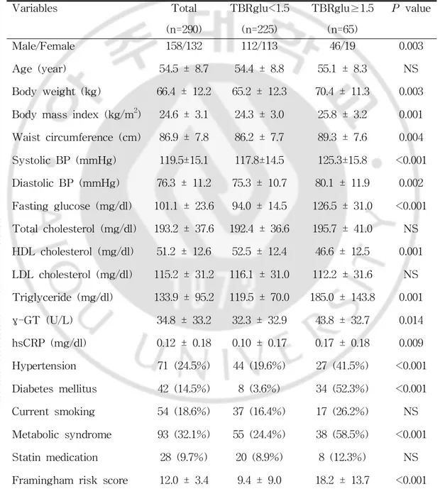
Variables Total (n=290) TBRglu<1.5 (n=225) TBRglu≥1.5 (n=65) P value Male/Female 158/132 112/113 46/19 0.003 Age(year) 54.5± 8.7 54.4± 8.8 55.1± 8.3 NS Bodyweight(kg) 66.4± 12.2 65.2± 12.3 70.4± 11.3 0.003 Bodymassindex(kg/m2) 24.6± 3.1 24.3± 3.0 25.8± 3.2 0.001 Waistcircumference(cm) 86.9± 7.8 86.2± 7.7 89.3± 7.6 0.004
SystolicBP (mmHg) 119.5±15.1 117.8±14.5 125.3±15.8 <0.001 DiastolicBP (mmHg) 76.3± 11.2 75.3± 10.7 80.1± 11.9 0.002 Fastingglucose(mg/dl) 101.1± 23.6 94.0± 14.5 126.5± 31.0 <0.001 Totalcholesterol(mg/dl) 193.2± 37.6 192.4± 36.6 195.7± 41.0 NS HDL cholesterol(mg/dl) 51.2± 12.6 52.5± 12.4 46.6± 12.5 0.001 LDL cholesterol(mg/dl) 115.2± 31.2 116.1± 31.0 112.2± 31.6 NS Triglyceride(mg/dl) 133.9± 95.2 119.5± 70.0 185.0± 143.8 0.001 γ-GT (U/L) 34.8± 33.2 32.3± 32.9 43.8± 32.7 0.014 hsCRP (mg/dl) 0.12± 0.18 0.10± 0.17 0.17± 0.18 0.009 Hypertension 71(24.5%) 44(19.6%) 27(41.5%) <0.001 Diabetesmellitus 42(14.5%) 8(3.6%) 34(52.3%) <0.001 Currentsmoking 54(18.6%) 37(16.4%) 17(26.2%) NS
Metabolicsyndrome 93(32.1%) 55(24.4%) 38(58.5%) <0.001 Statinmedication 28(9.7%) 20(8.9%) 8(12.3%) NS Framingham riskscore 12.0± 3.4 9.4± 9.0 18.2± 13.7 <0.001
I
I
I
.RESULTS
Theclinicalcharacteristicsofthe290subjectsaresummarizedinTable1.
Table1.ClinicalVariablesofStudy Subjects
blood pressure;HDL,high density lipoprotein;LDL,low density lipoprotein; hsCRP, high sensitive C-reactive protein; γ-GT, gamma glutamyl transpeptidase;NS,notsignificant.Dataarepresentasmean± SD.
Themeanagewas54.5years.Ofallsubjects,71(24.5%)hadhypertension, 42 (14.5%)diabetes,123 (42.4%)were currentsmokers,and 28 (9.7%)took statins.Themean FRS was12.0± 3.4%.Themean TBR and TBRglu were 1.3± 0.1and1.6± 1.2,respectively.Inthepresentstudy,wedidnotaim to compare TBR and TBRglu, thus we focus on TBRglu in the article. Ninety-three subjects (32.1%)were diagnosed with metabolic syndrome by health screening.Theirmean TBRglu was significantly greaterthan thatof subjectswithoutthesyndrome(1.5± 0.3vs.1.3± 0.2,P < 0.001).The75th percentile ofTBRglu in allsubjects was 1.5,and we chose the value as threshold fordataanalysis.Twogroups,showing high and low FDG uptake by the carotid arteries,could be distinguished using a TBRglu threshold of 1.5(Table1).
More members ofthe high uptake group were male (70.8 vs.49.7%,P = 0.003)and moreobese(high body weight,P = 0.003;BMI,P = 0.001;waist circumference,P = 0.004)than the low uptake group.The formersubjects had significantly higher BP (P < 0.005);elevated levels offasting plasma glucose(P < 0.001),triglycerides(P = 0.001),γ-GT (P = 0.014),and hsCRP (P =0.009);andlowerlevelsofHDL cholesterol(P =0.001).
Also,thesesubjectsexhibitedmorehypertension(P < 0.001),diabetes(P < 0.001),and metabolic syndrome (P < 0.001),than the low uptake group.In addition,theirFRSsweresignificantlyhigher(P < 0.001).
Correlation analysisshowed thatTBRglu was significantly associated with body weight(P = 0.001);BMI(P < 0.001);BP (P < 0.001);the levels of
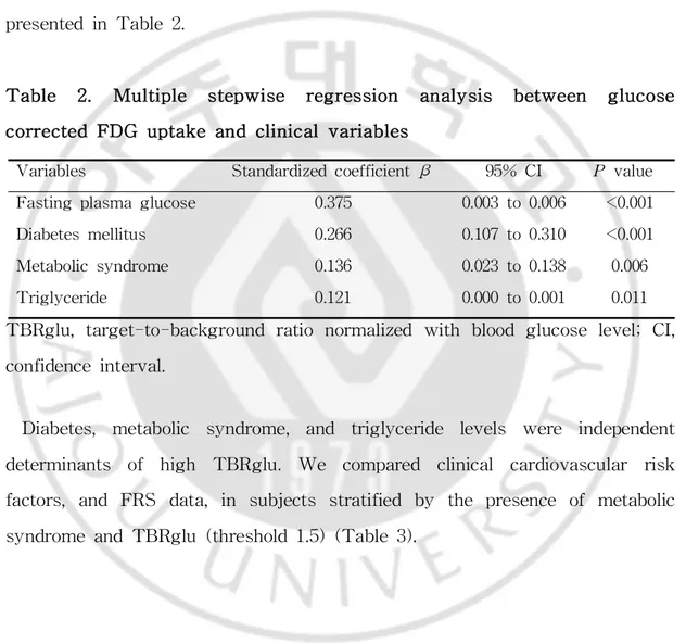
Variables Standardizedcoefficientβ 95% CI P value Fastingplasmaglucose 0.375 0.003to0.006 <0.001 Diabetesmellitus 0.266 0.107to0.310 <0.001 Metabolicsyndrome 0.136 0.023to0.138 0.006 Triglyceride 0.121 0.000to0.001 0.011
triglycerides(P < 0.001),and γ-GT (P < 0.001);and FRS (P < 0.001);and negatively associated with the levelof HDL cholesterol(P < 0.001).In addition,malegender(P < 0.001),diabetes(P < 0.001),metabolicsyndrome (P < 0.001),and hypertension (P < 0.001)weresignificantly associated with high TBRglu.Theresultsofmultiplestepwiselinearregression analysisare presentedinTable2.
Table 2. Multiple stepwise regression analysis between glucose correctedFDG uptakeandclinicalvariables
TBRglu,target-to-background ratio normalized with blood glucose level;CI, confidenceinterval.
Diabetes, metabolic syndrome, and triglyceride levels were independent determinants of high TBRglu. We compared clinical cardiovascular risk factors,and FRS data,in subjects stratified by the presence ofmetabolic syndromeandTBRglu(threshold1.5)(Table3).
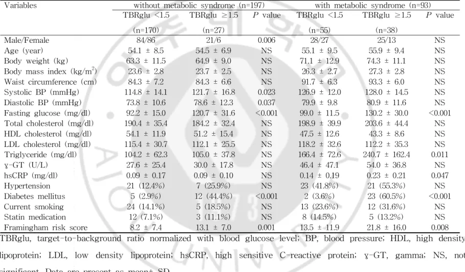
Variables withoutmetabolicsyndrome(n=197) withmetabolicsyndrome(n=93) TBRglu<1.5 (n=170) TBRglu≥1.5 (n=27) P value TBRglu<1.5 (n=55) TBRglu≥1.5 (n=38) P value Male/Female 84/86 21/6 0.006 28/27 25/13 NS Age(year) 54.1± 8.5 54.5± 6.9 NS 55.1± 9.5 55.9± 9.4 NS Bodyweight(kg) 63.3± 11.5 64.9± 9.0 NS 71.1± 12.9 74.3± 11.1 NS Bodymassindex(kg/m2) 23.6± 2.8 23.7± 2.5 NS 26.3± 2.7 27.3± 2.8 NS Waistcircumference(cm) 84.3± 7.2 84.3± 6.6 NS 91.7± 6.3 93.3± 6.0 NS SystolicBP (mmHg) 114.8± 14.1 121.7± 16.8 0.023 126.9± 12.0 128.0± 14.5 NS DiastolicBP (mmHg) 73.8± 10.6 78.6± 12.3 0.037 79.9± 9.8 80.9± 11.6 NS Fastingglucose(mg/dl) 92.2± 15.0 120.7± 31.6 <0.001 99.0± 11.5 130.2± 30.0 <0.001 Totalcholesterol(mg/dl) 190.4± 35.4 184.2± 32.4 NS 198.9± 39.9 203.6± 44.4 NS HDL cholesterol(mg/dl) 54.1± 11.9 51.2± 15.4 NS 47.5± 12.6 43.3± 8.6 NS LDL cholesterol(mg/dl) 115.4± 30.7 112.1± 25.5 NS 118.2± 32.6 112.2± 35.3 NS Triglyceride(mg/dl) 104.2± 62.3 105.0± 37.8 NS 166.4± 72.6 240.7± 162.4 0.011 γ-GT (U/L) 27.6± 25.4 30.0± 17.8 NS 46.4± 47.1 54.0± 36.8 NS hsCRP (mg/dl) 0.09± 0.17 0.09± 0.10 NS 0.14± 0.19 0.23± 0.21 0.047 Hypertension 21(12.4%) 7(25.9%) NS 23(41.8%) 21(55.3%) NS Diabetesmellitus 5(2.9%) 12(44.4%) <0.001 2(3.6%) 23(60.5%) <0.001 Currentsmoking 24(14.1%) 5(18.5%) NS 13(23.6%) 12(31.6%) NS Statinmedication 12(7.1%) 3(11.1%) NS 8(14.5%) 5(13.2%) NS Framingham riskscore 8.2± 7.4 13.1± 7.0 0.001 13.5± 11.9 21.8± 16.0 0.008
Table3.Comparison ofClinicalVariablesbetween Subjectswith andwithoutMetabolicSyndrome
TBRglu,target-to-background ratio normalized with blood glucose level;BP,blood pressure;HDL,high density lipoprotein; LDL,low density lipoprotein; hsCRP,high sensitive C-reactive protein; γ-GT,gamma; NS,not significant.Dataarepresentasmean± SD.
Ofthosewithoutmetabolicsyndrome,thesubgroupwith high TBRglu had more males (P = 0.006) and a higher frequency of diabetes (P < 0.001). Among thosewith metabolicsyndrome,thesubgroup with high TBRglu had higher fasting plasma glucose (P < 0.001),triglyceride (P = 0.011),and hsCRP (P = 0.047)levels;anddiabeteswasmorecommon (P < 0.001).High uptake subjects with metabolic syndrome had significantly higher levels of hsCRP compared to low uptakesubjectswith thesyndrome(0.23± 0.21vs. 0.14 ± 0.19),whereas the hsCRP leveldid notdiffer significantly between subgroupswithoutthesyndrome(Fig1a.).
Fig 1.(a) High sensitivity C-reactive protein (hsCRP) concentrations and (b)Framingham risk scores(FRSs)in study subjectsstratified by target-to-background ratio corrected forblood glucose level(TBRglu), and presence or absence ofmetabolic syndrome (MS).Bars:means ± standarderrors.
TheFRS wassignificantlyhigherinsubjectswithhighuptakecomparedto both non-syndromic and syndromic subjects with low uptake (8.2 ± 7.4 vs.
13.1 ± 7.0;13.5 ± 11.9 vs.21.8 ± 16.0,respectively;Fig 1b.).The FRS of subjectswithhighuptakebutnometabolicsyndromewascomparabletothat ofsubjects with low uptake and metabolicsyndrome (13.1 ± 7.0 vs.13.5 ± 11.9).Figure 2 demonstrates the representative PET images of a subject without metabolic syndrome but high carotid uptake and a subject with syndromebutlow uptake.
Fig 2. Representative maximum intensity of projection images of 18F-FDG PET/CT. (a) A 66-year-old man without metabolic syndrome shows mildly increased FDG uptake along bilateralcommon carotid arteries (arrows).(b)A 63-year-oldmanwithmetabolicsyndromehasnoremarkable FDG uptakeinbilateralcarotidarteries.Framingham riskscoreofthesubject with high uptake butno metabolic syndrome was comparable with thatof subjectwithlow uptakewithmetabolicsyndrome(11.6vs.13.4).
I
V.DI
SCUSSI
ON
In the present study, we found that carotid artery FDG uptake was significantly associated with clinical cardiovascular risk factors, and the 10-year general cardiovascular risk (assessed using the FRS), in an asymptomatic population.As expected,both carotid artery FDG uptake and FRS were elevated in subjects with metabolic syndrome.Ofsuch subjects, the hsCRP levelwas significantly higherin the high uptake than the low uptake group.Interestingly,among those with metabolic syndrome,the high uptake group had a significantly higher FRS than the low uptake group, whereas the FRS ofthe low uptake group with metabolic syndrome was comparabletothatofthehighuptakegroupwithoutthesyndrome.
We analyzed TBRglu along the full lengths of both common carotid arteries.Severalstudies have found thattumor FDG uptake is diminished during hyperglycemia(Wahletal,1992;Shepherd andKahn,1999).Although the need to apply a glucose correction to FDG uptake when noncancerous lesionsarebeing studied isnotwellunderstood,onerecentstudy evaluating carotid artery FDG uptake in type 2 diabetes patients observed that glucose-corrected FDG uptake parameters increased significantly as fasting blood glucose levels rose (Bucerius et al, 2012). Consistent with such findings, we noted that glucose-corrected FDG uptake values were significantly higherin hyperglycemicsubjects.In addition,wefound a weak (r < 0.2) and unexpected negative correlation between the presence of diabetesandglucose-uncorrectedcarotidarteryFDG uptake.Inaddition,60of oursubjects(20.7%)hadhighpre-scanglucoselevels(≥ 120mg/dl),andwe thus used the TBRglu,a glucose-corrected FDG uptake parameter,in our work.Additionally,whenweanalyzeddatausingSUVglu,itwassignificantly
associated with FRS (P < 0.001), fasting plasma glucose (P < 0.001), hypertension (P < 0.001),diabetes(P < 0.001),andmetabolicsyndrome(P < 0.001). Multiple stepwise regression analysis showed that diabetes and metabolicsyndromewereindependentdeterminantsofhighSUVglu.
We found thathigh carotid FDG uptake was significantly associated with clinicalcardiacrisk factorsincluding obesity,dyslipidemia,hypertension,male gender, and diabetes. This is in line with the results of previous investigations(Kanekoetal,2013;Buceriusetal,2012;Yun etal,2002;Kim etal,2010).We also found a significantassociation between carotid FDG accumulation,andtheFRS andmetabolicsyndrome.
Recently,Tahara etal.described a positive association between metabolic syndromecomponentsandFDG uptakeinpatientswithcarotidatherosclerosis (Taharaetal,2007).Consistentwith thesedata,wefound thatcarotid FDG accumulation was significantly higher in those with metabolic syndrome. Moreover, the hsCRP levels of subjects with high carotid uptake and metabolic syndrome weresignificantly higherthan thoseofsubjects without the syndrome,and those ofsubjects with the syndrome butexhibiting low carotid uptake.The hsCRP levelis an indicator ofatherosclerotic activity during the initialstages ofatherosclerosis (Hashimoto etal,2001).Previous studies found that arterialFDG uptake increased significantly in areas of atherosclerotic plaques thatwere histologically macrophage-rich (Rudd etal, 2002;Tawakolet al,2006).Thus,carotid artery FDG uptake and hsCRP concentration, both of which are inflammatory markers, reflect active inflammation during early stagecarotid atherosclerosis.Wethussuggestthat development of atherosclerosis may accelerate more markedly in metabolic syndrome subjects with higher rather than lower uptake,as hsCRP levels rise.Interestingly,thegeneralcardiovascularFRSsdifferedsignificantly when
metabolic syndrome subjects were stratified by carotid artery FDG activity. Thosewithhighuptakescoredsignificantlyhigher.Thus,carotidFDG uptake may help to stratify asymptomatic patients,identifying those needing active treatmentsuchasanti-inflammatorypharmacotherapy.
The present study had severallimitations.First,although we evaluated cardiovascular risk based on the FRS,our study was cross-sectional,and retrospective in nature. A longitudinal study with follow-up is needed. Second,our study population was heterogeneous.We included 42 patients with diabetes and 28 on statin medication.Among those with diabetes,28 (9.7%)patientsweretaking oralhypoglycemicagentsatthetimeofPET/CT, and 14 (4.8%)were newly diagnosed on screening.Although diabetes has been reported to significantly elevate arterial FDG uptake, we included diabetes patients in ourcurrentstudy because we wished to survey carotid artery FDG uptakein an asymptomaticgeneralpopulation.Thisisalso why we did notexclude those taking statins which may attenuate arterialFDG uptake.Finally,we used only TBRglu forevaluation ofcarotid artery FDG uptake in the presentstudy.The index may be affected by blood glucose leveland the clinicalsignificance of TBRglu has been limited.We now perform another study in a larger cohort to evaluate carotid artery FDG uptakeusingTBR,SUVgluandTBRglu.
V.CONCLUSI
ON
High carotid FDG uptake in asymptomatic subjects was significantly associated with the presence ofclinicalcardiovascular risk factors and an elevated FRS.Ofsubjects with metabolic syndrome,those exhibiting high carotid uptake had significantly elevated hsCRP levels and FRSs.Therefore, carotid artery FDG activity may serve as a possible biomarker allowing cardiovascularriskstratificationofasymptomaticpopulations.
REFERENCES
1.Berger JS,Jordan CO,Lloyd-Jones D,Blumenthal RS: Screening for cardiovascularrisk in asymptomatic patients.J Am CollCardiol55(12): 1169-1177,2010
2.Boellaard R,O'Doherty MJ,Weber WA,Mottaghy FM,Lonsdale MN, Stroobants SG,Oyen WJ,Kotzerke J,Hoekstra OS,Pruim J:FDG PET and PET/CT: EANM procedure guidelines for tumour PET imaging: version1.0.EurJNuclMedMolImaging 37(1):181-200,2010
3.BuceriusJ,Duivenvoorden R,ManiV,MoncrieffC,Rudd JH,CalcagnoC, Machac J,FusterV,Farkouh ME,Fayad ZA:Prevalenceand risk factors of carotid vesselwallinflammation in coronary artery disease patients: FDG-PET andCT imaging study.J Am CollCardiolImg 4(11):1195-1205, 2011
4.BuceriusJ,ManiV,MoncrieffC,Rudd JH,MachacJ,FusterV,Farkouh ME,Fayad ZA:Impactofnoninsulin-dependenttype2diabeteson carotid wall18F-fluorodeoxyglucose positron emission tomography uptake.J Am
CollCardiol59(23):2080-2088,2012
5.CockerMS,McArdleB,SpenceJD,Lum C,Hammond RR,Ongaro DC, McDonald MA,Tardif JC,Beanlands RS: Imaging atherosclerosis with hybrid [(18)F]fluorodeoxyglucose positron emission tomography/computed tomography imaging: what Leonardo da Vinci could not see. J Nucl Cardiol,19(6):1211-1225,2012
6. D'Agostino RB, Vasan RS, Pencina MJ, Wolf PA, Cobain M, Massaro JM,KannelWB:Generalcardiovascularrisk profile foruse in primary care:the Framingham Heart Study.Circulation 117(6):743-753, 2008
7.DekkerJM,Girman C,Rhodes T,Nijpels G,StehouwerCD,BouterLM, Heine RJ:Metabolic syndromeand 10-yearcardiovasculardisease risk in theHoornStudy.Circulation112(5):666-673,2005
8.Ford ES,LiC,Zhao G:Prevalenceand correlates ofmetabolicsyndrome based on a harmonious definition among adults in the US.J Diabetes
2(3):180-193,2010
9.Grundy SM,Cleeman JI,DanielsSR,DonatoKA,EckelRH,Franklin BA, GordonDJ,KraussRM,SavagePJ,SmithSC:Diagnosisandmanagement ofthemetabolicsyndrome:an American HeartAssociation/NationalHeart, Lung, and Blood Institute Scientific Statement. Circulation
112(17):2735-2752,2005
10.HanssonGK:Inflammation,atherosclerosis,andcoronaryarterydisease.N
EnglJMed352(16):1685-1695,2005
11.Hashimoto H,Kitagawa K,Hougaku H,Shimizu Y,SakaguchiM,Nagai Y,Iyama S,YamanishiH,Matsumoto M,HoriM:C-reactiveprotein is an independent predictor of the rate of increase in early carotid atherosclerosis.Circulation104(1):63-67,2001
12.KanekoK,KawasakiT,MasunariS,YoshidaT,OmagariJ:Determinants of extraaortic arterial 18F-FDG accumulation in asymptomatic cohorts: sex differences in the association with cardiovascular risk factors and coronaryarterystenosis.JNuclMed54(4):564-570,2013
13.Kim TN,Kim S,Yang SJ,YooHJ,SeoJA,Kim SG,Kim NH,BaikSH, Choi DS, Choi KM: Vascular inflammation in patients with impaired glucose tolerance and type 2 diabetes: analysis with 18F-fluorodeoxyglucose positron emission tomography. Circ Cardiovasc Imaging 3:142-148,2010
18F-FDG uptake with plasma high-density lipoprotein elevation by atherogenicriskreduction.JNuclMed49(8):1277-1282,2008
15.OhM,Kim JY,ShinKH,ParkSH,RyuJS,Kim JS,Kim HJ,KangDW, Moon DH: Imaging atherosclerosis in the carotid arteries with F-18-fluoro-2-deoxy-D-glucose positron emission tomography: effect of imaging timeafterinjection on quantitativemeasurement.NuclMed Mol Imaging 44(4):261-266,2010
16.Rudd JH,Warburton EA,Fryer TD,Jones HA,Clark JC,Antoun N, Johnström P, Davenport AP, Kirkpatrick PJ, Arch BN: Imaging atheroscleroticplaqueinflammationwith[18F]-fluorodeoxyglucosepositron emissiontomography.Circulation105(23):2708-2711,2002
17. Shepherd PR, Kahn BB: Glucose transporters and insulin action -implications for insulin resistance and diabetes mellitus. N Engl J
Med341:248-257,1999
18.TaharaN,KaiH,IshibashiM,NakauraH,KaidaH,BabaK,Hayabuchi N,ImaizumiT:Simvastatinattenuatesplaqueinflammation:evaluation by fluorodeoxyglucose positron emission tomography.J Am Coll Cardiol
48(9):1825-1831,2006
19.Tahara N,KaiH,YamagishiS,MizoguchiM,Nakaura H,IshibashiM, Kaida H,Baba K,Hayabuchi N,Imaizumi T: Vascular inflammation evaluated by [18F]-fluorodeoxyglucose positron emission tomography is associated with the metabolic syndrome. J Am Coll Cardiol 49(14): 1533-1539,2007
20.TawakolA,Migrino RQ,Bashian GG,BedriS,Vermylen D,Cury RC, Yates D, LaMuraglia GM, Furie K, Houser S: In vivo 18F-fluorodeoxyglucose positron emission tomography imaging provides a noninvasive measure of carotid plaque inflammation in patients.JAm CollCardiol48(9):1818-1824,2006
21.WahlRL,Henry CA,EthierSP:Serum glucose:effects on tumorand normal tissue accumulation of 2-[F-18]-fluoro-2-deoxy-D-glucose in rodentswithmammarycarcinoma.Radiology183(3):643-647,1992
22.Wilson PW,D'Agostino RB,Parise H,Sullivan L,Meigs JB:Metabolic syndrome as a precursorofcardiovasculardisease and type 2 diabetes mellitus.Circulation112(20):3066-3072,2005
23.WuYW,KaoHL,Huang CL,ChenMF,Lin LY,Wang YC,LinYH,Lin HJ,Tzen KY,Yen RF:The effects of3-month atorvastatin therapy on arterialinflammation,calcification,abdominaladiposetissueandcirculating biomarkers.EurJNuclMedMolImaging 39(3):399-407,2012
24.YooHJ,Kim S,ParkMS,YangSJ,Kim TN,SeoJA,Kim SG,Kim NH, Seo HS,Baik SH:Vascularinflammation stratified by C-reactiveprotein and low-density lipoprotein cholesterol levels: analysis with 18F-FDG PET.JNuclMed52(1):10-17,2011
25.YunM,Jang S,CucchiaraA,Newberg AB,AlaviA:18F FDG uptakein the large arteries:a correlation study with the atherogenic risk factors.
국문요약
-심혈관 질환 증상이 없는 성인에서 목동맥 FDG 섭취와
심혈관계 질환 위험인자 간의 관계 고찰
아주대학교 대학원의학과 이 동 현 (지도교수 이 수 진) 심혈관 질환은 동맥경화반이 염증과정의 진행에 따라 파열되어 생성된 혈색전 에 의한 것으로 알려져 있다.질환의 심각성에 대한 기존 방법인 내경의 협착 평 가나 증상 분석에 비해 F-18FDG PET/CT는 취약성 경화반의 상태를 더욱 잘 반영하는 것으로 보고되고 있다.이에 본 연구에서는 경동맥 경화증에서의 FDG 섭취 정도와 Framingham risk score를 기반으로 한 심혈관 위험인자 사이의 관 계를 분석하였고,F-18FDG PET/CT가 향후 증상이 없는 성인을 대상으로 심 혈관 질환의 예방적인 평가수단이 될 수 있는지 알아보고자 하였다.건강검진 대 상자 290명에 대해 FDG PET/CT를 시행하였다.PET/CT 영상은 양쪽 온목동맥 의 Target-to-backgroundratio(TBR)을 공복혈당으로 보정한 TBRglu값을 얻었 다.각각의 대상자들에 대해 FRS와 대사증후군의 유무가 분석되었고,TBRglu 값과 대사증후군 상태,임상적인 인자들 간의 통계분석이 이루어졌다.온목동맥 의 FDG 섭취 정도는 심혈관 위험인자와 유의한 관계가 있었다.다중회귀분석 결과 중성지방,당뇨,대사증후군은 높은 TBRglu 값의 독립적인 결정인자였다. 대사증후군 있는 집단에서 온목동맥의 높은 FDG 섭취 정도는 높은 hsCRP값과 유의한 관계가 있었다.대사증후군 없는 집단에서의 FRS 값은 온목동맥에서 낮 은 FDG 섭취 정도를 보이는 집단에 비해 높은 FDG 섭취 정도를 보이는 집단에 서 유의하게 높았으며 (13.1 ± 7.0 vs.8.2 ± 7.4),대사증후군 있는 집단에서도 같은 경향을 보였다 (21.8± 16.0vs.13.5± 11.9).온목동맥의 높은 FDG 섭취정도는 심혈관 위험인자와 상관관계가 있었으며 10년 후 심혈관 질환 위험도에 도 높은 경향을 보였다.대사증후군이 있는 집단에서는 온목동맥의 높은 FDG 섭취 정도가 높은 hsCRP과 FRS값과 유의한 관계를 보였다.그러므로 온목동맥 의 FDG 섭취 정도는 무증상의 일반인에서 심혈관 위험을 예측하는 하나의 생물 학적 인자 역할을 할 수 있을 것이다. 핵심어:동맥경화증,온목동맥,18F-FDG,위험도 평가,대사증후군,심혈관계질환