저작자표시-비영리-변경금지 2.0 대한민국 이용자는 아래의 조건을 따르는 경우에 한하여 자유롭게 l 이 저작물을 복제, 배포, 전송, 전시, 공연 및 방송할 수 있습니다. 다음과 같은 조건을 따라야 합니다: l 귀하는, 이 저작물의 재이용이나 배포의 경우, 이 저작물에 적용된 이용허락조건 을 명확하게 나타내어야 합니다. l 저작권자로부터 별도의 허가를 받으면 이러한 조건들은 적용되지 않습니다. 저작권법에 따른 이용자의 권리는 위의 내용에 의하여 영향을 받지 않습니다. 이것은 이용허락규약(Legal Code)을 이해하기 쉽게 요약한 것입니다. Disclaimer 저작자표시. 귀하는 원저작자를 표시하여야 합니다. 비영리. 귀하는 이 저작물을 영리 목적으로 이용할 수 없습니다. 변경금지. 귀하는 이 저작물을 개작, 변형 또는 가공할 수 없습니다.
Factors influencing on esophageal baseline impedance levels
in patients with suspected symptoms of gastroesophageal
reflux disease
by
Young GeonKim
Major in Medicine
Department of Sciences in medicine
The Graduate School, Ajou University
Factors influencing on esophageal baseline impedance levels
in patients with suspected symptoms of gastroesophageal
reflux disease
by
Young GeonKim
A Dissertation Submitted to The Graduate School of
Ajou University in Partial Fulfillment of the Requirements
for the Degree of
Master of Medicine
Supervised by
Kwang Jae Lee, M.D.
Major in Medicine
Department of Sciences in medicine
The Graduate School, Ajou University
This certifies that the dissertation
ofYoung Geon Kim is approved.
SUPERVISORY COMMITTEE
Kwang Jae Lee
Byung Moo Yoo
Jae Chul Hwang
The Graduate School, Ajou University
June, 19th, 2015
i
-ABSTRACT-
Factors influencing on esophageal baseline impedance levels in
patients with suspected symptoms of gastroesophageal reflux disease
Background/Aims: Measurements of mucosal impedance can detect GERD with highlevels of specificity and positive predictive values. However, it remains unclear whether mucosal impedance changes frequently during a day and is influenced by the time from themeal period and sleeping time.The aim of the study was to investigate factors influencing on esophageal baseline impedance levels,such as time from the meal period, sleep onset, and the location of measurement, in patients with suspected symptoms of GERD.
Methods:Consecutive endoscopy-negative patients with suspected symptoms of GERD such as heartburn, chest pain, and/or globus who underwent 24h esophageal multichannel intraluminal impedance-pH (MII-pH) monitoring were eligible for inclusion in this study. Baseline impedance was measured in the proximal esophagus (the average of 15 and 17 cm) and the distal esophagus (the average of 3 and 5 cm) at 5 min before meal, 5 min after the meal period, 30 min after the meal period, 30 min before sleep, 30 min after sleep onset and 60 min after sleep onset. Each single baseline value represents the average value of a 30-s time period around each time point.
Results:Baseline impedance levels did not significantly differ according to gender or age. Baseline impedance levels at 5 min and 30 min after meal were significantly lower in the proximal (p<0.001) and distal esophagus (p<0.001), compared with those at 5 min before meal. Significantly lower baseline impedance levels were observed at 30 min (p<0.005) and 60 min (p<0.005) after sleep onset in the proximal esophagus, compared with those at 30 min before sleep onset, but not in the distal esophagus. Baseline impedance levels in the distal esophagus weresignificantly lower in patients with pathologic reflux, compared with the GERD-unrelated group. No significant difference in the baseline impedance levels was observed between patients with positive symptom index alone and the GERD-unrelated
ii
group.
Conclusions: The time from the meal period or sleep onset, and the presence of pathological acid reflux or pathological bolus reflux appear to influence on esophageal baseline impedance levels.However, sex, age, and the presence of esophageal hypersensitivity do not seem to be associated with esophageal baseline impedance levels.
Key words: Gastroesophageal reflux disease, Combined Multichannel intraluminal impedance, Non-erosive reflux disease
iii
TABLE OF CONTENTS
ABSTRACT ···i
TABLE OF CONTENTS ··· iii
LIST OF FIGURES ··· iv
LIST OF TABLES ··· v
Ⅰ. INTRODUCTION ··· 1
Ⅱ. PATIENTS AND METHODS ··· 2
A. Study Population ··· 2 B. 24hr MII-pH monitoring ··· 2 C. Statistical Analysis··· 3 Ⅲ. RESULTS ··· 4 Ⅳ. DISCUSSION ··· 13 Ⅴ. CONCLUSION ··· 15 REFERENCES ··· 16 국문요약 ··· 20
iv
LIST OF FIGURES
Figure 1.Baseline impedance levels in the proximal and distal esophagus according to the time from the meal period ··· 8 Figure 2. Baseline impedance levels in the proximal and distal esophagus according to
v
LIST OF TABLES
Table 1.GERD-related and GERD-unrelated symptoms ··· 4 Table 2. Comparison of baseline impedance levels in the distal esophagus between males
and females ··· 5 Table 3. Comparison of baseline impedance levels in the distal esophagus between the
age group < 60 years old and the age group ≥60 years old ··· 6 Table 4. Comparison of baseline impedance levels in the proximal esophagus between
males and females ··· 7 Table 5. Comparison of baseline impedance levels in the proximal esophagus between
the age group < 60 years old and the age group ≥60 years old ··· 7 Table 6. Baseline impedance levels of the proximal esophagus in the GERD-related and
GERD-unrelated groups ··· 10 Table 7. Baseline impedance levels of the distal esophagus in the GERD-related and
GERD-unrelated groups ··· 10 Table 8. Baseline impedance levels of the proximal esophagus in the pathologic reflux
(PR), positive symptom index alone (PSI), and GERD-unrelated groups··· 11 Table 9. Baseline impedance levels of the distal esophagus in the pathologic reflux (PR),
- 1 -
I.INTRODUCTION
Gastro-esophageal reflux disease (GERD) is common, and its prevalence is increasing in Asian countries including South Korea. Based on the presence of erosions in the esophagus, GERD is classified into erosive reflux disease (ERD) and non-erosive reflux disease (NERD). NERD is known to be more common than ERD. When there is no evidence of ERD, symptoms may or may not be related to GERD (Dent J et al, 1999).The ambulatory 24hr esophageal pH-impedance monitoring to confirm the causal relationship between symptoms and gastroesophageal reflux is not considered to be satisfactory, because itcan only measure reflux activity. Therefore, patients with suspected GERD symptoms who do not have reflux events during the test period have falsely negative results.
Intra-luminal baseline impedance measurement can be used to evaluate changes in the integrity of the esophageal mucosa. Alterations in the integrity of the esophageal mucosa are associated with trans-epithelial resistance. Accordingly, changes in intra-luminal baseline impedance levels are suggested to be evidence of GERD. The esophageal lumen is collapsed except reflux episodes and swallowing episodes, thus the resulting baseline impedance level is determined by the surrounding esophageal wall. Studies have shown the association between changes of baseline impedance levels and GERD symptoms. A recent study demonstrated that measurements of mucosal impedance can detect GERD with higherlevels of specificity and positive predictive values than wirelesspH monitoring(Ates et al, 2015). However, it remains unclear whether mucosal impedance changes frequently during a day and is influenced by the meal time and sleeping time.Moreover, it is still uncertain whether mucosal impedance differs according to the location of measurement.
Thus, the aim of this study was to investigate factors influencing on esophageal baseline impedance levels,such as time from the meal period, sleep onset, and the location of measurement, in patients with suspected symptoms of GERD.
- 2 -
II. PATIENTS AND METHODS
A. Study Population
Consecutive endoscopy-negative patients with suspected symptoms of GERD such as heartburn, chest pain, and/or globus who underwent 24h esophageal multichannel intraluminal impedance-pH (MII-pH) monitoring were eligible for inclusion in this study. TheirMII-pH data performed at Ajou University Hospital were analyzed. Patients were excluded if they were taking medication which could influence esophageal motor function or gastric acid secretion, or if they had erosive or ulcerative esophagitis on endoscopy, achalasia, or a history of upper gastrointestinal surgery.
B. 24hr MII-pH monitoring
Each study was done using stationary MII-pH system (Sandhill, Scientific, Denver, CO). MII-pH monitoring was performed after discontinuing therapy such as acid-suppressing or motility agents one week before the monitoring. We calibrated the MII-pH catheter in buffer solutions of pH 4.0 and 7.0, and passed the catheter through the nose after the lower esophageal sphincter (LES) location was detected by esophageal manometry. The pH electrode was located at 5cm above the upper margin of the LES, and segments monitoring intraluminal impedance were positioned at 3, 5, 7, 9, 15, and 17cm above the LES. Patients were discharged and were asked to maintain normal activities, sleep schedule and eat their usual meals at their normal times. They were encouraged to keep a diary in which they recorded the times for meals, postural changes and vice versa, and symptoms. Impedance and pH information was analyzed using a dedicated software program (BioView Analysis, Sandhill Scientific, Inc.) and visual analysis.
An acid reflux episode was defined as a drop in pH to less than 4.0, and pathological acid reflux was defined as the % of time with an acid reflux episode above 4.2%.A reflux episode was defined as a retrograde drop in impedance by more than 50% of baseline in the distal two consecutive channels, and pathological reflux was defined as the % of time with a reflux
- 3 -
episode above 1.4%. Symptom index (SI) was defined as the number of symptoms associated with reflux divided by the total number of symptoms during 24 hours multiplied by 100, and SI more than or equal to 50% was defined as positive SI. When there was pathological acid reflux, pathological reflux, or positive symptom index, patients were considered to have GERD-related symptoms.
Baseline impedance was measured in the proximal esophagus (the average of 15 and 17 cm) and the distal esophagus (the average of 3 and 5 cm) at 5 min before meal, 5 min after the meal period, 30 min after the meal period, 30 min before sleep, 30 min after sleep onset and 60 min after sleep onset. Each single baseline value represents the average value of a 30-s time period around each time point avoiding 30-swallow30-s, refluxe30-s, and pH drop30-s. MII-pH monitoring was performed and tracing data were reviewed manually by investigator blinded for the information of the patients.
C. Statistical Analysis
Data are expressed asmeans ± standard deviations (SDs). Continuous variableswere compared using Student’st-test or ANOVA. Chi-square test was applied to compare categorical variables between groups. Statistical analyses were conducted using SPSS version 18.0 (IBM Corp., Armonk, NY, USA). A P-value < 0.05 was taken as statistically significant.
- 4 -
III. RESULTS
GERD-related and GERD-unrelated symptoms
A total of 104 patients (57 males, mean age 47.9 ± 13.9 [Range, 20-74] years) were enrolled in thestudy. The chief complaints were heartburn in 39 patients, chest pain in 28 patients, globus in 20 patients, and chronic cough in 17 patients. Each symptom groupshowed no statistical difference in gender ratio and age (Table 1). Seventy-four percent of subjects who complained heartburn were considered to be GERD-related. GERD-related symptoms were confirmed in 74% of the heartburn group, 53% of the chest pain group, 40% of the globus group, and 35% of the chronic cough group. Heartburn was most correlated with GERD.A total of 42 patients showed pathologic acid reflux or bolus reflux. Positive symptom index was observed in 16 patients. A total of 46patients showed negative results for 24-h MII-pH monitoring, and was diagnosed to be GERD-unrelated.
Table 1.GERD-related and GERD-unrelated symptoms Predominant
symptom Sex (M:F) Age (<60 : ≥60)
GERD-related, n (%) Heartburn (n=39) 23:16 30:9 29 (74%) Chest pain (n=28) 15:13 19:9 15 (53%) Globus (n=20) 11:9 17:3 8 (40%) Chronic cough (n=17) 8:9 12:5 6 (35%) P value 0.872 0.556 0.015
- 5 -
Comparison of baseline impedance levels in the distal esophagus according to sex and age
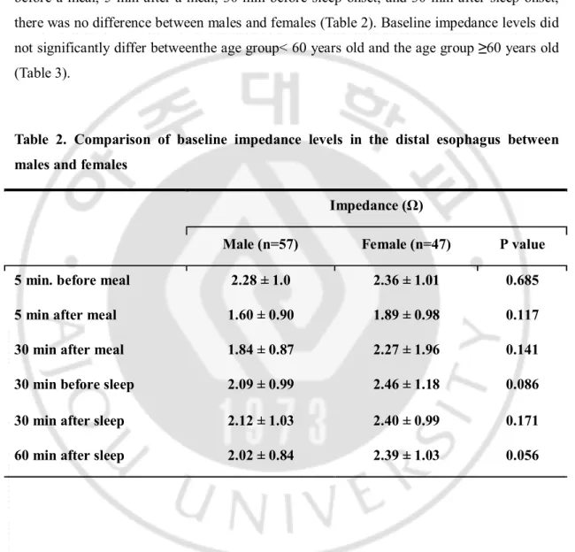
When comparing the baseline impedance levels measured in the distal esophagus at 5 min before a meal, 5 min after a meal, 30 min before sleep onset, and 30 min after sleep onset, there was no difference between males and females (Table 2). Baseline impedance levels did not significantly differ betweenthe age group< 60 years old and the age group ≥60 years old (Table 3).
Table 2. Comparison of baseline impedance levels in the distal esophagus between males and females
Impedance (Ω)
Male (n=57) Female (n=47) P value
5 min. before meal 2.28 ± 1.0 2.36 ± 1.01 0.685
5 min after meal 1.60 ± 0.90 1.89 ± 0.98 0.117
30 min after meal 1.84 ± 0.87 2.27 ± 1.96 0.141
30 min before sleep 2.09 ± 0.99 2.46 ± 1.18 0.086
30 min after sleep 2.12 ± 1.03 2.40 ± 0.99 0.171
- 6 -
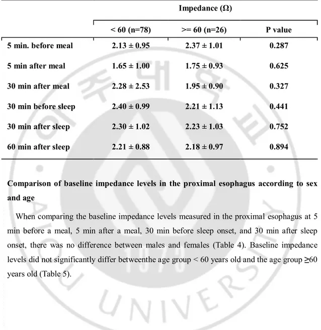
Table 3. Comparison of baseline impedance levels in the distal esophagus between the age group < 60 years old and the age group ≥60 years old
Impedance (Ω)
< 60 (n=78) >= 60 (n=26) P value
5 min. before meal 2.13 ± 0.95 2.37 ± 1.01 0.287
5 min after meal 1.65 ± 1.00 1.75 ± 0.93 0.625
30 min after meal 2.28 ± 2.53 1.95 ± 0.90 0.327
30 min before sleep 2.40 ± 0.99 2.21 ± 1.13 0.441
30 min after sleep 2.30 ± 1.02 2.23 ± 1.03 0.752
60 min after sleep 2.21 ± 0.88 2.18 ± 0.97 0.894
Comparison of baseline impedance levels in the proximal esophagus according to sex and age
When comparing the baseline impedance levels measured in the proximal esophagus at 5 min before a meal, 5 min after a meal, 30 min before sleep onset, and 30 min after sleep onset, there was no difference between males and females (Table 4). Baseline impedance levels did not significantly differ betweenthe age group < 60 years old and the age group ≥60 years old (Table 5).
- 7 -
Table 4. Comparison of baseline impedance levels in the proximal esophagus between males and females
Impedance (Ω)
Male (n=57) Female (n=47) P value
5 min. before meal 2.76 ± 0.96 2.67 ± 0.95 0.626
5 min after meal 1.91 ± 0.45 1.94 ± 0.86 0.867
30 min after meal 2.26 ± 0.88 2.04 ± 0.94 0.222
30 min before sleep 2.97 ± 1.09 2.90 ± 1.15 0.755
30 min after sleep 2.21 ± 0.99 2.26 ± 1.08 0.787
60 min after sleep 2.13 ± 0.96 2.27 ± 1.06 0.482
*p<0.05, **p<0.01 and ***p<0.005, compared with the GERD-unrelated group
Table 5. Comparison of baseline impedance levels in the proximal esophagus between the age group < 60 years old and the age group ≥60 years old
Impedance (Ω)
Age < 60 (n=78) Age ≥ 60 (n=26) P value
5 min. before meal 2.50 ± 0.91 2.80 ± 0.95 0.164
5 min after meal 1.90 ± 0.97 1.94 ± 0.88 0.864
30 min after meal 2.18 ± 0.92 1.95 ± 0.91 0.902
30 min before sleep 2.64 ± 1.19 3.04 ± 1.08 0.117
30 min after sleep 2.05 ± 0.88 2.29 ± 1.07 0.313
- 8 -
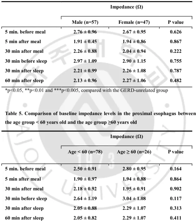
Comparison of baseline impedance levels according to the time from the meal period Baseline impedance levels at 5 min and 30 min after meal were significantly lower in the proximal (p<0.001) and distal esophagus (p<0.001), compared with those at 5 min before meal (Figure 1).
Figure 1.Baseline impedance levels in the proximal (P-esophagus) and distal esophagus (D-esophagus) according to the time from the meal period
- 9 -
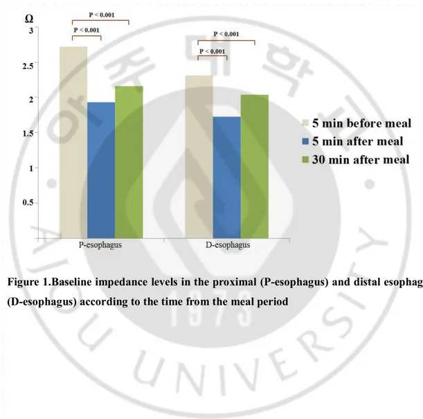
Comparison of baseline impedance levels according to the time from sleep onset Significantly lower baseline impedance levels were observed at 30 min (p<0.005) and 60 min (p<0.005) after sleep onset in the proximal esophagus, compared with those at 30 min before sleep onset, but not in the distal esophagus (Figure 2).
Figure 2.Baseline impedance levels in the proximal (P-esophagus) and distal esophagus (D-esophagus) according to the time from sleep onset
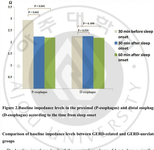
Comparison of baseline impedance levels between GERD-related and GERD-unrelated groups
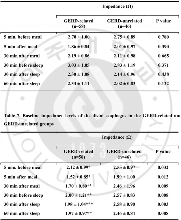
The baseline impedance levels of the proximal esophagus did not show a significant difference at any measurement time between the GERD-related and GERD-unrelated groups (Table 6). However, the baseline impedance levels of the distal esophagus were significantly lower at all measurement times in the GERD-related group than in the GERD-unrelated group (Table 7).
- 10 -
Table 6. Baseline impedance levels of the proximal esophagus in the GERD-related and GERD-unrelated groups Impedance (Ω) GERD-related (n=58) GERD-unrelated (n=46) P value
5 min. before meal 2.70 ± 1.00 2.75 ± 0.89 0.780
5 min after meal 1.86 ± 0.84 2.01 ± 0.97 0.390
30 min after meal 2.19 ± 0.86 2.11 ± 0.98 0.665
30 min before sleep 3.03 ± 1.05 2.83 ± 1.19 0.371
30 min after sleep 2.30 ± 1.08 2.14 ± 0.96 0.438
60 min after sleep 2.33 ± 1.11 2.02 ± 0.83 0.122
Table 7. Baseline impedance levels of the distal esophagus in the GERD-related and GERD-unrelated groups Impedance (Ω) GERD-related (n=58) GERD-unrelated (n=46) P value
5 min. before meal 2.12 ± 0.99* 2.55 ± 0.97 0.032
5 min after meal 1.52 ± 0.85* 1.99 ± 1.00 0.012
30 min after meal 1.70 ± 0.80** 2.46 ± 1.96 0.009
30 min before sleep 2.00 ± 1.21** 2.57 ± 0.83 0.008
30 min after sleep 1.98 ± 1.04*** 2.58 ± 0.90 0.003
60 min after sleep 1.97 ± 0.97** 2.46 ± 0.84 0.008
- 11 -
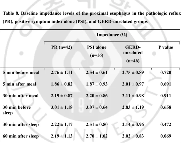
Comparison of baseline impedance levels among the pathologic reflux (PR), positive symptom index alone (PSI), and GERD-unrelated groups
In the proximal esophagus, there was no difference in the baseline impedance levels among the PR, PSI and GERD-unrelated groups (Table 8).However, baseline impedance levels in the distal esophagus weresignificantly lower in the PR group at all measurement times, compared with the GERD-unrelated group (Table 9). No significant difference in the baseline impedance levels was observed between the PSI and GERD-unrelated groups.
Table 8. Baseline impedance levels of the proximal esophagus in the pathologic reflux (PR), positive symptom index alone (PSI), and GERD-unrelated groups
Impedance (Ω) PR (n=42) PSI alone (n=16) GERD-unrelated (n=46) P value
5 min before meal 2.76 ± 1.11 2.54 ± 0.61 2.75 ± 0.89 0.720
5 min after meal 1.86 ± 0.82 1.87 ± 0.93 2.01 ± 0.97 0.691
30 min after meal 2.19 ± 0.87 2.20 ± 0.86 2.11 ± 0.98 0.911
30 min before sleep
3.01 ± 1.18 3.07 ± 0.64 2.83 ± 1.19 0.658
30 min after sleep 2.22 ± 1.17 2.51 ± 0.80 2.14 ± 0.96 0.472
60 min after sleep 2.19 ± 1.13 2.70 ± 1.02 2.02 ± 0.83 0.069
- 12 -
Table 9. Baseline impedance levels of the distal esophagus in the pathologic reflux (PR), positive symptom index alone (PSI), and GERD-unrelated groups
Impedance (Ω) PR (n=42) PSI alone (n=16) GERD-unrelated (n=46) P value
5 min before meal 1.89 ± 0.94* 2.73 ± 0.90 2.55 ± 0.97 0.001
5 min after meal 1.31 ± 0.69* 2.07 ± 1.01 1.99 ± 1.00 0.001
30 min after meal 1.46 ± 0.69* 2.34 ± 0.72 2.46 ± 1.96 0.004
30 min before sleep
1.76 ± 1.18* 2.64 ± 1.07 2.57 ± 0.83 0.001
30 min after sleep 1.67 ± 0.93* 2.79 ± 0.88 2.58 ± 0.90 <0.001
60 min after sleep 1.69 ± 0.83* 2.70 ± 0.96 2.46 ± 0.84 <0.001
*p<0.005 compared with the GERD-unrelated group pathologic reflux (PR), positive symptom index alone (PSI)
- 13 -
IV. DISCUSSION
Previous studies have shown that patients with GERD or several esophageal disorders havelow distal baseline impedance during MII-pH monitoring. (Conchillo et al, 2006; Blonskiet al, 2008) The reason why the distal baseline impedance in patients with GERD is low includes the disruption of mucosal integrity of the esophagus, such as that seen inesophagitis or Barrett esophagus. (Domingues et al, 2005) Recent studies have shown that mucosal impedance values are significantly lower at the distal esophagus in patients with GERD than in those without GERD, suggesting the possibility of the measurement of mucosal impedance in diagnosing GERD. (Sarutas et al, 2012; Ates et al, 2015) Our results of the present study demonstrated that the measurement time from the meal period or sleep onset affected the baseline impedance of the esophagus. Although the baseline impedance did not differ according to gender and age, it was decreased after the meal ingestion and after the sleep onset. These findings indicate that factors including meal and sleep should be considered in measuring the baseline impedance for the diagnosis of GERD.
When the subjects with suspected GERD symptoms do not have erosive changes in the esophagus, the confirmation of the relationship between GERD and symptoms is generally difficult. In that case, ambulatory 24hr MII-pH monitoring is considered to be a gold standard test. However, a catheter-based study is troublesome and its false negative rate is relatively high. The baseline impedance reflects mucosal integrity. An impaired mucosal integrity in patients with erosiveesophagitis has been demonstrated. Dilated intercellular spaces (DIS) are a condition similar to an impaired mucosal integrity. DIS can be induced by stress(Cho et al, 2011). Moreover, it is observed in the non-affected mucosa of patients with erosive esophagitis and in non-erosive reflux disease patients.(Caviglia et al, 2007, Solcia et al, 2000; Hopwood et al, 1979; Neumann et al, 2011; Ribolsi et al, 2009; Malenstein et al, 2008; Vela et al, 2011; Ravelli et al, 2006). The presence of an impaired mucosal integrity is associated with heartburn perception in NERD patients (Barlow et al, 2005; Orlando et al, 2009).Proton pump inhibitors reverse DIS, which is accompanied by improvementof heartburn (Calabrese et al, 2005; Xue et al, 2008).Similarly, PPI therapy improves mucosal integrity (Loots et al, 2011). The baseline impedance correlates well with acid exposure time, number of acidic, and weakly acidic reflux episodes and total number of reflux
- 14 -
events(Borrelli et al, 2012; Loot et al, 2012). These studies are in keeping with our results.The present study revealed that the baseline impedance levels of the distal esophagus were significantly lower in the GERD-related group than in the GERD-unrelated group. Particularly, lower baseline impedance levels in the distal esophagus were observed in patients with pathologic gastroesophageal reflux, compared with the GERD-unrelated group, but not in patients with positive symptom index alone. The latter is called as hypersensitive esophagus. Therefore, abnormally increased reflux appears to decrease baseline impedance in the esophagus. It is also possible that microscopic or minimal changes of esophagitis induced by gastroesophageal reflux cause the reduction of baseline impedance.
Baseline impedance was decreased after the meal period. This finding may be attributed to the postprandial increase of gastroesophageal reflux. Increased retention of stomach contents in the postprandial period may decrease baseline impedance. Exposure to gastric contents including acid, pepsin, and/or bile acid can damage the esophageal epithelium, leading to impaired mucosal integrity (Farré et al, 2011; Ribolsi et al, 2012).Thus, baseline impedance appears to be influenced by the time from the meal period. In the present study, sleep also affected the baseline impedance. Supine position and/or prolonged retention of reflux contents might contribute to the decrease in the baseline esophageal impedance levels. The change of esophageal baseline impedance in the postprandial period and sleep period was observed in both the GERD-related and GERD-unrelated groups (data not shown).Studies using esophageal impedance data excluded meal time (Borrelli et al, 2012; Farré et al, 2011; Martinucci et al, 2014). However, our results of the present study suggest that the time from the meal period and from sleep onset should be considered in measuring baseline impedance in the esophagus.
Since this study was performed retrospectively, there was noasymptomatic control group. GERD was not able to be completely excluded in symptomatic subjects with GERD-unrelated esophageal symptoms, because false negative results may be produced by the 24h MII-pH study. Thosearethe limitations of our study.
- 15 -
V. CONCLUSION
Baseline impedance levels in the esophagus were significantly lower in the postprandial period and in the sleep period. Baseline impedance levels in the distal esophagus were significantly lower in patients with pathological reflux, compared with GERD-unrelated patients, but this was not observed in patients with positive symptom index alone. Therefore, the time from the meal period or sleep onset, and the presence of pathological acid reflux or pathological bolus reflux appear to influence on esophageal baseline impedance levels.However, sex, age, and the presence of esophageal hypersensitivity do not seem to be associated with esophageal baseline impedance levels.
- 16 -
REFERENCE
1. Ates F, Yuksel ES, Higginbotham T, Slaughter JC, Mabary J, Kavitt RT, Garrett CG, Francis D, Vaezi MF: Mucosal impedance discriminates GERD from non-GERD conditions.Gastroenterology. 148(2):334-43, 2015
2. Barlow WJ,Orlando RC: The pathogenesis of heartburn in nonerosive reflux disease: a unifying hypothesis. Gastroenterology.128(3): 771-778, 2005
3. Borrelli O, Salvatore S, Mancini V, Ribolsi M, Gentile M, Bizzarri B, Cicala M, Lindey KJ, de’Angelis GL: Relationship between baseline impedance levels and esophageal mucosal integrity in children with erosive and non‐erosive reflux disease.
Neurogastroenterology & Motility.24(9):828-e394, 2012
4. BlonskiW, Hila A, VelaM, Castell DO: An analysis of distal esophageal impedance in individuals with and without esophageal motility abnormalities. J Clin Gastroenterol. 42:776–781, 2008
5. Calabrese C, Bortolotti M, Fabbri A, Areni A, Cenacchi G, Scialpi C, Miglioli M, Di Febo G: Reversibility of GERD ultrastructural alterations and relief of symptoms after omeprazole treatment. Am J Gastroenterol.100: 537–42, 2005
6. Caviglia R, Ribolsi M, Gentile M, Rabitti C, Emerenziazi S, Guarino MPL, Petitti T, Cicala M: Dilated intercellular spaces and acid reflux at the distal and proximal oesophagus in patients with non-erosive gastro-oesophageal reflux disease. Aliment
Pharmacol Ther. 25: 629–36, 2007
7. Cho YJ, Kim JH, Yim HE, Lee da M, Im SK, Lee KJ: Role of corticotrophin-releasing factor in the stress-induced dilation of esophageal intercellular spaces.J Korean Med
- 17 -
8. Conchillo JM, Selimah M, Bredenoord AJ, Samsom M, Smout AJ: Assessment ofoesophageal emptying in achalasia patients by intraluminalimpedance monitoring. Neurogastroenterol Motil. 18:971–977, 2006
9. Domingues GR, Winograd R, Lemme EM,Lammert F, Silny J, Matern S, Nguyen HN: Characteristicsof oesophageal bolus transport in patients with mildoesophagitis. Eur J Gastroenterol Hepatol. 17:323–332, 2005
10. Dent J, Brun J, Fendrick AM, Fennerty MB, Janssens J, Kahrilas, PJ,Lauritsen K,Reynolds JC, Shaw M,Talley NJ: An evidence-based appraisal of reflux disease management—the Genval Workshop Report. Gut.44 (Suppl 2):S1-16, 1999
11. Farré R, Blondeau K, Clement D, Vicario M, Cardozo L, Vieth M, Mertens V, Pauwels A, Silny J, Jimenez M, Tack J, Sifrim D: Evaluation of oesophageal mucosa integrity by the intraluminal impedance technique. Gut. 60(7): 885-892, 2011
12. Hopwood D, Milne G, Logan KR: Electron microscopic changes in human oesophageal epithelium in oesophagitis. J Pathol, 129:161–7, 1979
13. Kessing BF, Bredenoord AJ, Weijenborg PW, Hemmink GJ, Loots CM, Smout AJPM: Esophageal acid exposure decreases intraluminal baseline impedance levels. The
American journal of gastroenterology. 106(12): 2093-2097, 2011
14. Loots CM, Van Wijk MP, Smits MJ, Wenzl TG, Benninga MA, Omari TI:
Measurement of mucosal conductivity by MII is a potential marker of mucosal integrity restored in infants on acid-suppression therapy. Journal of pediatric gastroenterology
- 18 -
15. Loots CM, Wijnakker R, van Wijk MP, Davidson G, Benninga MA, Omari TI: Esophageal impedance baselines in infants before and after placebo and proton pump inhibitor therapy. Neurogastroenterol Motil. 24: 758–2, 2012
16. Malenstein H, Farre R, Sifrim D: Esophageal dilated intercellular spaces (DIS) and nonerosive reflux disease. Am J Gastroenterol. 103: 1021–8, 2008
17. Martinucci I, Bortoli N, Savarino E, Piaggi P, Bellini M, Antonelli A,Savarino V, Frazzoni M, Marchi S: Esophageal baseline impedance levels in patients with pathophysiological characteristics of functional heartburn. Neurogastroenterology &
Motility.26(4):546-555, 2014
18. Neumann H, Monkemuller K, Fry LC, Dombrowski F, Kuester D, Meyer M, Malfertheiner P: Intercellular space volume is mainly increased in the basal layer of esophageal squamous epithelium in patients with GERD. Dig Dis Sci. 56:1404–11, 2011 19. Orlando LA, Orlando RC: Dilated intercellular spaces as a marker of GERD. Curr
Gastroenterol Rep.11:190–4, 2009
20. Ravelli AM, Villanacci V, Ruzzenenti N, Grigolato P, Tobanelli P, Klersy C, Rindi G: Dilated intercellular spaces: a major morphological feature of esophagitis. J Pediatr Gastroenterol Nutr. 42:510–5, 2006
21. Ribolsi M, Perrone G, Caviglia R, Gentile M, Emerenziani S, Luca Guarino MP, Petitti T, Cicala M: Intercellular space diameters of the oesophageal epithelium in NERD patients: head to head comparison between light and electron microscopy analysis. Dig Liver Dis. 41:9–14, 2009
22. Ribolsi M, Emerenziani S, Borrelli O, Balestrieri P, Addarii MC, Petitti T, Cicala M: Impedance baseline and reflux perception in responder and non-responder non-erosive
- 19 -
reflux disease patients. Scandinavian journal of gastroenterology. 47(11):1266-1273, 2012
23. Saritas Yuksel E, Higginbotham T, Slaughter JC, Mabary J, Kavitt RT, Garrett CG, Vaezi MF: Use of direct, endoscopic-guided measurements of mucosal impedance in diagnosis of gastroesophageal reflux disease.Clin Gastroenterol Hepatol. 10:1110-6, 2012.
24. Solcia E, Villani L, Luinetti O, Trespi E, Strada E, Tinelli C, Fiocca R: Altered intercellular glycoconjugates and dilated intercellular spaces of esophageal epithelium in reflux disease. Virchows Arch. 436:207–16, 2000
25. Vela MF, Craft BM, Sharma N, Freeman J, Hazen-Martin D: Refractory heartburn: comparison of intercellular space diameter in documented GERD vs. functional heartburn. Am J Gastroenterol. 106:844–50, 2011
- 20 - - 국문요약 -
위식도 역류 질환이 의심되는 증상을 가진 환자에서 식도 기저
임피던스에 영향을 주는 인자
목적: 위식도역류질환에 있어 다채널 강내 임피던스 측정은 특이도와 양성 예측도가 높은 것으로 알려져 있다. 하지만 식도 강내 임피던스가 식사, 잠 등의 요인들로 인해 하루 중 어떠한 변화를 보이는 지에 대해서는 아직 알려진 바가 없다. 따라서 이 연구는 위식도 역류 질환이 의심되는 증상을 갖은 환자 군에서 임피던스가 하루중 식사, 잠, 측정 위치 등의 요인에 의해 어떠한 변화를 보이는 지에 대해 밝히고자 하였다. 방법:가슴 쓰림, 흉통, 목의 이물감 등 위식도 역류 질환의 증상으로 의심되는 환자 중 상부 위장관 내시경상 미란 등의 특이 소견을 보이지 않는 환자 군에게 시행한 24 시간 다채널 식도 강내 임피던스-pH 검사를 시행한 사람들이 연구 대상에 포함되었다. 상부 식도 임피던스는 카테터의 15cm 과 17cm 의 임피던스의 평균으로 구했으며, 하부 식도 임피던스는 3cm 과 5cm 에서 평균을 구하였으며, 각각 식사 5 분전, 5 분후, 30 분후 그리고 수면 30 분전, 30 분후 60 분후의 값을 구하였다. 하나의 임피던스 값은 각 시간대에서 안정된 상태에서 30 초간의 임피던스의 평균으로 구하였다. 결과:기저 임피던스는 성별이나 나이에 따라 다르지 않았다. 식후 5 분, 30 분의 임피던스는 식사 5 분전 임피던스에 비해 상부 식도(p<0.001)와 하부 식도(p<0.001) 모두에서 낮았다. 수면 30 분 후와 60 분후의 상부 식도 임피던스는 수면 30 분전에 비해 통계적으로 유의하게 낮아졌다 (p<0.005). 하지만 하부 식도의 임피던스는 통계적인 차이를 보이지 않았다 (p>0.05). GERD 와 비연관성을 보인 환자군에 비해 병적 역류를 보인 환자군에서 하부- 21 - 식도에서의 임피던스는 식전, 식후, 수면 전, 후에 있어 모두 통계적으로 감소 소견을 보였다. 하지만 증상지수에서 양성을 보인 환자는 GERD 와 비연관성을 보인 환자군에 비해 통계적으로 임피던스의 차이를 보이지 않았다. 결론:식사와 수면 그리고 병적 역류에 의해 식도의 임피던스를 낮춘다고 결론지을 수 있겠다. 그러나 성별, 나이, 식도 과민성은 식도의 기저 임피던스에 유의한 영향을 주지 못한다는 결론을 얻을 수 있었다. 핵심어 :위식도역류질환, 비 미란성 역류질환, 다채널 강내 임피던스-pH 검사