저작자표시-비영리-변경금지 2.0 대한민국 이용자는 아래의 조건을 따르는 경우에 한하여 자유롭게 l 이 저작물을 복제, 배포, 전송, 전시, 공연 및 방송할 수 있습니다. 다음과 같은 조건을 따라야 합니다: l 귀하는, 이 저작물의 재이용이나 배포의 경우, 이 저작물에 적용된 이용허락조건 을 명확하게 나타내어야 합니다. l 저작권자로부터 별도의 허가를 받으면 이러한 조건들은 적용되지 않습니다. 저작권법에 따른 이용자의 권리는 위의 내용에 의하여 영향을 받지 않습니다. 이것은 이용허락규약(Legal Code)을 이해하기 쉽게 요약한 것입니다. Disclaimer 저작자표시. 귀하는 원저작자를 표시하여야 합니다. 비영리. 귀하는 이 저작물을 영리 목적으로 이용할 수 없습니다. 변경금지. 귀하는 이 저작물을 개작, 변형 또는 가공할 수 없습니다.
농학박사학위논문
피리독신과 유도체들의 식후 혈당상승억제 효능
The Postprandial Anti-Hyperglycemic Effect of
Pyridoxine and Its Derivatives
2019 년 2 월
서울대학교 대학원
농생명공학부 농생명공학전공
A Dissertation for the Degree of Doctor of Philosophy
The Postprandial Anti-Hyperglycemic Effect of
Pyridoxine and Its Derivatives
피리독신과 유도체들의 식후 혈당상승억제 효능
February, 2019
Hyuk Hwa Kim
MAJOR IN AGRICULTURAL BIOTECHNOLOGY
DEPARTMENT OF AGRICULTURAL
The Postprandial Anti-Hyperglycemic Effect of
Pyridoxine and Its Derivatives
피리독신과 유도체들의 식후 혈당상승억제 효능
지도교수 이 기 원
이 논문을 농학박사학위논문으로 제출함
2019 년 2 월
서울대학교 농생명공학부 농생명공학전공
김 혁 화
김혁화의 농학박사학위논문을 인준함
2019 년 2 월
위 원 장: 유 상 열
(인)
부위원장: 이 기 원
(인)
위 원: 하 남 출 (인)
위 원: 최 형 균
(인)
위 원: 권 영 인
(인)
The Postprandial Anti-Hyperglycemic Effect of
Pyridoxine and Its Derivatives
Advisor : Professor Ki Won Lee
A dissertation submitted in partial fulfillment of
The requirements for the degree of
DOCTOR OF PHILOSOPHY
to the Faculty of Department of Agricultural Biotechnology
at
SEOUL NATIONAL UNIVERSITY
by
Abstract
Type 2 Diabetes Mellitus (T2DM), a common disorder of glucose metabolism, is linked to insulin resistance and high calorie diets. T2DM is associated with changes in dietary pattern towards high calorie sweetened foods with disaccharides such as maltose and sucrose. T2DM accounts for the vast majority of diabetes cases in adults, and expecting to reach 366million diabetes patients by the year 2030. Specifically in the United States, it is expected that 1/3 of all adults will have diabetes by 2030. High blood glucose levels are typical in type 2 diabetes patients, and more specifically this is characterized by a postprandial increase of glucose levels following food consumption, due to digestion of carbohydrates by pancreatic α-amylase and small intestinal α-glucosidases, resulting in elevated liberation of glucose. A strategy to reduce high blood glucose levels following meal administration, is through the inhibition of enzymes that hydrolyze carbohydrates, in the small intestine. Therefore, commercial available α-glucosidase inhibitors, Acarbose® (Bayer AG, Leverkusen, Germany), Voglibose® (Takeda, Tokyo, Japan), and Miglitol® (Bayer AG, Leverkusen, Germany) are generally prescribed either alone, or in combination with insulin sensitizers in T2DM patients, clinically. Pyridoxine (Vitamin B6) has been used for managing normal cognitive function and in lowering the incidence of coronary heart disease (CHD) among the seniors. In addition, vitamin B6 administration has been shown to decrease complications of diabetes and the incidence of neurodegenerative diseases. Reduced levels of vitamin B6 have been associated with the existence of both type 1 and type 2 diabetes. However, pyridoxine and its derivatives have not been thoroughly evaluated for their possible blood glucose lowering effect and there is no study suggesting the exact
mechanism of vitamin B6 for this specific health benefit. Therefore, the aim of this study is to investigate mode of action and effect of pyridoxine and its derivatives on postprandial hyperglycemia. This work will contribute towards the understanding of the activity and the mechanism of action of vitamin B6 and its derivatives, specifically towards the management and prevention of type 2 diabetes. At first, I investigated the inhibitory activity of pyridoxine, pyridoxal, and pyridoxamine, against various digestive enzymes such as α-glucosidase, sucrase, maltase, and glucoamylase. Inhibition of these enzymes involved in the absorption of disaccharide can improve postprandial hyperglycemia due to a carbohydrate-based diet. Pyridoxal (4.14 mg/mL of IC50) had the highest rat intestinal α-glucosidase inhibitory activity, followed by pyridoxamine and pyridoxine (4.85 and 5.02 mg/mL of IC50, respectively). Pyridoxal demonstrated superior inhibition against maltase (0.38 mg/mL of IC50) and glucoamylase (0.27 mg/mL of IC50). In addition, pyridoxal showed significant higher α-amylase inhibitory activity (10.87 mg/mL of IC50) than that of pyridoxine (23.18 mg/mL of IC50). This indicates that pyridoxal can also inhibit starch hydrolyzing by pancreatic α-amylase in small intestine. Based on these in vitro results, the deeper evaluation of the anti-hyperglycemic potential of pyridoxine and its derivatives using Sprague-Dawley (SD) rat models was initiated. The post-prandial blood glucose levels were tested two hours after sucrose/starch administration, with and without pyridoxine and its derivatives. In the animal trial, pyridoxal (p < 0.05) had a significantly reduction to the postprandial glucose levels,
0.05) in sucrose and starch loading tests, respectively, when compared to the control in pharmacodynamics study. The pyridoxal administration significantly decreased the minimum, maximum, and mean level of postprandial blood glucose at 0.5 h after meals. These results indicate that water-soluble vitamin pyridoxine and its derivatives can decrease blood glucose level via the inhibition of carbohydrate-hydrolyzing and absorption-linked enzymes. Therefore, pyridoxal may have the potential to be used as a food ingredient for the prevention of prediabetes progression to type 2 diabetes. I extended my findings to evaluate whether long-term supplementation of pyridoxal lowers blood glucose level in SD rat model. The SD rats were randomly assigned to high-carbohydrate diets (66.1 % corn starch) with and without pyridoxal (4% in the diet) for 36 days. Changes in body weight, blood glucose level, and food intake were measured daily for 36 days. Dietary supplementation of pyridoxal resulted in a significant decrease of blood glucose level (p < 0.001) and body weight gaining (p < 0.001). The level of HbA1c, a better indicator of plasma glucose concentration over prolonged periods of time, was also significantly decreased for 5-week period (p < 0.001). Dietary treatment of Acarbose® (0.04 % in diet), a positive control, also significantly alleviated the level of blood glucose, HbA1c, and body weight. These results indicate that pyridoxal decreases weight gaining and improves postprandial hyperglycemia by suppressing glucose absorption as well as decreasing HbA1c level. Postprandial blood glucose lowering effect of vitamin B6 (pyridoxine) was evaluated in healthy individuals with normal blood glucose levels. Blood glucose levels were measured every 30 minutes for 2 hours after oral sugar administration with or without 50 mg of pyridoxine. Pyridoxine significantly lowered the postprandial blood glucose levels at 30 min
(from 165.95 ± 17.19 to 138.36 ± 20.43, p < 0.01) and 60 min (from 131.40 ± 17.20 to 118.50 ± 15.95) after administration. In addition, the area under the concentration-time curve (AUCt) was reduced by about 8.3% (from 257.08 ± 22.38 to 235.71 ± 12.33, p < 0.05) and the maximum concentration of blood glucose (Cmax) was reduced by about 13.8% (from 165.95 ± 17.19 to 143.07 ± 11.34, p < 0.01) when compared with those of the control group. The findings suggest that pyridoxine supplementation may be beneficial for controlling postprandial hyperglycemia.
Key words: vitamin B6, anti-hyperglycemia, postprandial hyperglycemia,
type 2 diabetes,, blood glucose, α-glucosidase inhibition, pyridoxal, pyridoxine, pyridoxamineContents
Chapter 1. Literature review ………...1
1.1.Diabetes mellitus ….………..………2
1.2.Trends in type 2 diabetes (T2D) ………8
1.2.1.α-Glucosidase inhibitors ……..…………...………11
1.2.2.Side-effects of α-glucosidase inhibitors ………..13
1.3.New Approach for α-glucosidase inhibition from soluble
vitamins ……….…….……14
1.3.1.Pyridoxine ..….………19
1.4.Objectives ….………20
Chapter 2. The Postprandial Anti-Hyperglycemic
Effect of Pyridoxine and Its Derivatives Using In Vitro
and In Vivo Animal Models ……….………21
Abstract ………..22
2.2. Materials and Methods ………26
2.2.1. Materials ………..……….………..26
2.2.2. Carbohydrate-Hydrolyzing Enzyme Inhibition Assay 26
2.2.2.1. Rat Intestinal α-Glucosidase Inhibition Assay ….….26
2.2.2.2. Porcine Pancreatic α-Amlyase Inhibition Assay ..….27
2.2.2.3. Maltase, Sucrase, and Glucoamylase Inhibition Assay
……….…27
2.2.3. The Sucrose/Starch Loading Test …..……….…...……28
2.2.4. Statistical Analysis ………...…….…..….……..29
2.3. Result ………..……….…30
2.3.1. Rat Intestinal α-Glucosidase Inhibitory Activity of
Water-Soluble Vitamins ...……….30
2.3.2. Porcine pancreatic α-amlyase inhibition assay ……..…33
2.3.5.Pharmacodynamics Parameters …………..……….……43
2.4. Discussion ………..…….………....……45
Chapter 3. Effect of Long-Term Supplementation
Pyridoxal on the Blood Glucose level and Weight
Gaining in Animal Model ………...
47
Abstract ……….……….48
3.1. Introduction ……….49
3.2. Materials and Methods ………51
3.2.1. Materials ……….…..………..51
3.2.2. Animal and Study Design ..……….………….……..…51
3.2.3. Blood Analysis ..……….52
3.2.4. Statistical Analysis ...…...………53
3.3. Result ………..54
3.3.1. SD Rats Trial ………...……..………54
3.3.2. Effects of Pyridoxal supplementation on Total cholesterol,
Triglyceride, HDL-cholesterol, LDL-cholesterol, GOT and GPT
content ………...……….58
3.4. Discussion ………...…..………..60
Chapter 4. Postprandial Anti-hyperglycemic Effect of
Vitamin B6 (Pyridoxine) Administration in Healthy
Individuals ……….…………..……62
Abstract ……….…….………63
4.1. Introduction ……….…………64
4.2. Materials and Methods ………66
4.2.1. Preparation of pyridoxine and sucrose ……..………….66
4.2.2. Assessment of postprandial blood glucose regulation ...66
4.3. Result ……….…..68
4.3.1. Effects of pyridoxine on postprandial blood glucose
levels ..………..68
5.1. Conclusion . ……….…………74
5.2. Future directions ………..………75
References ………..….…….…77
List of Tables
Table 1. Diabetes prevalence in Korea ………4
Table 2. Prospective randomized placebo-controlled trials of the
efficacy of α-glucosidase inhibitors on glycemic control in
diet-treated patients with
T2D ………...12
Table 3. Dietary Reference Intakes for Koreans (KDRis) …….18
Table 4. The half maximal inhibitory concentration (IC
50) of
pyridoxine and its derivatives on rat intestinal α-glucosidase,
sucrase, maltase, glucoamylase, and porcine pancreatic α-amylase
activities ……….…………..………..39
Table 5. Pharmacodynamic (PD) parameters of control,
pyridoxine, pyridoxal, pyridoxamine, and Acarbose in SD rats
ingested with starch or sucrose ……….…....…….……44
Table 8. Postprandial blood glucose levels (BGL; mg/dL) and
blood glucose increase (BGI; mg/dL) in control or after
administration of pyridoxine ………..71
Table 9. Pharmacodynamics (PD) parameters in control or after
List of Figures
Figure 1. Total number of people living with diabetes by IDF
region, 2017 and 2014 (18~99 years) ………...6
Figure 2. Chemical structure of α-glucosidase inhibitors .……10
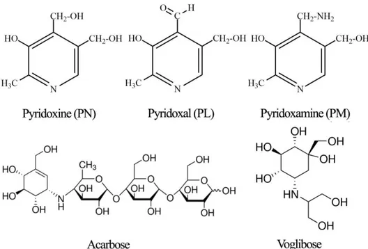
Figure 3. Structure of pyridoxine and its derivatives (pyridoxal
and pyridoxamine), and commercial a-glucosidase inhibitors
(Acarbose
®and Voglibose
®) ………..31
Figure 4. Dose dependent changes in rat intestinal α-glucosidase
(% inhibition) of pyridoxine, pyridoxal, and pyridoxamine. The
results are expressed as mean ± S.D. with three independent
experiments in triplicate ………32
Figure 5. Dose dependent changes in porcine pancreatic α
-amylase inhibitory activities (% inhibition) of pyridoxine,
pyridoxal, and pyridoxamine ……….……..……….34
Figure 7. Dose-dependent changes in rat intestinal maltase
inhibitory activities (% inhibition) of pyridoxine, pyridoxal, and
pyridoxamine ……….…………..……...37
Figure 8. Dose-dependent changes in rat intestinal glucoamylase
inhibitory activities (% inhibition) of pyridoxine, pyridoxal, and
pyridoxamine ……….…………..……...38
Figure 9. Postprandial blood glucose-lowering effects of
pyridoxine, pyridoxal, and pyridoxamine in the starch loading test
……….………...……….41
Figure 10. Postprandial blood glucose-lowering effects of
pyridoxine, pyridoxal, and pyridoxamine in the sucrose loading
test ………..42
Figure 11. Changes in body weight gains after administration of
pyridoxal. Male SD rats were free access to a high
carbohydrate-diet with pyridoxal (4 %), acarbose (0.04 %), and vehicle for 6
weeks. Each point represents mean ± standard deviation (SD)
……….55
Figure 12. Changes in food intake after administration of
carbohydrate-diet with pyridoxal (4 %), acarbose (0.04 %) or vehicle for 6 weeks.
Each
point
represents
mean
±
SD
(n
=
10)
…….………...….56
Figure 13. Changes in blood glucose levels after administration of
pyridoxal. Male SD rats were free access to a high
carbohydrate-diet with pyridoxal (4%), acarbose (0.04%) or vehicle for 6 weeks.
Each
point
represents
mean
±
SD
(n
=
10)
……….57
Figure 14. Changes in postprandial blood glucose levels in control
and pyridoxine treated groups ………...………….69
Figure 15. The extent of blood glucose increase in control and
pyridoxine treated groups ………...70
Figure 16. Pyridoxine binding to human lysosomal
alpha-glucosidase ………...……….…….92
Figure 17. Model of Pyridoxine bound to human lysosomal
Chapter 1
1.1.Diabetes mellitus
The worldwide prevalence of diabetes mellitus has risen rapidly over the past 2 decades, from an estimated 177 million cases in 2000 to 451 million in 2017. The prevalence of diabetes mellitus, especially in Korea has increased 4.8 million Koreans (13.7%), aged 30 years or older, had diabetes in 2014. In addition, nearly a quarter of Korean adults had prediabetes (Table 1). Furthermore, diabetic patients in Korea suffered from various diabetic complications and diabetes-related mortality has rapidly increased over the last decades. International diabetes federation (IDF) reported that estimated prevalence individuals will be increased until 2045 (Figure 1) [1].
Diabetes is a condition in which the body either does not produce enough insulin, or does not properly respond to insulin. Diabetes is classified as type 1 (insulin-dependent diabetes mellitus, IDDM) and type 2 (non insulin-dependent diabetes mellitus, NIDDM). Type 1 diabetic individuals can’t effectively produce insulin. The 5%-10% of patient who are diagnosed with diabetes have type 1 diabetes. Type 2 diabetic individuals have an insulin resistance condition in various tissues (muscle, liver, and adipose) and/or deficiency. The exact molecular mechanism of insulin resistance is not clearly understood. Most of individuals who are diagnosed with diabetes have type 2 diabetes [3].
Type 2 diabetes is strongly associated with diets high in calories and linked to changes in dietary pattern towards high calorie foods sweetened with maltose and
are the enzymes which digest disaccharides such as maltose, sucrose and lactose in small intestine, and inhibition of these enzymes could suppress postprandial hyperglycemia [5]. The intestinal absorption of dietary carbohydrates such as maltose and sucrose are carried out by a group of α-glucosidases which include intestinal maltase and sucrase. Inhibition of these enzymes can significantly decrease the postprandial increase of blood glucose level after a mixed carbohydrate diet and can be a key strategy in the control of diabetes mellitus.
Table 1. Diabetes prevalence in Korea [2]
Year 2012 2013 2015 2016
Date source
KNHANES KNHANES NHIS KNHANES
2007-2010 2011 2002-2013 2013-2014
Year applied to Korean census
2010 2011 2006-2013 2014 Co-working Institute CDC CDC NHIS CDC Prevalence of diabetes (%, n)§ 10.1% (3.2M) 12.4% (4.0 M) 8.0% (2.7 M) 13.7% (4.8M) Prevalence of IFG (%, n) 19.9% (6.2 M) 19.3% (6.1 M) 25% (8.4M) 24.8% (8.3 M) Awareness of diabetes (%) 73.4% 72% 70.7% No treatment for diabetes
(%)
14.1% 11% 10.8%
Treatment with insulin (%) 7.4% 11% 16.4% 8.9% Glycemic control (< 6.5%, %) 29.5% 27.9% 23.3% Hypertension (%)§§ 54.6% 62.5% 54.7 Hypertension control (%)§§ 37% 39.5% 69.1% Dyslipidemia (%)† 79.6% 49.5% 31.6% Dyslipidemia control (%)† 17.4% 49.8% Albuminuria (%)‡ 27.3% 23.9%
Chronic kidney disease (%)‡ 10.0% 12.5% Diabetes in ESRD (%)†† 38.8%
Database from January 2002 through to December 2013. §Diagnostic of diabetes, based on fasting plasma glucose (≥ 126 mg/dL), current taking of anti-diabetic medication, or previous diagnosis in 2012, and addition of HbA1c ≥ 6.5% in 2013 and 2016. In 2015, based on ICD-10 code (E11-E14) and prescription of antidiabetic medications. §§Definition and control rate of hypertension (systolic and diastolic blood pressure, mmHg), ≥ 140/90 or taking anti-hypertensive medication and < 130/80 in 2012 and 2013, ICD-10 code and taking anti-hypertensive medication in 2015, ≥ 140/90 or taking anti-hypertensive medication(s) and < 140/85 in 2016. †Definition and control rate of dyslipidemia, one or more than of following; hypercholesterolemia (total cholesterol ≥ 240 mg/dL or medication(s)), hypertriglyceridemia (TG) (≥ 150 mg/dL), hyper-low density lipoprotein (LDL)-cholesterolemia ≥ 160 mg/dL), hypo-high density lipoprotein (HDL)-cholesterolemia (<40 mg/dL for men; < 50 mg/dL for women), or taking medication, and all of all following; < 100 mg/dL for LDL-C, < 150 mg/dL for TG, and > 40 mg/dL (men)/50 mg/dL (women)for HDL-C in 2015; hypercholesterolemia (total cholesterol ≥ 240 mg/dL) and < 100 mg/dL for LDL-C in 2016. ‡The definition of albuminuria or chronic kidney disease in person with diabetes are increased albuminuria determined by albumin-creatinine ratio > 30 ug/mg of creatinine or estimated glomerular filtration rate (eGFR) < 60 mL/min/1.73 m2. GFR (mL/min/1.73 m2) = 175 × (SCr)-1.154 × (Age)-0.203 × (0.742 if female), respectively. ††Definition of ESRD, ICD-10 code of renal failure (N18, N19) or treated with renal replacement therapy. ‡‡ Diabetic neuropathy, Questionnaire (Michigan Neuropathy Screening Instrument, score ≥ 3) and 10 g monofilament exam. ∫Diabetic retinopathy, presence of at least one definite retinal blot hemorrhage
and/or microaneurysm with or without more severe lesions (hard exudates, soft exudates, intraretinalmicrovascular abnormalities, venous bleeding, new retinal vessels, fibroproliferations) Based on the diagnosis in the more severely affected eye in 2013. ∬Body mass index, 23.0~24.9 kg/m2 for overweight and ≥ 25.0 kg/m2 for obesity, overweight and obese in 2012, and obese in 2013 and 2016.
Figure 1. Total number of people living with diabetes by IDF region, 2017 and 2045 (18-99years) [1].
1.2.Trends in type 2 diabetes (T2D)
Type 2 diabetes mellitus (T2D) is a metabolic dysfunction of glucose and fat metabolism and is the most common form of diabetes mellitus[7,8]. The estimated prevalence of T2D has increased dramatically during past two decades. According to the World Health Organization (WHO), the prevalence of diabetes in the adult population around the world was 8.5% in 2014 which is about twice higher than the prevalence in 1980 [9]. Furthermore, it is estimated that more than 640 million people will be diagnosed as T2D by 2040 [10]. Increased morbidity of T2D is particularly prominent in low and middle income developing countries [10]. According to Asia database report, developing countries in Asia have increased by a factor of three to five, accounting for approximately 60% of the world's diabetes population [11,12]. The rise in T2D is largely observed in adult populations, the increase in the prevalence of T2D in adolescents is a serious new T2D epidemic in the 21st century. The prevalence of T2D in youth in 1990 considered as a rare case. However, increasing number of T2D prevalence has risen significantly within 10 years [13,14]. Although there are several contributing factors of T2D, the dramatic increase of T2D prevalence in the low middle income and youth is closely related to lifestyle changes with high caloric diets [15,19,20]. Plasma glucose concentration after meals under normal physiological conditions is regulated by the action main hormone, insulin and glucagon [16-18]. In the nursing state, islet β cells secrete insulin in response to plasma glucose levels. As a result of insulin secretion, plasma
[18-20].
On the other hand, excess carbohydrate and high fat-containing food intake causes hyperglycemia and decreased insulin sensitivity [21,22]. Prolonged hyperglycemia ultimately causes insulin resistance and insulin beta cell dysfunction in peripheral tissues. Also, the state of insulin resistance increases the secretion of inflammatory cytokines from adipose tissue and exacerbates insulin resistance in muscle and liver [23]. Many studies have demonstrated that elevated plasma free fatty acids (FFAs) impair insulin-stimulated glucose uptake in skeletal muscle as a result of increased lipolysis. Similarly, elevated FFA also stimulates hepatic gluconeogenesis and form endogenous glucose production [24]. Declining glucose utilization by peripheral tissues in the insulin resistant state increases plasma glucose levels but can also lead to postprandial hyperglycemia [25]. Therefore, high carbohydrates diets are considered as a major contributing factor of T2D.
In humans, complex carbohydrate α-1-4 glycosidic linkages are digested into disaccharides and oligosaccharides by the action of saliva and pancreatic amylase. The disaccharides are then further hydrolyzed to monosaccharides by the action of two subunits α-glucosidases, sucrase-isomaltase (SI) and maltase-glucoamylase (MG), in the apical membrane of the small intestine [26]. Monosaccharides are then absorbed into our body through glucose transporters SGLT1 and GLUT2. Therefore, one important therapeutic approach for treating postprandial hyperglycemia in T2D is to reduce digestion and absorption of dietary carbohydrates. The approach can be achieved by inhibiting carbohydrate hydrolase in the small intestine.
Acarbose
®Miglitol
®Voglibose
®1.2.1.α-Glucosidase inhibitors
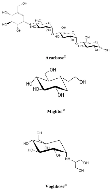
Acarbose, Voglibose, Miglitol (Figure 2) are α-glucosidase inhibitors prescribed for postprandial blood glucose control in pre-diabetic and people with diabetes [27]. Intestinal α-glucosidase inhibitors act as competitive and reversible enzyme inhibitors, which delay the intestinal digestion and absorption of carbohydrates and alleviate the rapid rise in blood sugar levels after meals (Table 2). α-Glucosidase inhibitors are considered to be safe and effective because they are excreted out of the body after inhibiting carbohydrate digesting enzymes [28,29].
Table 2. Prospective randomized placebo-controlled trials of the efficacy of α-glucosidase inhibitors on glycemic control in diet-treated patients with T2D [30-39] Study Number of Patients Dose (mg/day) Duration (weeks) Change in Plasma Glucose (mg/dL) Change in HbA1c (%) Fasting Post- prandial Hanefeld et al. [30] 94 300 24 -15 -53 -0.6 Hotta et al. [31] 37 300 24 - -65 -1.0 Santeusanio et al. [32] 62 150 16 - - -0.60 300 16 - - -0.74 Hoffman and Spengler [33] 58 300 24 -25 -40 -1.1 Chiasson et al. [34] 67 150-600 -39 -81 -0.9 Coniff et al. [35] 189 150-900 24 -16 -50 -0.77 Coniff et al. [36] 129 600 24 -22 -49 -0.71 Coniff et al. [37] 122 300 16 -27 -76 -0.78 Braun et al. [38] 86 300 24 -23 -32 -1.4 Lindstrom et al. [39] 75 300 24 -22 -52 -0.9
1.2.2.Side-effects of α-glucosidase inhibitors
The main side-effects of α-glucosidase inhibitors are gastro-intestinal abnormalities such as bloating, abdominal discomfort, diarrhea, and flatulence [40-43]. Glucosidase inhibitors also have the excessive inhibition of pancreatic α-amylase, which results in the abnormal bacterial fermentation of undigested carbohydrates in the colon. Dehghan-Kooshkghazi et al [44] reported that Acarbose-treatment (250 and 500 mg/kg body weight) in rats showed a fewer activity of starch digestion. Moreover, cecum size, colon length, and stomach weight of Acarbose-treatment groups were significantly increased but liver weight decreased in Wistar rat model [44].Therefore, identifying natural sources that inhibit carbohydrate digesting enzymes and do not result in excessive starch digestion inhibition is an important strategy to prevent the prevalence of T2D.
1.3.New Approach for α-glucosidase inhibition from soluble
vitamins
There are many types of water-soluble vitamins such as ascorbic acid, thiamine, riboflavin, niacin, carnitine, pantothenic acid, pyridoxine, biotin, folic acid, and amygdalin (Table 3). Ascorbic acid is an important antioxidant in human, capable of scavenging oxygen-derived free radicals. Ascorbic acid is structurally similar to glucose and can replace it in many chemical reactions, and thus is effective in prevention of non-enzymatic glycosylation of proteins. In addition, ascorbic acid acts as a regulator of catabolism of cholesterol to bile acid in guinea pig and has been demonstrated to be an important factor in lipid regulation [45]. Thiamine is that one key organic cofactor of thiamine diphosphate (ThDP), which is the active form of thiamine. The early evolutionary emergence of this molecule is suggested by its essential role in most, if not all, organisms and its requirement at several central points of anabolic and catabolic intermediary metabolism, such as the pentose-phosphate pathway and the Krebs cycle, also known as the tricarboxylic acid (TCA) cycle. In its varied metabolic roles, ThDP assists in making and breaking bonds between carbon and sulfur, oxygen, hydrogen, and nitrogen; and most remarkably, the breaking and making of carbon carbon bonds [46]. In association with a wide variety of apoenzymes, the flavocoenzymes derived from riboflavin are probably the most chemically versatile cofactors. They have long been known to enable a wide
decrease lipids and LDL, VLDL, increase HDL, novel non-lipid-related action of niacin to influence vascular inflammatory and oxidative processes involved in atherogenesis, and induce an adverse flush response [48]. The importance of carnitine in cardiac metabolism and function is emphasized by the growing number of studies demonstrating a close association between systemic and myopathic carnitine deficiency and both hypertrophic and congestive cardiomyopathies, which in some cases can be reversed by carnitine treatment. Thus, the observation that diabetic hearts have a deficiency in the total carnitine pool and the correlation between carnitine deficiency and cardiomyopathy suggested that carnitine therapy may ameliorate alterations in cardiac contractile performance seen during diabetes by decreasing accumulation of lipid intermediates [49]. Pantothenic acid (PA) has been shown to be of interest in improving surgical wound healing. A great deal of information is now available on PA steady state, body pool, urinary excretion and availability in humans. Information is however, scanty on the effect of both PA supplementation and deficiency on wound healing [50]. Pyridoxine has been shown to be important for normal cognitive function and in lowering the incidence of coronary heart disease among the elderly. In addition, pyridoxine supplementation has been shown to reduce diabetic complications and the incidences of neurodegenerative diseases in varying degrees [51]. The biotin carboxylase involved in four components of the main steps of the vitamin as glucose, amino acids, fatty acid containing sulfur [52]. Folate (as methyltetrahydrofolate) is required for methylation of homocysteine to methionine catalysed by methionine synthase with methylcobalamin as a cofactor. 5’-deoxyadenosylcobalamin is an essential cofactor in the enzymatic conversion of methylmalonyl-CoA to succinyl-CoA.5 The
conversion of methylmalonyl-CoA to Methylmalonic acid results in an elevated serum Methylmalonic acid concentration in cobalamin deficiency [53]. Recently, a new prodrug system, known as antibody-guided enzyme nitrile therapy (AGENT), has been developed. This system exploits the activation of amygdalin, a naturally occurring cyanogenic glucoside, instead of using a modified chemotherapeutic agent. The hydrolysis of amygdalin by the enzyme β-glucosidase results in the release of the powerful metabolic poison cyanide [54].
The mutual relation between diabetes and water-soluble vitamins and minerals is characterized by a high degree of reciprocity. Chronic uncontrolled hyperglycemia can cause significant alterations in the status of these nutrients, and conversely, some of these substances, especially those that have been characterized as micronutrients, can directly modulate glucose homeostasis. Especially, case of ascorbic acid and pyridoxine have been known to effect of antidiabetics. Ascorbic acid is an excellent hydrophilic antioxidant in plasma because it disappears faster than other antioxidants when plasma is exposed to ROS. It is widely consumed in food. There is a report showing that oral administration of ascorbic acid reduces blood sugar levels and improves glucose tolerance test in STZ induced diabetic rats [55]. In the other study have reported that intraperitoneal administration of ascorbic acid decreases the levels of thiobarbituric acid reactive substances (TBARS) and improves enzymic and nonenzymic antioxidants in STZ induced diabetic rats [55]. Another report showed that ascorbic acid reduces plasma cholesterol and
high oxidative stress. The major role of l-carnitine is in the transport of long-chain fatty acids into the mitochondrial matrix for b-oxidation and subsequent energy production, and it has been reported to decline in aging mice and humans. Of late, the role of carnitine as an antioxidant has been implicated in adriamycin-induced membrane damage, diphtheria toxins, and ischemia-reperfusion injury. Studies on the antioxidant effect of carnitine on aging are sparse and yet to be elucidated. Because nutritional supplementation with antioxidants has been found to delay the onset of aging and ageassociated degenerative diseases, a study of l-carnitine supplementation (a naturally occurring and conditionally essential nutrient) was undertaken [58].
Table 3. Dietary Reference Intakes for Koreans (KDRIs) Vitamin Tolerance (19~29 man UL1) Tolerance (19~29 man RNI2) Effect Vitamin C (Ascorbic
acid) 2,000 mg/d 100 mg/d Antioxidant effect Vitamin B₁ (Thiamine) - 1.2 mg/d Beriberi efficacy Vitamin B₂ (Riboflavin) - 1.5 mg/d Acceleration of
growth Vitamin B₃ (Niacin)
35 mg NE/d (nicotinic acid)
16 mg NE/d A medical action Vitamin B₄
(L-carnitine) 2,000 mg/d 20~200 mg/d Antiaging Vitamin B₅
(Pantothenic acid) 5 mg/d(AI
3) - Solution to stress Vitamin B₆ (Pyridoxine) 100 mg/d (~67 mg/d*) 1.5 mg/d Diabetes prevention (Help to maintain normal conc. of homocysteine and utilize protein/A.A*)
Vitamin B₇ (Biotin) 30 µg/d(AI3) - Hair loss, gray hair
prevention Vitamin B₉ (Folic
acid) 1,000 µg DFE/d 400 µg DFE/d Hematogenesis Vitamin B₁₇
(Amygdalin) - - Anticancer effect
Ul1: Tolerate Upper intake Limit level
1.3.1.Pyridoxine
Vitamin B6 is widely distributed in foods in both its free and bound forms. Cooking, storage, and processing losses of vitamin B6 vary and in some foods may be more than 50% depending on the form of vitamin present in the food. Plant foods lose the least during processing, as they contain mostly pyridoxine, which is far more stable than the pyridoxal or pyridoxamine found in animal foods. For example, milk can lose 30–70% of its vitamin B6 content when dried [60]. Vitamin B6 is found in the germ and aleurone layer of grains, and milling results in the reduction of this vitamin in white flour. The heating that occurs before most freezing and canning processes are other methods that may result in the loss of vitamin B6 in foods [60]. Vitamin B6 is absorbed in the jejunum and ileum by passive diffusion. With the capacity for absorption being so great, animals are able to absorb quantities much greater than necessary for physiological demands. The absorption of pyridoxal phosphate and pyridoxamine phosphate involves their dephosphorylation catalyzed by a membrane-bound alkaline phosphatase. Those products and nonphosphorylated forms in the digestive tract are absorbed by diffusion, which is driven by trapping of the vitamin as 5′-phosphates through the action of phosphorylation (by a pyridoxal kinase) in the jejunal mucosa. The trapped pyridoxine and pyridoxamine are oxidized to pyridoxal phosphate in the tissue [60]. The products of vitamin B6 metabolism are excreted in the urine, the major product of which is 4-pyridoxic acid. An estimated 40–60% of ingested vitamin B6 is oxidized to 4-pyridoxic acid. Several studies have shown that 4-pyridoxic acid is undetectable in the urine of vitamin B6-deficient subjects, making it a useful clinical marker to assess the vitamin B6 status
of an individual [61]. Other products of vitamin B6 metabolism excreted in the urine when high doses of the vitamin have been given include pyridoxal, pyridoxamine, pyridoxine and their phosphates. A small amount of vitamin B6 is also excreted in the feces [61].
Many studies of pyridoxine have reported the existence of pyridoxine deficiency in both type 1 and type 2 diabetic patients and experimental diabetes. Clinical trials have demonstrated that supplementation with pyridoxine has beneficial effects on the clinical symptoms of neuropathy and retinopathy in diabetic patients. Supplementation with pyridoxine has also been shown to lower blood glucose levels in streptozocin-treated diabetic animals and glycosylated hemoglobin levels in type 2 diabetic patients. Recent in vitro studies have shown that pyridoxamine can inhibit formation of glycation end products [57,59]. However, pyridoxine and its derivatives have not been thoroughly evaluated for their possible blood glucose lowering effect and there is no study suggesting the exact mechanism of vitamin B6 for this specific health benefit.
1.4.Objectives
I) To investigate the enzyme inhibition pattern and evaluate anti-hyperglycemic effects of several forms of vitamin B6 in vitro and in vivo models.
Chapter 2
The Postprandial Anti-Hyperglycemic Effect of
Pyridoxine and Its Derivatives Using In Vitro and In
Vivo Animal Models
Abstract
In the current study, I investigated the inhibitory activity of pyridoxine, pyridoxal, and pyridoxamine, against various digestive enzymes such as α-glucosidases, sucrase, maltase, and glucoamylase. Inhibition of these enzymes involved in the absorption of disaccharide can improve postprandial hyperglycemia due to a carbohydrate-based diet. Pyridoxal (4.14 mg/mL of IC50) had the highest rat intestinal α-glucosidase inhibitory activity, followed by pyridoxamine and pyridoxine (4.85 and 5.02 mg/mL of IC50, respectively). Pyridoxal demonstrated superior inhibition against maltase (0.38 mg/mL IC50) and glucoamylase (0.27 mg/mL of IC50). In addition, pyridoxal showed significant higher α -amylase inhibitory activity (10.87 mg/mL of IC50) than that of pyridoxine (23.18 mg/mL of IC50). This indicates that pyridoxal can also inhibit starch hydrolyzing by pancreatic α -amylase in small intestine. Based on these in vitro results, the deeper evaluation of the anti-hyperglycemic potential of pyridoxine and its derivatives using Sprague-Dawley (SD) rat models, was initiated. The postprandial blood glucose levels were tested two hours after sucrose/starch administration, with and without pyridoxine and its derivatives. In the animal trial, pyridoxal (p < 0.05) had a significantly reduction to the postprandial glucose levels, when compared to the control. The maximum blood glucose levels (Cmax) of pyridoxal administration group were decreased by about 18% (from 199.52 ± 22.93 to 164.10 ± 10.27, p < 0.05) and 19% (from 216.92 ± 12.46 to 175.36 ± 10.84 p < 0.05) in sucrose and starch loading tests,
water-soluble vitamin pyridoxine and its derivatives can decrease blood glucose level via the inhibition of carbohydrate-hydrolyzing and absorption-linked enzymes. Therefore, pyridoxal may have the potential to be used as a food ingredient for the prevention of prediabetes progression to type 2 diabetes.
Key words: pyridoxine, anti-hyperglycemia, postprandial, α-glucosidase,
inhibition
2.1. Introduction
Type 2 Diabetes Mellitus (T2DM), a common disorder of glucose metabolism, is linked to insulin resistance and high calorie diets. T2DM is associated with changes in dietary pattern towards high calorie sweetened foods with disaccharides such as maltose and sucrose [62]. T2DM accounts for the vast majority of diabetes cases in adults [63], and expecting to reach 366million diabetes patients by the year 2030 [64]. Specifically in the United States, it is expected that 1/3 of all adults will have diabetes by 2030 [65]. High blood glucose levels are typical in type 2 diabetes patients, and more specifically this is characterized by a postprandial increase of glucose levels following food consumption, due to digestion of carbohydrates by pancreatic α-amylase and small intestinal α-glucosidases, resulting in elevated liberation of glucose [66]. A strategy to reduce high blood glucose levels following meal administration, is through the inhibition of enzymes that hydrolyze carbohydrates, in the small intestine [67, 68]. Therefore, commercial available α-glucosidase inhibitors, Acarbose® (Bayer AG, Leverkusen, Germany), Voglibose® (Takeda, Tokyo, Japan), and Miglitol® (Bayer AG, Leverkusen, Germany) are generally prescribed either alone, or in combination with insulin sensitizers in T2DM patients, clinically. Pyridoxine (Vitamin B6) has been used for managing normal cognitive function and in lowering the incidence of coronary heart disease (CHD) among the seniors [69-71]. In addition, vitamin B6 administration has been shown to decrease complications of diabetes and the incidence of neurodegenerative
diabetic patients [75,76]. When the effect of vitamin B6 was evaluated on STZ-mice, a blood glucose level reduction was observed [77], and in type 2 diabetes patients HbA1c levels were reduced [78]. Other in vitro evaluations of the vitamin B6 derivative pyridoxamine have indicated the effect on the inhibition of formation of AGE products [79]. However, pyridoxine and its derivatives have not been thoroughly evaluated for their possible blood glucose lowering effect and there is no study suggesting the exact mechanism of vitamin B6 for this specific health benefit. Therefore, the aim of this study is to investigate mode of action and effect of pyridoxine and its derivatives on postprandial hyperglycemia. To determine the above, inhibitory activities of pyridoxine, pyridoxal, and pyridoxamine are investigated against the α-glucosidase and α-amylase (anti-hyperglycemia potential). Anti-hyperglycemic effect of these compounds was also evaluated in Sprague-Dawley (SD) rat models. This work will contribute towards the understanding of the activity and the mechanism of action of vitamin B6 and its derivatives, specifically towards the management and prevention of type 2 diabetes.
2.2. Materials and Methods
2.2.1. Materials
Water-soluble pyridoxine and its derivatives was used from reagent companies in Sigma-Aldrich (Sigma-Aldrich Co. LLC., St. Louis, MO, USA), Daejung (Daejung Chemicals & Metals Co., Ltd., Gyeonggi-do, Korea), Duksan (Duksan Pure Chemical Co., Ltd., Gyeonggi-do, Korea), and Junsei (Junsei Chemical Co., Ltd., Tokyo, Japan). Rat intestinal acetone powder, p-nitrophenyl-α-D-glucopyranoside (pNPG), porcine pancreatic α-amlyase enzyme powder, starch, sucrose, and maltose were purchased from Sigma-Aldrich (St. Luis, MO, USA). Unless noted, all chemicals were purchased from Sigma-Aldrich (St. Luis, MO, USA).
2.2.2. Carbohydrate-Hydrolyzing Enzyme Inhibition Assay
2.2.2.1. Rat Intestinal α-Glucosidase Inhibition Assay
The rat intestinal α-glucosidase assay was administered as per the method of Kwon et al. [80] with slight modification. A total of 1 g of rat-intestinal acetone powder was suspended in 3 mL of 0.1 M sodium phosphate buffer (pH 6.9), and the suspension was sonicated 12 times for 30 s at 4 ◦C. After centrifugation (10,000x g, 30 min, 4 ◦C), the resulting supernatant was used for the assay. Sample solution (50 µL) and 0.1 M phosphate buffer (pH 6.9, 100 µL) containing α -glucosidase
(Sigma-The reaction mixtures were incubated at 37 ◦C for 30 min. Before and after incubation, absorbance was read at 405 nm and compared to a control, which had 50 µL of buffer solution in place of the extract by micro-plate reader (SUNRISE; Tecan Trading AG, Salzburg, Austria). The α-glucosidase inhibitory activity was expressed as inhibition % and was calculated as follows:
Inhibition(%) = (
∆𝐴 405 (𝐶𝑜𝑛𝑡𝑟𝑜𝑙) − ∆𝐴 405 (𝑠𝑎𝑚𝑝𝑙𝑒)
∆𝐴 405 (𝐶𝑜𝑛𝑡𝑟𝑜𝑙)
) × 100
2.2.2.2.
Porcine Pancreatic α-Amlyase Inhibition Assay
The porcine pancreatic α-amlyase inhibition assay was administered as per the method of Kwon et al. [81] Sample solution (200 µL) and 0.02 M sodium phosphate buffer (pH 6.9 with 0.006 M sodium chloride, 500 µL) containing α-amylase solution (0.5 mg/mL, 5.0 MU/mL) were incubated at 25 ℃ for 10 min. After preincubation, 500 µL of a 1% starch solution in 0.02 M sodium phosphate buffer was added. The reaction mixture was then incubated at 25 ℃ for 10 min. The reaction was stopped with 1.0 mL of dinitrosalicylic acid (DNS). The reaction mixture was then incubated in a boiling water bath for 5 min and cooled to room temperature. The reaction mixture at 540 nm with ELISA microplate reader (SUNRISE; Tecan Trading AG, Saltzburg, Austria).
2.2.2.3. Maltase, Sucrase, and Glucoamylase Inhibition Assay
The crude enzyme solution prepared from rat intestinal acetone powder (Sigma-Aldrich Co. St. Louise, MO, USA) was used as the small intestinal maltase, sucrose, and glucoamylase, showing specific activities of 0.70, 0.34, and 0.45
units/mL respectively. Rat intestinal acetone powder (1.0g) was suspended in 3 mL of 0.1 M sodium phosphate buffer, and the suspension was sonicated 12 times for 30 s at 4 ℃. After centrifugation (10,000x g, 30 min, 4 ℃), the resulting supernatant was used for the assay. Maltase, sucrose, and glucoamylase inhibitory activity were assay by modifying a method developed by Dahlqvist [82]. The inhibitory activity was determined by incubating a solution of an enzyme (50 µL), 0.1 M phosphate buffer (pH 7.0, 100 µL) containing 0.4 mg/mL sucrose or maltose or 1% soluble starch, and a solution(50 µL) with various concentrations of sample solution (between 0.05 and 1.0 mM) at 37 ℃ for 30 min. The reaction mixture was heated in a boiling water bath to stop the reaction for 10 min, and the amount of liberated glucose was then measured by the glucose oxidase [83]. The inhibitory activity was calculated from the formula as follows. Inhibition (%) = (C − T)/C × 100, where C is the enzyme activity without inhibitor, and T is the enzyme activity with inhibitor.
2.2.3. The Sucrose/Starch Loading Test
Effect on hyperglycemia induced by carbohydrate loads in Sprague-Dawley (SD) rats was determined by an inhibitory action of water-soluble vitamins and Acarbose on postprandial hyperglycemia. All animal procedures were approved by Institutional Animal Care and Use Committee (IACUC) of the Hannam University (Approval number: HNU2017-003). Four-week-old male SD rats were purchased from Raon Bio Co. (Gyeonggi-do, Korea) and fed a solid diet (Raon Bio
2.0 g/kg of sucrose or starch were orally administrated concurrently with 0~100 mg/kg inhibitors (water-soluble vitamins and Acarbose). The blood samples were then taken from the tail after administration and blood glucose levels were measured at 0, 0.5, 1, 2, and 3 h. The glucose level in blood was determined by glucose oxidase method and compared with that of the control group, which had not taken the inhibitors.
2.2.4. Statistical Analysis
All data are presented as mean ± Standard deviation (SD). Statistical analysis was carried out using statistical package SPSS 10 (Statistical Package for Social Science; SPSS Inc., Chicago, IL, USA), and the significance of each group was verified with the analysis of one-way analysis of variance (ANOVA) followed by Duncan’s test of p < 0.05.
2.3. Result
2.3.1. Rat Intestinal α-Glucosidase Inhibitory Activity of Water-Soluble
Vitamins
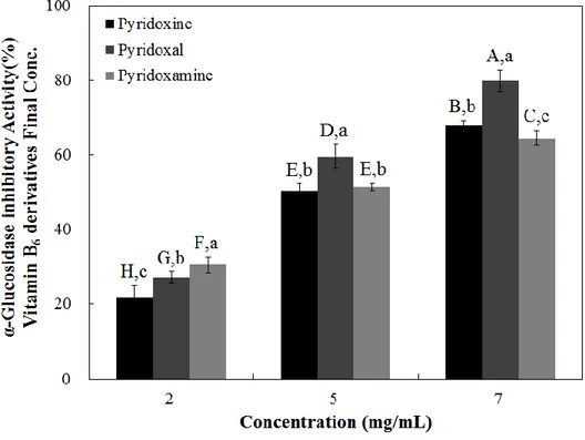
α -Glucosidase inhibitors, such as Acarbose® and Voglibose® , delay the digestion of oligosaccharide and disaccharide to monosaccharide by inhibiting α-glucosidases on the small intestinal brush-border, and reduce the rate of glucose absorption [67]. Inhibition of these enzymes involved in the absorption of disaccharide can improve postprandial hyperglycemia due to the consumption of carbohydrate-based diet. As a result, administration of such inhibitors prior to meal consumption result in reduced postprandial blood glucose concentrations. To screen the glucosidase inhibitory effects of vitamin B6 and its derivatives, I examined α-glucosidase activity using rat acetone powder (Figure 3). Pyridoxal exhibited the highest inhibitory effect among the tested compounds, resulting in a 79.83 % inhibition at the highest tested dose (7 mg/mL) (Figure 4). Pyridoxamine and pyridoxine appeared to have similar inhibitory activities, but showed significantly less activity compared with pyridoxal (Figure 4). When the IC50 values were calculated, I observed that pyridoxal had the lowest value (4.15 mg/mL), while pyridoxine had the highest (5.02 mg/mL) (Table 4). My observations suggest that pyridoxal has the highest inhibitory potential against α-glucosidase while pyridoxine had the lowest (Figure 4 and Table 4).
Figure 3. Structure of pyridoxine and its derivatives (pyridoxal and pyridoxamine), and commercial a-glucosidase inhibitors (Acarbose® and
Figure 4. Dose dependent changes in rat intestinal α-glucosidase (%
inhibition) of pyridoxine, pyridoxal, and pyridoxamine. The results are
expressed as mean ± S.D. with three independent experiments in
triplicate. Different corresponding letters indicate significant differences at p
< 0.05 by Duncan’s test. The first letters in uppercase (
A–H) indicate significant
differences among all samples. The second letters in lowercase (
a– c) are
different among types of vitamin within the same concentration.
2.3.2. Porcine pancreatic α-amlyase inhibition assay
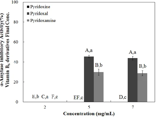
When the pancreatic α-amlyase inhibitory activity was evaluated, I observed no inhibitory effect at 2 mg/mL concentrations in all samples. Pyridoxine showed no inhibitory effects, even at the highest tested dose (7 mg/mL) (Figure 5). Pyridoxal and pyridoxamine showed α-amylase inhibitory effects at the two highest tested doses (5 and 7 mg/mL) with pyridoxal appearing to have a higher inhibitory effect (Figure 5). When the IC50 values were calculated, I observed that pyridoxal had the lowest value (12.92 mg/mL), while pyridoxine had the highest (23.17 mg/mL) (Table 4). My observations are similar to the previous results, suggesting that pyridoxal has the highest inhibitory potential against α-amylase, while pyridoxine had the lowest (Figure 5 and Table 4).
Figure 5. Dose dependent changes in porcine pancreatic α -amylase inhibitory activities (% inhibition) of pyridoxine, pyridoxal, and pyridoxamine. The results
are expressed as mean ± S.D. with three independent experiments in triplicate. Different corresponding letters indicate significant differences at p < 0.05 by Duncan’s test. The first letters in uppercase (A–H) indicate significant differences
among all samples. The second letters in lowercase (a– c) are different among types
2.3.3. Maltase, Sucrase, and Glucoamylase Inhibition Assay
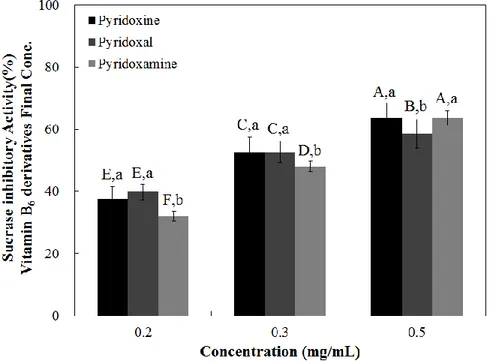
Since α-glucosidase inhibitory effects of the vitamin B6 group was shown, I examined whether vitamin B6 group could selectively inhibit enzymes in the small intestine, namely sucrase, maltase, and glucoamylase. The sucrase inhibitory effect of the tested compounds revealed that all of them had similar inhibitory effects (Figure 6, Table 4). All samples demonstrated maltase inhibitory activity in a dose-dependent manner (Figure 5) and pyridoxal resulted in the highest maltase inhibitory activity at all tested doses (0.5 mg/mL 48.39 %, 1 mg/mL 70.10 %, and 2 mg/mL 83.29 %) (Figure 7). Similar to maltase inhibitory activity, all tested vitamin B6 structures resulted in dose-dependent glucoamylase inhibition (Figure 8), and pyridoxal had the highest inhibitory effect at all tested doses (42.84% at 0.2 mg/mL, 66.07% at 0.5 mg/mL, and 78.59% at 1 mg/mL) (Figure 8). Based on these dose-dependent results, half maximal concentration (IC50) of samples in vitro system was shown in Table 4. Pyridoxal yielded to the lower IC50 value for maltase and glucoamylase (0.38 and 0.27 mg/mL, respectively), suggesting higher inhibition potential. Against sucrase all tested samples yielded similar and not significant different IC50 values (Table 4), suggesting that all tested compounds have similar inhibitory potential against this carbohydrate-hydrolyzing enzyme.
Figure 6. Dose-dependent changes in rat intestinal sucrase inhibitory activities (% inhibition) of pyridoxine, pyridoxal, and pyridoxamine. The results are
expressed as mean ± S.D. with three independent experiments in triplicate. Different corresponding letters indicate significant differences at p < 0.05 by Duncan’s test. The first letters in uppercase (A–F) indicate significant differences among all samples.
The second letters in lowercase (a–c) are different among types of vitamin with in the
Figure 7. Dose-dependent changes in rat intestinal maltase inhibitory activities (% inhibition) of pyridoxine, pyridoxal, and pyridoxamine. The results are
expressed as mean ± S.D. with three independent experiments in triplicate. Different corresponding letters indicate significant differences at p < 0.05 by Duncan’s test. The first letters in uppercase (A–H) indicate significant differences among all samples.
The second letters in lowercase (a–c) are different among types of vitamin with in the
Figure 8. Dose-dependent changes in rat intestinal glucoamylase inhibitory activities (% inhibition) of pyridoxine, pyridoxal, and pyridoxamine. The results
are expressed as mean ± S.D. with three independent experiments in triplicate. Different corresponding letters indicate significant differences at p < 0.05 by Duncan’s test. The first letters in uppercase (A–G) indicate significant differences
among all samples. The second letters in lowercase (a–c) are different among types of
Table 4. The half maximal inhibitory concentration (IC50) of pyridoxine and its
derivatives on rat intestinal α-glucosidase, sucrase, maltase, glucoamylase, and porcine pancreatic α-amylase activities
IC50 (mg/mL) Rat intestinal α-glucosidase Porcine pancreatic α-amylase Rat intestinal sucrase Rat intestinal maltase Rat intestinal glucoamylase Pyridoxine 5.02 ± 0.05a 23.17 ± 0.29a 0.32 ± 0.03a 0.87 ± 0.08a 0.63 ± 0.04a Pyridoxal 4.15 ± 0.17b 12.92 ± 1.22c 0.32 ± 0.03a 0.37 ± 0.13b 0.26 ± 0.08b Pyridoxamine 4.85 ± 0.06a 14.93 ± 0.96b 0.35 ± 0.01a 1.08 ± 0.10a 0.60 ± 0.06a
2.3.4. Starch/Sucrose Loading Test
A sucrose loading test using SD rat models was established, during which I evaluated the changes in the postprandial blood glucose levels, as described in the materials and methods. In the pyridoxal-treated group with starch, the blood glucose level was 175.36 ± 10.84 mg/dL at 30 min after administration. Compared to the pyridoxine-treated group (201.78 ± 18.99 mg/dL) and starch group (216.92 ± 12.46 mg/dL) (Figure 9), pyridoxal-treated group suppresses the rising of plasma glucose level by 26.41 mg/dL, and 41.56 mg/dL, respectively, after administration. At 30 min after administration, I found that the pyridoxal-treated group showed the lowest increase in blood glucose level (79.78 ± 10.84 mg/dL), which was 27.91 mg/dL less than that of the pyridoxine-treated group (107.69 ± 18.99 mg/dL) and 44.45 mg/dL less than that of the starch group (124.23 ± 12.46 mg/dL).
Among the three tested compounds, change of blood glucose level in the pyridoxal-treated group with sucrose was increased by 64.88 ± 12.06 mg/dL at 30 min after administration (Figure 10). This is lower than the pyridoxine-treated group (92.86 ± 10.10 mg/dL) and the sucrose group (101.27 ± 20.18 mg/dL) by 27.98 mg/dL and 36/39 mg/dL, respectively. At 30 min after administration, I found that the blood glucose level of the pyridoxal-treated group was 164.74 ± 12.06 mg/dL, which was 27.76 mg/dL less than that of the pyridoxine-treated group (192.50 ± 10.10 mg/dL) and 35.89 mg/dL less than that of the sucrose-treated group (200.63 ± 20.18 mg/dL).
Figure 9. Postprandial blood glucose-lowering effects of pyridoxine, pyridoxal, and pyridoxamine in the starch loading test. After fasting for 24 h, six-week-old,
male SD rats were orally administered with a starch solution (2.0 g/kg) with or without samples (pyridoxine, pyridoxal, pyridoxamine, and Acarbose). Each point represents mean ± S.D. (n = 5). The letters in lowercase (a–d) indicate significant
Figure 10. Postprandial blood glucose-lowering effects of pyridoxine, pyridoxal, and pyridoxamine in the sucrose loading test. After fasting for 24 h, six-week-old,
male SD rats were orally administered with a sucrose solution (2.0 g/kg) with or without samples (pyridoxine, pyridoxal, pyridoxamine, and Acarbose). Each point represents mean ± S.D (n = 5). The letters in lowercase (a–c) indicate significant
2.3.5.Pharmacodynamic Parameters
Pharmacodynamic parameters of the sucrose and starch loading test are shown in Table 5. In terms of Tmax, there is no significance between sucrose/starch and vitamin B6-treated groups. In contrast, vitaminB6-treated groups resulted in significantly decreased Cmax and AUCt values (both with sucrose and starch loading), but less effective than the Acarbose group. In sucrose loading, pyridoxal and pyridoxamine resulted to the highest reduction both in terms of Cmax and AUCt (Table 5). In the case of starch loading, pyridoxal resulted in the greatest reduction of Cmax, while all tested compounds had similar AUC values (Table 5). In terms of absolute values, the maximum blood glucose levels (Cmax) of the pyridoxal administration group were decreased by about 18% (from 199.52 ± 22.93 to 164.10 ± 10.27, p < 0.05) and 19 % (from 216.92 ± 12.46 to 175.36 ± 10.84, p < 0.05) in the sucrose and starch loading tests, respectively, when compared to the control in the pharmacodynamics study. My findings suggest that vitamin B6 and its derivatives can inhibit the breakdown of disaccharide to monomer in brush-border and suppress the entrance of blood glucose into the bloodstream.
Table 5. Pharmacodynamic (PD) parameters of control, pyridoxine, pyridoxal, pyridoxamine, and Acarbose in SD rats ingested with starch or sucrose
Groups
PD parameters
Cmax(mg/dL) Tmax(h) AUCt(h∙mg/dL)
BGL (mg/dL) Starch 2.0g/kg 216.92±11.58d 0.50±0.00a 299.02±12.10c Acarbose 0.005 g/kg 126.60±9.44a 0.50±0.00a 228.88±13.09a Pyridoxine 0.1 g/kg 200.00±20.89cd 0.50±0.00a 274.28±15.62b Pyridoxal 0.1 g/kg 172.67±11.45b 0.50±0.00a 268.27±8.80b Pyridoxamine 0.1 g/kg 191.54±13.70c 0.50±0.00a 275.10±7.31b BGL (mg/dL) Sucrose 2.0 g/kg 199.52±22.93c 0.50±0.00a 303.57±14.71c Acarbose 0.005 g/kg 126.92±6.87a 0.86±0.24b 237.94±8.82a Pyridoxine 0.1 g/kg 192.48±9.03c 0.50±0.00a 290.64±15.55c Pyridoxal 0.1 g/kg 164.10±10.27b 0.50±0.00a 272.99±5.96b Pyridoxamine 0.1 g/kg 174.50±18.29b 0.50±0.00a 269.14±13.40b
The results are expressed as mean ± S.D. The letters in lowercase (a–d)
indicate statistically significant differences between groups one-way ANOVA
followed by Duncan’s test of p < 0.05.
2.4. Discussion
In this manuscript, I am presenting the first report of vitamin B6 and its derivatives, for the potential management of type 2 diabetes, via the inhibition of small-intestinal α -glucosidases. The significant in vitro and in vivo findings indicate that pyridoxine (vitamin B6) has α -glucosidase inhibitory activity, resulting in reduced postprandial blood glucose levels following sucrose and starch ingestion.
The purpose of this research was to evaluate the mode of action and type 2 diabetes-relevant effect of vitamin B6 and its derivatives. More specifically, I evaluated the effect on in vivo postprandial hyperglycemia via the in vitro inhibition of carbohydrate-hydrolyzing enzymes. Vitamin B6 (pyridoxine, pyridoxal, and pyridoxamine) forms are readily absorbed by passive diffusion in the jejunum and ileum [84], while alpha-glucosidases are expressed and located in the duodenum [85]. This studies suggest that the blood glucose lowering effect of pyridoxine is possibly due to the α-glucosidase inhibitory activities. Furthermore, this studies show that pyridoxine, one of essential vitamins has high activity of enzyme inhibition (on sucrase, maltase, and glucoamylase). These results could be attributed to the structural similarities between pyridoxine and Acarbose and Voglibose, the pharmacological agents used for the inhibition of carbohydrate-hydrolyzing enzymes (Figure 3). Compared to Acarbose, pyridoxine resulted in an enhanced inhibitory effect against α-glucosidases, but the inhibitory effect against α-amylase was significantly reduced, suggesting fewer side-effects.
When the postprandial blood glucose levels in adult, normal SD rats were measured, I observed that pyridoxine administration resulted in a significant
reduction in postprandial blood glucose levels after 30 min (Figures 9 and 10). More specifically, I observed a blood glucose reduction around 18 % at 30 min when pyridoxal administration was compared to control. Taking into consideration that pyridoxine is present in a wide variety of food products, knowledge of this additional health benefit of pyridoxine can assist in the development of efficacious anti-hyperglycemia supplements and food products.
Chapter 3
Effect of Long-Term Supplementation Pyridoxal on
the Blood Glucose level and Weight Gaining in
Abstract
I have previously reported that Pyridoxine and its derivatives exert anti-diabetic effects by lowering post-prandial hyperglycemia via inhibition of carbohydrate hydrolyzing enzymes in normal Sprague-Dawley (SD) rats. In the present study I extended my recent findings to evaluate whether long-term supplementation of Pyridoxal lowers blood glucose level in SD rat model. The SD rats were randomly assigned to high-carbohydrate diets (66.1 % corn starch) with and without Pyridoxal (4% in the diet) for 36 days. Changes in body weight, blood glucose level, and food intake were measured daily for 36 days. Dietary supplementation of pyridoxal resulted in a significant decrease of blood glucose level (p < 0.001) and body weight gaining (p < 0.001). The level of HbA1c, a better indicator of plasma glucose concentration over prolonged periods of time, was also significantly decreased for 5-week period (p < 0.001). Dietary treatment of Acarbose® (0.04 % in diet), a positive control, also significantly alleviated the level of blood glucose, HbA1c, and body weight. These results indicate that pyridoxal decreases weight gaining and improves postprandial hyperglycemia by suppressing glucose absorption as well as decreasing HbA1c level.
Key words: type 2 diabetes; pre-diabetes; blood glucose; α-glucosidase inhibition;
![Table 1. Diabetes prevalence in Korea [2]](https://thumb-ap.123doks.com/thumbv2/123dokinfo/4723103.9373/23.808.125.683.113.958/table-diabetes-prevalence-in-korea.webp)
![Figure 1. Total number of people living with diabetes by IDF region, 2017 and 2045 (18-99years) [1]](https://thumb-ap.123doks.com/thumbv2/123dokinfo/4723103.9373/26.808.138.668.78.424/figure-total-number-people-living-diabetes-region-years.webp)
![Table 2. Prospective randomized placebo-controlled trials of the efficacy of α- α-glucosidase inhibitors on glycemic control in diet-treated patients with T2D [30-39] Study Number of Patients Dose (mg/day) Duration (weeks) Change in Plasma Glucose](https://thumb-ap.123doks.com/thumbv2/123dokinfo/4723103.9373/31.808.126.682.197.826/prospective-randomized-controlled-glucosidase-inhibitors-glycemic-patients-duration.webp)