대한내분비외과학회지:제 11 권 제 1 호
□ 증 례 □ Vol. 11, No. 1, March 2011
31 Correspondence: Kee-Hyun Nam, Department of Surgery, Yonsei
University College of Medicine C.P.O. Box 8044, 250 Seongsan- no, Seodaemun-gu, Seoul 120-752, Korea
Tel: +82-2-2228-2100, Fax: +82-2-313-828925 E-mail: khnam@yuhs.ac
접수일:2010년 9월 29일, 게재승인일:2011년 1월 1일
A Neurogenic Tumor as a Rare Differential Diagnosis of a
Perithyroidal Masses
Jae Hyun Park, M.D., Choong Bai Kim, M.D., Hyae Min Jeon, M.D.
1, Sang-Wook Kang, M.D., Jong Ju
Jeong, M.D., Yong Sang Lee, M.D., Kee-Hyun Nam, M.D., Hang Seok Chang, M.D., Woong Youn Chung,
M.D. and Cheong Soo Park, M.D.
Departments of Surgery, 1Pathology, Yonsei University College of Medicine, Seoul, Korea
We report here on a case of a neurogenic tumor of the neck with an uncertain origin on the preoperative evalu-ation. A 67-year-old woman with a palpable mass in the left side of the neck was referred to our hospital. The mass had slowly grown over 7 years and her dyspnea had gradu-ally become more severe over the recent 6 months. Computerized tomography and magnetic resonance imaging showed an 8 cm sized solid mass that abutted the trachea and the esophagus without invasion, but the origin of the mass was not clearly identified. During surgical exploration, we identified that the tumor was located in the esophageal muscle layer. Immunohistochemical staining revealed that the tumor cells were positive for S-100 protein, which con-firmed a diagnosis of schwannoma. (Korean J Endocrine Surg 2011;11:31-34)
Key Words: Neural tumors, Esophagus, Schwannoma, Thy-roid, S-100 protein
INTRODUCTION
Primary neural tumors in the cervical region may originate from several structures of neck.
These lesions can then present as thyroid nodules and it may be virtually impossible to detect the extra thyroidal origin of these lesions based on either clinical or radiologic findings.(1) Moreover, resection of these lesions may be challenging, and the risks of misdiagnosis and of using an incorrect surgical approach are high. Furthermore, such errors can result in high morbidity.(2) Esophageal schwannoma is a rare neoplasm, which is difficult to diagnose based on endoscopic or radiologic evaluations. The
au-thors report a neurogenic tumor of the neck with uncertain origin in preoperative evaluation.
CASE REPORT
A 67-year-old woman with a palpable mass in the left side of the neck and a pathologic finding of a neurogenic tumor based on ultrasound-guided core needle biopsy findings was referred to our hospital. According to the patient, the mass had grown slowly over 7 years and dyspnea had gradually become more severe over recent 6 months.
Approximately 6 years before this presentation, fine-needle as-piration biopsy on a palpable mass in the left side of the neck performed at a community hospital showed benign looking fol-licular cells. Her medical and family histories were unremarkable, and a physical examination revealed her to be well nourished without any neurogenic symptoms.
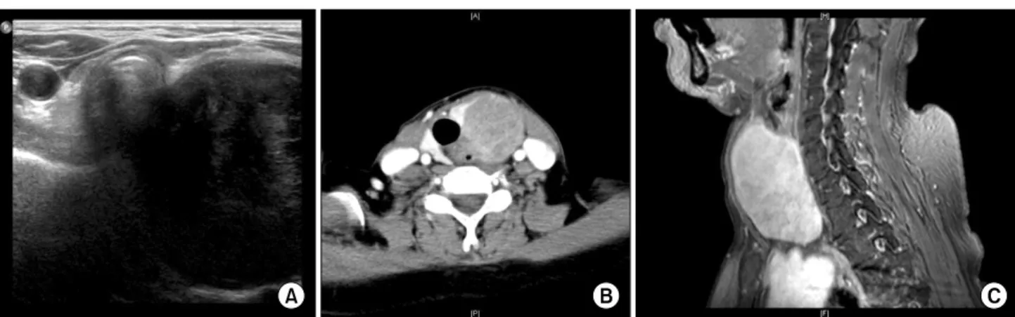
An ultrasound examination (Fig. 1A) depicted a 4.5×4.3×7.2 cm well-defined, solid, hypoechoic mass in the inferior portion of the left thyroid. CT (computerized tomography) and MRI (magnetic resonance imaging) (Fig. 1B, 1C) showed an 8 cm sized solid mass in the left visceral neck space from the upper margin of the C6 spine to the lower margin of the T3 spine that mass abut-ted the trachea and the esophagus without invasion. Radiologi-cally, the origin of mass was not clearly identified.
We decided on surgery to treat the dyspnea and substantiate the diagnosis. During surgical exploration, we identified the ex-tra-thyroidal mass, but found no anatomic relationship between the tumor and thyroid, trachea, recurrent laryngeal nerve, or va-gus nerve. During stepwise dissection of the mass, it was con-firmed that the tumor was located in the esophageal muscle layer. After mass excision, a Levin-tube was inserted and a defect in the esophageal wall was confirmed (Fig. 2B). The esophageal mucosal layer and the muscle layer were then securely repaired (Fig. 2C).
32 대한내분비외과학회지:제 11 권 제 1 호 2011
Fig. 1. Preoperative imaging study. (A) Preoperative ultrasonography depicted a well-defined solid, hypoechoic mass in the inferior portion
of the left thyroid. (B, C) CT and MR images showed an 8 cm sized solid mass in the left visceral neck space from the upper margin of the C6 spine to the lower margin of the T3 spine that mass abutted the trachea and the esophagus without invasion.
Fig. 2. Operative findings. (A) The resected tumor was found to be an encapsulated solid, elastic mass. (B) After mass removal, a levin-tube
was inserted and the presence of an esophageal wall was confirmed. (C) The esophageal mucosa layer and muscle layer were securely repaired.
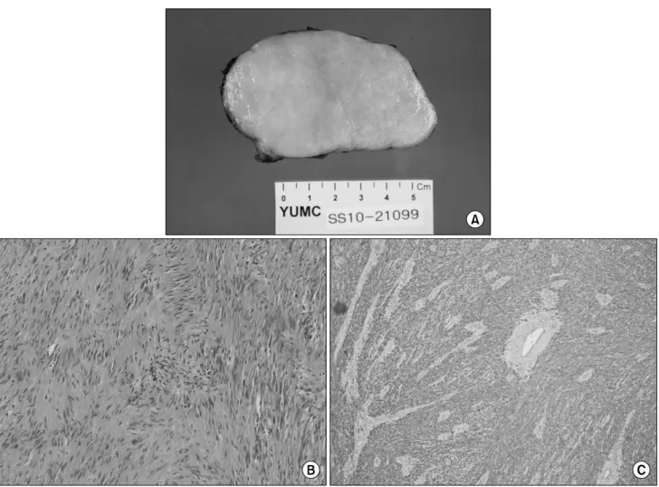
Macroscopically, the resected tumor appeared to be an encap-sulated solid, elastic mass, 8×5×4 cm in size (Fig. 2A). In cross section, it was found to have a homogeneous yellow color without hemorrhage or necrosis (Fig. 3A). Microscopically, the tumor was composed of oval to spindle-shaped cells arranged in a pallisad-ing pattern with peritumoral lymphocytic infiltration. Furthermore, tumor cell nuclei showed mild variations in size and shape (Fig. 3B). Immunohistochemical staining revealed that tumor cells were positive for S-100 protein (Fig. 3C) and negative for smooth mus-cle actin, which confirmed a diagnosis of schwannoma. The patient was maintained on total parenteral nutrition (TPN) for 5 days postoperatively. On the postoperative 6th day, esoph-agography confirmed that the operative site was well preserved without ulceration and leakage. The patient recovered without any postoperative complications, and was discharged 10 days after surgery.
DISCUSSION
Schwannomas are found most commonly in the posterior me-diastinum and rarely originate in the esophagus. Since Chatelin and colleagues(3) first reported this entity in 1967, about 30 cas-es have been reported in English-language literature. A review of previous reports showed that esophageal schwan-nomas occur most frequently in middle-aged women and are usu-ally located at the thoracic level of the esophagus.(4) Esophageal schwannomas arise exclusively from the neural plexus of the esophagus, and result in esophageal luminal narrowing, which usually causes dysphagia, although a few cases have reported that presented with dyspnea caused by airway compression.(5) On the other hand, schwannomas of the gastrointestinal tract are submucosal tumors, and are commonly covered by normal muco-sa and involve the submucomuco-sa and muscularis propria.(6) Typically, esophageal schwannoma is diagnosed preoperatively
Jae Hyun Park, et al:Esophageal Schwannoma 33
Fig. 3. Pathologic finding. (A) In cross section, the excised mass had a homogeneous yellow color without hemorrhage or necrosis. (B)
The tumor was composed of oval to spindle-shaped cells arranged in a pallisading pattern with peritumoral lymphocytic infiltration. (C) Immunohistochemical staining of tumor cells revealed positivity for S-100 protein
as an esophageal submucosal tumor, leiomyoma, leiomyosarco-ma, or another type of tumor, and it is not possible to establish a definitive preoperative diagnosis based on the findings of imag-ing studies.(7) Accordimag-ingly, an immunohistochemical findimag-ing of S-100 protein positivity, which is specific for its neurogenic origin, is regarded necessary to confirm the diagnosis.(8)
Generally, the prognosis of esophageal schwannomas is good after surgical removal.
However, a few cases of esophageal schwannoma with malig-nant potential or with lymph node metastasis have been reported.(5) A diagnosis of malignancy depends on histological findings of mitotic figures, invasion of esophageal muscle, and cellular atypia.(9) Local recurrence after surgical resection has al-so been reported.(10) Thus, all patients with eal-sophageal schwan-noma are recommended for surgical intervention and long-term follow-up.
In our case, esophageal schwannoma was not expected based
on considerations of its clinical manifestations and preoperative findings. However, if esophageal schwannoma is suspected, esophageal mucosa might be preserved. A careful approach is required to resect neurogenic tumors in the cervical space, due the possibility of damaging surrounding structures. Although rare, the possibility of esophageal schwannoma should be borne in mind and surgical dissection should be undertaken cautiously to prevent injury of esophageal mucosa.
REFERENCES
1) Badwai RA, Scott-Coombes D. Ancient schwannoma masquerading as a thyroid mass. Eur J Surg Oncol 2002;28:88-90.
2) De Paoli F, Giugliano G, Casadio C, Tredici P, Bruschini R, De Fiori E. Schwannoma of thyroid bed. A case report and considerations on interdisciplinary collaboration. Acta Otorhinola-ryngol Ital 2005;25:250-2.
34 대한내분비외과학회지:제 11 권 제 1 호 2011
Confront Radio Anat Clin 1967;7:114.
4) Park BJ, Carrasquillo J, Bains MS, Flores RM. Giant benign esophageal schwannoma requiring esophagectomy. Ann Thorac Surg. 2006;82:340-2.
5) Murase K, Hino A, Ozeki Y, Katagiri Y, Onitsuka A, Sugie S. Malignant schwannoma of the esophagus with lymph node meta-stasis: literature review of schwannoma of the esophagus. J Gastroenterol 2001;36:772-7.
6) Miettinen M, Lasota J. Gastrointestinal stromal tumors (GISTs): definition, occurrence, pathology, differential diagnosis and molecular genetics. Pol J Pathol 2003;54:3-24.
7) Yoon HY, Kim CB, Lee YH, Kim HG. An obstructing large
schwan-noma in the esophagus. J Gastrointest Surg 2008;12:761-3. 8) Prévot S, Bienvenu L, Vaillant JC, de Saint-Maur PP. Benign
schwannoma of the digestive tract: a clinicopathologic and immuno-histochemical study of five cases, including a case of esophageal tumor. Am J Surg Pathol 1999;23:431-6.
9) Iwata H, Kataoka M, Yamakawa Y, Kawabara Y, Kureyama Y, Masaoka A. Esophageal schwannoma. Ann Thorac Surg 1993;56: 376-7.
10) Eberlein TJ, Hannan R, Josa M, Sugarbaker DJ. Benign schwan-noma of the esophagus presenting as a giant fibrovascular polyp. Ann Thorac Surg 1992;53:343-5.