THE JOURNAL OF KOREAN ACADEMY OF OSSEOINTEGRATION http://www.kaoimplant.org
Vol. 9 No. 2, December 2017
The Journal of Korean Academy of Osseointegration 2017, 9(2) 7-10
Ridge preservation in the posterior maxilla to preserve vertical dimension
from pneumatization
Jin-Young Park, Hyung-Chul Pae, Joo-Hyun Kang, Jae-Kook Cha, Ui-Won Jung, Seong-Ho Choi
Department of Periodontology, Research Institute for Periodontal Regeneration, College of Dentistry, Yonsei University, Seoul, Korea
INTRODUCTION
많은 임상가들이 상악 구치부에 임플란트를 식립할 때 어려움을 겪는 다. 가장 큰 이유 중 하나는 치아 상실이 유발하는 수직적 골 소실로 인해 해부학적으로 이상적인 위치에 임플란트를 식립하는 것이 어려워지는 것 이다. 수직적인 골 소실의 원인은 두 가지로 추정해 볼 수 있는데 첫 번째 는 치조골의 흡수이며 두 번째는 상악동의 함기화다. 발치 후 일어나는 치조골의 흡수에 대해서는 많은 연구가 이루어져 왔 다. 구강 내에서 발치 후 치조골의 흡수는 수평, 수직적으로 동시에 일어 난다. 발치 후 치조골 소실 양에 대한 systematic review에 의하면 수평 적으로는 3.87 mm, 수직적으로는 1.57 mm의 흡수가 일어난다고 보고 되었다1). 하지만 발치와의 변화에 대한 대부분의 연구는 전치부와 소구치 부위에서 진행되었으며 상악 구치부에서의 변화에 대해서는 아직 추가적 인 연구가 필요한 상황이다.상악 대구치 발치 후 치조제보존술을 통한 상악동 함기화의 감소 및 치조골
높이의 보존
박진영, 배형철, 강주현, 차재국, 정의원, 최성호
연세대학교 치과대학병원 치주조직재생연구소Aim: The aim of the current study is to present a bilateral clinical case in the posterior maxilla, in which alveolar ridge preservation (RP) was performed to prevent sinus pneumatization and allow one-stage implant placement.
Material and Methods: In 2007, the patient was presented with a hopeless left second maxillary premolar and a maxillary first molar due to advanced periodontitis. Extraction was performed; however, RP was not performed. On the opposite side, in 2016, the right second maxillary premolar and the maxillary first molar were extracted for the same reason. Extraction and RP was performed using collagenated DBBM and a collagen plug.
Results: In the left maxilla, pneumatization of the sinus floor occurred, resulting in residual ridge height of 1 mm. Consequently, si-nus lift by lateral approach had to be performed. 8 months after surgery, two 10 mm length implants were placed simultaneously with further sinus lift by crestal approach. The total treatment period was 14 months. In the right maxilla, the cone-beam computed tomography (CBCT) taken 3 months after RP, revealed residual ridge height of 6.27 mm at the right maxillary second premolar site, and 7.46 mm at the first molar site. Two 8 mm length implants were placed with simultaneous sinus lift procedure by the crestal ap-proach. The total treatment period was 6 months.
Conclusion: Ridge preservation in the posterior maxilla is a useful therapeutic option to prevent vertical intra-antral resorption, there-by improving the feasibility of one-stage implant placement and reducing the total treatment period. (THE JOURNAL OF KOREAN ACADEMY OF OSSEOINTEGRATION 2017;9(2):7-10)
Key words: Ridge preservation, Posterior maxilla, Maxillary sinus augmentation, Bone graft, Dental implant
Received November 15, 2017, Revised November 15, 2017, Accepted November 20, 2017
cc This is an open access article distributed under the terms of the Creative Commons Attribution Non-Commercial License (http://creativecommons.org/licenses/ by-nc/4.0/) which permits unrestricted non-commercial use, distribution, and reproduction in any medium, provided the original work is properly cited. 교신저자: 최성호, 03722, 서울시 서대문구 연세로 50-1, 연세의료원 치과대학병원 치주과
Corresponding Author: Seong-Ho Choi, Department of Periodontology, Research Institute for Periodontal Regeneration, College of Dentistry, Yonsei University, Seoul 03722, Korea, Tel: +82-2-2228-3189, Fax: +82-2-392-0398, E-mail: shchoi726@yuhs.ac
박진영 등: 상악 대구치 발치 후 치조제보존술을 통한 상악동 함기화의 감소 및 치조골 높이의 보존
The Journal of Korean Academy of Osseointegration 2017, 9(2) 7-10
8
상악동의 함기화는 상악동 부피를 증가시키는 정상적인 생리적 현상이 다. 상악동 함기화의 정확한 이유나 기전은 아직 밝혀지지 않았으나 유전 적 요인2), 골 밀도3), 과거 상악동 수술의 유무4), 연령 등이 영향을 미치는 것으로 알려져 있다. 한 연구에 따르면 상악 대구치의 발치가 상악동의 함 기화를 야기할 수 있다고 하였고, 특히 상악동저의 외형이 상악동 내부로 돌출돼있는 경우, 다수의 대구치를 발치하는 경우, 상악 제 2대구치를 발 치하는 경우 함기화의 정도가 확연하게 나타난다고 하였다5). 치조제 보존술을 시행하면 골이식재가 발치와 내부의 공간을 유지하 여 치조골의 체적 수축을 최소화하며, 치유되는 기간동안 신생골의 자라 들어갈 수 있는 스캐폴드로써의 역할을 한다. Nevins 등은 상악 전치부 에서 치조제 보존술을 시행하였을 때 79% 이상의 사이트에서 20% 미만 의 순측 골 소실을 보인 반면, 아무런 처치를 하지 않은 경우 71% 이상의 사이트에서 20% 이상의 순측 골 소실이 나타났다고 보고했다6). 치조제 보존술은 발치 전 치조제의 형태를 유지 시켜 줌으로써, 1-stage 임플란 트 식립을 가능하게 해주고, 임플란트 식립 시 골이식의 필요성을 줄여준 다. 이는 환자와 술자 모두에게 편리한 방법이다. 2010년 Rasperini 등은 상악 구치부에 치조제 보존술을 시행하여 임 플란트 식립 시 상악동 거상술의 필요도가 감소되었다는 결과를 발표하였 다7). 이 결과는 상악대구치의 치근이 상악동 내부로 돌출되어있는 경우 골이식재가 발치와 내부의 공간을 유지할 뿐만 아니라 상악동의 함기화로 인한 intra-antral 골 흡수에 저항하는 역할을 하는 것으로 해석될 수 있 다. 본 증례보고에서는 양쪽 상악 구치부에서 동일하게 발치를 시행한 후 한쪽은 치조제 보존술을 시행하고 반대쪽은 아무런 처치도 하지 않았을 때 치조골의 수직적 고경 변화, 임플란트 식립 전 측방법 상악동 거상술의 필요성, 임플란트 보철이 완성될 때까지 걸리는 총 치료기간을 비교 평가 하고자 한다.CASE REPORT
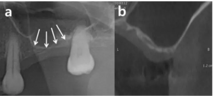
1. 좌측 상악 구치부 본 55세 여환은 2007년에 처음 내원 하였고 좌측 상악 구치부에 통증 을 호소했다. 임상 및 방사선 검사 시 좌측 상악 제 2소구치, 제 1대구치 부위 3도의 동요도가 관찰됐고 좌측 상악 제 1대구치의 경우 치근단 부위 를 둘러싼 방사선 투과성 병소가 관찰되었다(Fig. 1). 이 환자를 gener-alized advanced periodontitis로 진단했고 좌측 상악 제 2소구치와 제 1대구치는 가망 없는 치아로 판단하고 발치 했다. 환자는 발치 2년 후 에 재내원 했고 발치된 부위 상악동의 함기화가 일어난 것이 확인되었으며 좌측 상악 제 2대구치 부위의 잔존 치조골 높이는 1 mm였다(Fig. 2). 따 라서 lateral sinus augmentation을 시행하였는데 수술 중 mesial septum 부위에 7 mm 크기의 상악동막 천공이 발생하여 collagen tape (Collatape, Zimmer, USA)으로 보강한 후 골 이식을 시행하였고, 8개월 정도의 긴 치유 기간을 갖게 되었다. 임플란트 1차 수술 시 10 mm 임플란트(4.0×10 mm, 5.0×10 mm; Osseospeed; AstraTech;Swe-Fig. 1. The initial panoramic radiograph showed advanced periodontitis with apical involvement on the left maxillary second premolar and first molar, which were decided to be extracted.
Fig. 2. (a) After the extraction, the socket walls had been resorbed due to maxillary sinus pneumatization, resulting in the loss of height of the alveolar ridge. (b) The remaining height of the ridge was 1 mm, therefore, sinus elevation by the lateral approach and two-stage implant placement was necessary.
Fig. 3. (a) Sinus lift by lateral app-roach was performed. The sinus membrane was perforated during the surgery. (b) 8 months after, two implants were placed (OsseoSpeed, AstraTech, Sweden) with further sinus elevation by the crestal app-roach. (c) Final prosthesis was deli-vered after another 6 months.
Park JY, et al: Ridge preservation in the posterior maxilla to preserve vertical dimension from pneumatization
The Journal of Korean Academy of Osseointegration 2017, 9(2) 7-10
9
den)를 식립하기 위해 추가적인 sinus augmentation을 crestal법으로 시행했다(Fig. 3). 그로부터 6개월 후에 최종 보철물을 장착하여 총 14개 월의 치료기간이 소요됐다. 2. 우측 상악 구치부 2016년에 동일한 환자가 이번에는 우측 상악 구치부 통증을 호소하며 재내원 했다. 임상 및 방사선 검사 시 우측 상악 제 2소구치, 제 1대구치 부 위 깊은 탐침 깊이와 치근단 병소가 관찰되어 combined endo-perio lesion으로 진단하였고 가망 없는 치아로 판단됐다(Fig. 4). 따라서 해당 부위 발치 및 치조제보존술 시행한 후 임플란트 식립 계획하였다. 우측 상 악 제 2소구치, 제 1대구치 발치 후 블록형 콜라겐 함유 탈단백
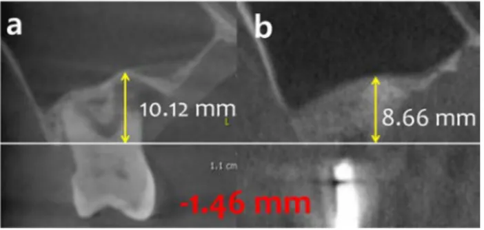
우골(Bio-Oss Collagen, Geistlich, Switzerland)을 이용하여 치조제 보존술을 시행하였으며 collagen plug (Teruplug, Olympus Terumo Bioma-terials, Japan)를 상부에 적용한 후 봉합하였다(Fig. 5). 발치 전 후 CBCT을 이용하여 상악동이 함기화 된 양을 측정하였다: 발치 전 후 우측 상악 제 1대구치의 중심을 관통하는 동일한 sagittal view에서 발치와의 치조정 부위에 임의의 기준선을 긋고 상악동 저의 가장 낮은 점까지의 거 리를 각각 측정했을 때 발치 전에는 10.12 mm, 발치 및 치조제보존술 후 에는 8.66 mm로 계측되었다(Fig. 6). 따라서 함기화로 인해 1.46 mm의 intra-antral 골 흡수가 일어난 것이 확인되었다. Crestal법 상악동 거상 술을 동반하여 우측 상악 제 2소구치, 제 1대구치 부위 임플란트를 식립하 였고(4.1×8 mm, Straumann, Switzerland), 수술 3개월 후 안정적인 periotest value가 측정되어 최종 보철 진행하였다. 발치 및 치조제 보존
Fig. 4. In 2016, the patient was presented with pain on the right maxi-llary area. Panoramic radiograph revealed advanced periodontitis with apical involvement on the right maxillary second premolar and first molar. Extraction and ridge preservation was planned for these teeth.
Fig. 5. (a) Pre-operative clinical photo. (b) Extraction of #25 and 26. (c) Socket grafted with collagenated deproteinized bovine bone substiute. (d) Placement of collagen plug and suture. (e) Stitch removal. (f) 3 Months post-operation. (g) Flap elevation for implant placement. (h) Implant placement (fixture size: 4.0×8 mm, Straumann). (i) Final prosthesis. Fig. 6. (a) Cone-beam computed tomography (CBCT) taken before extraction revealed ridge height of 10.12 mm from a reference line. (b) CBCT taken 3 months after extraction and ridge preservation revealed residual ridge height of 8.66 mm. 1.46 mm of pneumatization occurred.
박진영 등: 상악 대구치 발치 후 치조제보존술을 통한 상악동 함기화의 감소 및 치조골 높이의 보존
The Journal of Korean Academy of Osseointegration 2017, 9(2) 7-10
10
술을 시행한 이후로부터 6개월 만에 최종보철이 완성되었다.DISCUSSION
본 증례는 양쪽 상악 구치부 제 2소구치와 제 1대구치를 발치한 후 우 측은 치조제 보존술을 시행하고 좌측은 아무런 처치도 하지 않았을 때의 결과를 잔존 치조제의 수직적 높이, 추가적인 상악동 거상술의 여부, 치료 기간에 따라 비교 평가하였다. 좌측 상악 구치부는 발치 후 1∼2 mm의 잔존치조골만이 남게 되어 lateral sinus augmentation 후 2 stage로 임플란트 식립을 시행하였고, 보철완성까지 총 14개월의 시간이 걸렸다. 우측 상악 구치부는 치조제 보존술을 시행하여 8 mm의 치조골 높이를 확보할 수 있었고 crestal 법을 동반하여 1 stage로 임플란트를 식립할 수 있었다. 총 치료기간은 6개월로 좌측에 비해 절반이상 짧았다. Sharan 등은 파노라마 방사선 사진에 나타나는 상악 대구치 치근과 상악동 저와의 관계를 다섯가지로 분류하였다: (1) 상악대구치의 치근이 상악동 저의 피질골과 접촉하지 않는 경우, (2) 상악동 저가 하방으로 굽어 져 있으나 치근이 협설측으로 뻗어있어 상악동 밖에 있는 경우, (3) 상악동 저가 하방으로 굽어져 있고 치근의 측면이 상악동 내부로 돌출되어 있는 경우, (4) 상악동저가 하방으로 굽어져 있고 치근단첨이 상악동 내로 돌출 된 경우, (5) 상악동저가 상방으로 돌출돼 있고 치근을 포함하고 하는 경우5), 발치 전 후 방사선 사진을 비교하였을 때 (4)과 (5)의 경우 94%에서 상방 으로 돌출돼있던 상악동저가 발치 후 함기화로 인해 납작해지거나 하방으 로 굽어진 형태로 변형되었다. 본 증례 또한 발치된 양측 상악 제 1대구치 가 상악동 내부로 돌출된 경우였으며, 발치만 시행했을 때 함기화로 인해 상악동 저가 하방으로 굽어진 형태로 변형됐다는 점에서 Sharan의 논문 과 일치한다. 반면 우측 제 1대구치의 경우 치조제 보존술을 시행하므로써 함기화를 줄일 수 있었으며 lateral sinus augmentation 없이도 8 mm 길이의 임플란트를 식립할 수 있었다.Lateral sinus augmentation은 침습적인 술식으로써 술후 부종, 통 증, 감염이나 술중 상악동막의 천공과 같은 여러가지 합병증을 동반한다. 일반적으로 이식되는 골의 양이 많기 때문에 오랜 치유기간을 필요로 하 며, 상악동막의 천공이 심할 경우 골이식을 연기해야하기 때문에 더 오랜 치료기간을 야기한다. 상악동막의 천공과 같은 경우 상악동 내에 septum 이 존재할 경우 더 흔하게 발생한다. 최근 발표된 후향적 연구에 의하면, 79개의 상악동 수술 증례 중 48.1%의 경우 septum이 관찰되었다8). 상악 동막 천공의 발생률은 22.8%였으며 이는 septum의 유무와 밀접한 관계 가 있음이 통계학적으로 유의미하게 나타났다. 본 증례에서도 상악동의 septum으로 인해 상악동막이 수술중 천공되었고 이로 인해 치유기간이 연장되었다. 우측 상악구치부의 경우 치조제 보존술로 인해 치조제 높이 가 유지되었기 때문에 간단한 crestal법만을 동반하여 임플란트를 식립할 수 있었으며, 합병증을 예방하고 치유기간 또한 감소되었다. 상악 전치부에서 발치와 치유는 순측 골의 소실과 심미적인 영역에서 의 임플란트 수복과 관련되어 많은 연구가 이루어졌다. 구치부의 경우 협 측 골의 수평적 소실 보다는 높이의 감소가 더욱 임상적인 의미가 있으나 이에 대한 연구는 거의 없다. 한 무작위배정 임상시험에서 상악대구치를 발치한 후 시험 군은 치조제 보존술을 시행하고 대조군은 아무런 처치를 하지 않았다7). 시험 군은 6명 중 1명만 추가적인 상악동 거상술을 시행한 반면, 대조 군은 8명 중 3명이 측방법 상악동 거상술을 받음으로써 상악구 치부의 치조제 보존술은 함기화를 예방하고 상악동 거상술을 회피할 수 있다는 결론을 내렸으며 이는 본 증례보고의 결과와 일치한다.
CONCLUSION
본 증례보고에서는 양측 상악구치부의 동일한 치아 발치 및 치조제 보 존술을 시행한 후 비교하였다. 상악 구치부의 치조제 보존술은 상악동의 함기화 양을 감소시키고 치조제의 수직적 고경을 유지함으로써 임플란트 식립 전 상악동 거상술을 회피하고, 1-stage 임플란트 식립을 가능하게 해주며, 전반적인 치료기간을 단축시켜준다.REFERENCES
1. Van der Weijden F, Dell’Acqua F, Slot DE. Alveolar bone dimensional changes of post-extraction sockets in humans: a systematic review. J Clin Periodontol 2009;36(12):1048-58.
2. J. S. Morphologic characteristics of the sinuses. Arch Otolaryngol 1936;23:484-7.
3. Nowak R MG. Studies on the state of pneumatization of the sinus maxillaris. Anat Anz 1975:138-43.
4. Kosko J HB, Tunkel D. Acquired maxillary sinus hypoplasia: A conse-quence of endoscopic sinus surgery? Laryngoscope 1996;106: 1212-3.
5. Sharan A, Madjar D. Maxillary sinus pneumatization following extrac-tions: a radiographic study. Int J Oral Maxillofac Implants 2008;23(1): 48-56.
6. Nevins M, Camelo M, De Paoli S, Friedland B, Schenk RK, Parma-Benfenati S, Simion M, Tinti C, Wagenberg B. A study of the fate of the buccal wall of extraction sockets of teeth with prominent roots. Int J Periodontics Restorative Dent 2006;26(1):19-29.
7. Rasperini G, Canullo L, Dellavia C, Pellegrini G, Simion M. Socket grafting in the posterior maxilla reduces the need for sinus augmen-tation. Int J Periodontics Restorative Dent 2010;30(3):265-73. 8. Irinakis T, Dabuleanu V, Aldahlawi S. Complications During Maxillary
Sinus Augmentation Associated with Interfering Septa: A New Classi-fication of Septa. Open Dent J 2017;11:140-50.