Total elbow replacement was used first in treatment of rheuma-toid arthritis patients in 1970 [1]. Development of implants and better surgical techniques have led to widespread use of this method in various elbow diseases, such as osteoarthritis, post-traumatic arthritis, severe comminuted fracture with bone loss, chronic instability, and tumors [2,3]. However, total elbow re-placement can cause various complications, such as aseptic loos-ening, infection, and periprosthetic fractures, during long-term follow-up. To address these issues, revision surgery has been re-ported [4].
Periprosthetic fracture is widely recognized as a complication of total hip or knee replacement but may also occur following
to-Periprosthetic fracture after total elbow replacement surgery is a difficult complication to manage, especially when it comes together with implant loosening. If stem revision and internal fixation of the periprosthetic fracture are performed simultaneously, this would be a very challenging procedure. Most of total elbow replacement implants are cemented type. Cement usage at periprosthetic fracture site may inter-fere healing of fractured site. Authors underwent internal fixation with use of locking plate and cerclage wire for periprosthetic fracture, al-logenous fibular strut bone inserted into the humerus intramedullary canal allowing the fractured site to be more stable without cement us-age. At 10-month follow-up, the complete union and good clinical outcome was achieved. We present a novel technique for treating peri-prosthetic fracture with implant loosening after total elbow replacement surgery, using intramedullary allogenous fibula strut bone graft. Keywords: Elbow; Periprosthetic fracture; Arthroplasty
Technical Note
Clin Shoulder Elbow 2020;23(3):152-155 https://doi.org/10.5397/cise.2020.00213
Intramedullary fibula strut bone allograft in a periprosthetic
humeral shaft fracture with implant loosening after total elbow
arthroplasty
Young-Hoon Jo
1, Seung Gun Lee
2, Incheol Kook
2, Bong Gun Lee
21Department of Orthopedic Surgery, Hanyang University Guri Hospital, Guri, Korea 2Department of Orthopedic Surgery, Hanyang University College of Medicine, Seoul, Korea
Received: August 3, 2020 Revised: August 18, 2020 Accepted: August 18, 2020
Correspondence to: Bong Gun Lee
Department of Orthopedic Surgery, Hanyang University College of Medicine, 222-1 Wangsimni-ro, Seongdong-gu, Seoul 04763, Korea Tel: +82-2-2290-8485, Fax: +82-2-2290-3774, E-mail: bglee@hanyang.ac.kr, ORCID: https://orcid.org/0000-0002-4003-5529
IRB approval: None.
Financial support: None.
Conflict of interest: None.
tal elbow replacement [5]; the incidence has been reported to range from 5% to 29% [6,7]. Periprosthetic fractures after total elbow replacement are classified according to location, degree of bone loss around the implant, and presence of implant loosening [6]. If the periprosthetic fracture is accompanied by implant loosening, complex techniques such as revision surgery and bone grafting for bone defects are often required in addition to inter-nal fixation. Due to the high rate of complications after surgery with this procedure, it is vital to have an appropriate preoperative surgical plan.
Sanchez-Sotelo et al. [5] reported 11 cases of total elbow re-placement with allograft strut bone augmentation and cement
eISSN 2288-8721
Copyright© 2020 Korean Shoulder and Elbow Society. All Rights Reserved.
This is an Open Access article distributed under the terms of the Creative Commons Attribution Non-Commercial License (http://creativecommons.org/licenses/by-nc/4.0/) which permits unrestricted non-commercial use, distribution, and reproduction in any medium, provided the original work is properly cited.
www.cisejournal.org 152
fixation that required revision for periprosthetic fracture follow-ing total elbow replacement caused by implant loosenfollow-ing. How-ever, six of these 11 cases had poor prognosis with one or more complications, and technical difficulties were reported. If internal fixation and bone graft are performed after fixing the implant with cement, circulation at the fracture site may be compromised by the cement, which will likely delay bone union. In addition, it is also difficult to perform simultaneous internal fixation and bone grafting.
This report introduces a novel surgical technique using an al-logenous fibula strut bone graft inserted into the intramedullary canal to improve stability without cement to treat periprosthetic fracture around the humeral stem after total elbow arthroplasty.
TECHNIQUE
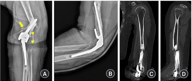
The patient was a 65-year-old woman who had been diagnosed with rheumatoid arthritis 10 years prior. She had recently under-gone total elbow replacement surgery using a Coonrad-Morrey (Zimmer, Warsaw, IN, USA) implant at another hospital after sustaining an injury while lifting a heavy object. She had under-gone regular bone mineral density testing once a year and did not need medication for osteopenia. Upon radiographic exam-ination, a periprosthetic fracture around the humeral stem corre-sponding to Mayo classification type H-II was identified, and implant loosening and osteolysis were also observed (Fig. 1).
The authors performed revision of total elbow replacement us-ing the Coonrad-Morrey implant (Zimmer). The previous oper-ative skin incision was used along with a triceps-preserving ap-proach. The incision was deepened to the subcutaneous tissue,
and then the triceps fascia was undermined to identify the medi-al and latermedi-al edges of the triceps muscle. On the medimedi-al side, the ulnar nerve was identified and confirmed to be located anterior-ly. A longitudinal incision was made on the exposed triceps ten-don on the radial side of the midline; care was taken to avoid the tip of the olecranon, and then the joint was exposed. To obtain an adequate view during the approach, the authors elevated the flexor carpi ulnaris and part of the pronator muscle group sub-periosteally and released the triceps partially. Loosening of the humeral stem was identified, and osteolysis and metallosis were present. Debridement was carried out with complete removal of all soft tissue around the humeral stem. The ulnar stem was sta-ble with no sign of loosening, so ulnar replacement was not re-quired. After removal of a link between a polyethylene insert and the humeral stem, thorough cement removal and irrigation of the intramedullary canal was performed to avoid influencing the revision surgery. Because the cortical bone around the fracture site was thin and the medullary canal was wide, we opted to per-form revision total elbow replacement with a novel technique us-ing an intramedullary fibula bone graft.
For the revision procedure, we used allogenous fibula strut bone (Community Tissue Services, Kettering, OH, USA). After determining the joint line and optimum size of the humeral stem, the diameter and length of the allogenous fibula strut bone were estimated according to the diameter and size of the bone defect and humeral stem. To resolve the difference in diameter between the humeral bone defect lesion and the allogenous fibu-la strut bone, the fibu-latter was trimmed using a high-speed burr to fit the outer diameter to better enter the humeral bone defect area. In addition, the inner diameter of the allogenous fibula
B
D
A
C
Fig. 1. Elbow anteroposterior (A) and lateral (B) radiographs, coronal (C) and sagittal (D) view of computed tomography images of total elbow arthroplasty with periprosthetic fracture. Previous fracture line are marked with arrow (A), and implant loosening areas are marked with ar-rowheads (A).
153 https://doi.org/10.5397/cise.2020.00213
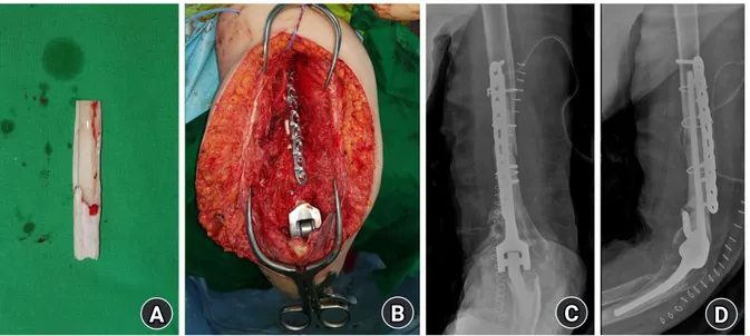
strut bone was enlarged using a burr to allow the humeral stem to pass (Fig. 2A). Next, it was necessary to confirm whether the allogenous fibula strut bone was located in the fracture site so that the trial stem could pass into the allogenous fibula strut bone before cementation. After closing the proximal portion of the distal fragment from the fracture site using a gauge, we in-serted the cement compactly from the distal portion and placed the humeral stem from the distal end. At this time, the cement at the fracture site was removed, and the allogenous fibula strut bone, which contained the humeral stem, was passed through the fracture site. The proximal part of the humeral stem was not performed infusion of bone cement which would inhibit bone union at the fracture site. The fracture site was additionally fixed using a locking plate, eight locking screws, and three cerclage wires (Fig. 2B). After confirming the appropriateness of fixation
and bone graft using simple radiographs (Fig. 2C, D), the surgery was complete.
Postoperatively, the elbow was positioned in a long arm splint at a 90° flexion position for 6 weeks. Two weeks after surgery, the previous long arm splint was replaced with a removable splint, and passive elbow joint motion was performed. Active elbow joint motion was allowed beginning at six weeks after surgery. No other physical therapy or additional fixation was performed. At three months postoperatively, the range of motion (ROM) of the affected elbow was confirmed at 0°–110°, and no other com-plications were observed. At 10 months after surgery, bone union was confirmed on radiographs (Fig. 3A, B), and a satisfactory clinical outcome was obtained with an ROM from 0° to 150° of the affected elbow without complications (Fig. 3C, D).
Fig. 2. Allograft fibula strut bone that was manipulated with a high-speed burr (A), and clinical photograph of the operative field after fixation with a locking plate and cerclage wire (B). Anteroposterior (C) and lateral (D) radiographs after operation.
B
D
A
C
Fig. 3. Anteroposterior (A) and lateral (B) radiographs at postoperative 10 months. Postoperatively, fibula strut-graft incorporation and union were observed with satisfactory clinical outcome and range of motion 0° to 15° (C, D).
B
D
A
C
https://doi.org/10.5397/cise.2020.00213 154
DISCUSSION
Periprosthetic fracture following total elbow replacement surgery is graded using the Mayo classification system according to frac-ture site, fixation status of the implant, and bone stock around the implant [5,6]. Different operative techniques may be required for different stages [5]. Treatment strategy can be chosen based on type of humeral shaft fracture [7]. Non-displaced peripros-thetic fracture without loosening of the implant is treated with conservative treatment, including long arm splint fixation [6], whereas fracture with implant loosening should undergo revision surgery with internal fixation. Various internal fixation tech-niques have been reported, including allogenous strut bone graft [5], locking plate and cerclage wire, and external fixation using the Ilizarov system [7]. To manage humeral bone loss, various methods including humeral shortening, use of a longer humeral stem or bone graft around the anterior flange, use of a tumor prosthesis system, or attachment of a humeral intramedullary nail to the humeral stem followed by autogenous bone graft have been reported [8].
In this case, which was Mayo classification type H-II, implant loosening and osteolysis with periprosthetic fracture prompted revision of the humeral stem, bone graft, and internal fixation. A previously reported technique used an allogenous strut bone graft obtained from outside of the fracture site or an on-lay tech-nique fixed with cerclage wire, which resulted in instability around the fracture site [5]. Even when additional plate fixation is used for stability, allogenous bone graft with cerclage wire may interfere with satisfactory positioning of the plate above the hu-meral shaft. This technique could cause the fracture site to be bulky and might produce surgical site discomfort. This approach can also give rise to complications such as nerve injury and ex-tensor mechanism rupture [5]. Cement leakage via a fracture gap may also occur, causing cement-related complications and in-creasing risk of non-union.
In this case, we manipulated the allogenous fibular strut bone into a cylinder shape and inserted it into the humerus shaft in an intramedullary manner to maintain the advantage of allogenous bone graft and to reduce possible complications. This approach improved stability without bone loss or shortening and prevented or reduced cement leakage or cement-related complications be-cause the cement was only injected into the distal area of the hu-meral stem. This technique also provided good positioning of the plate compared to the on-lay technique as no allogenous bone was present outside the humerus.
The most important and challenging component of this
tech-nique was shaping the allogenous fibular strut bone into the ap-propriate diameter to fit into the humeral intramedullary area and allowing the humeral stem to pass through the inner diame-ter of the fibular bone graft. Thinning the fibular bone while pre-serving its shape requires delicate handling and precautions, which may take up a substantial amount of time.
The authors’ case showed fracture site union, good elbow joint ROM, and pain relief at 10 months postoperatively, indicating that this novel technique can be considered as a treatment option for periprosthetic fracture with implant loosening after total el-bow replacement surgery.
ORCID
Young-Hoon Jo https://orcid.org/0000-0002-4299-2496 Seung Gun Lee https://orcid.org/0000-0001-7401-7620 Incheol Kook https://orcid.org/0000-0001-8691-5495 Bong Gun Lee https://orcid.org/0000-0002-4003-5529
REFERENCES
1. Dee R, Sweetnam DR. Total replacement arthroplasty of the el-bow joint for rheumatoid arthritis: two cases. Proc R Soc Med 1970;63:653-5.
2. Kozak TK, Adams RA, Morrey BF. Total elbow arthroplasty in primary osteoarthritis of the elbow. J Arthroplasty 1998;13:837-42.
3. Mighell MA, Dunham RC, Rommel EA, Frankle MA. Primary semi-constrained arthroplasty for chronic fracture-dislocations of the elbow. J Bone Joint Surg Br 2005;87:191-5.
4. DeBernardis DA, Horneff JG, Davis DE, Ramsey ML, Pontes MC, Austin LS. Revision total elbow arthroplasty failure rates: the impact of primary arthroplasty failure etiology on subse-quent revisions. J Shoulder Elbow Surg 2020;29:321-8. 5. Sanchez-Sotelo J, O'Driscoll S, Morrey BF. Periprosthetic
hu-meral fractures after total elbow arthroplasty: treatment with implant revision and strut allograft augmentation. J Bone Joint Surg Am 2002;84:1642-50.
6. O'Driscoll SW, Morrey BF. Periprosthetic fractures about the el-bow. Orthop Clin North Am 1999;30:319-25.
7. Seo H, Im JH, Lee JY. Treatment of periprosthetic fracture after total elbow replacement arthroplasty. J Korean Fract Soc 2020;33:110-7.
8. Kwak JM, Koh KH, Jeon IH. Total elbow arthroplasty: clinical outcomes, complications, and revision surgery. Clin Orthop Surg 2019;11:369-79.
155 https://doi.org/10.5397/cise.2020.00213