저작자표시-비영리-변경금지 2.0 대한민국 이용자는 아래의 조건을 따르는 경우에 한하여 자유롭게 l 이 저작물을 복제, 배포, 전송, 전시, 공연 및 방송할 수 있습니다. 다음과 같은 조건을 따라야 합니다: l 귀하는, 이 저작물의 재이용이나 배포의 경우, 이 저작물에 적용된 이용허락조건 을 명확하게 나타내어야 합니다. l 저작권자로부터 별도의 허가를 받으면 이러한 조건들은 적용되지 않습니다. 저작권법에 따른 이용자의 권리는 위의 내용에 의하여 영향을 받지 않습니다. 이것은 이용허락규약(Legal Code)을 이해하기 쉽게 요약한 것입니다. Disclaimer 저작자표시. 귀하는 원저작자를 표시하여야 합니다. 비영리. 귀하는 이 저작물을 영리 목적으로 이용할 수 없습니다. 변경금지. 귀하는 이 저작물을 개작, 변형 또는 가공할 수 없습니다.
Serum markers for predicting significant necroinflammatory
activity in patients with chronic hepatitis B
By
Hyo Jung ChoMajor in Medicine
Department of Medical Sciences
The Graduate School, Ajou University
Serum markers for predicting significant necroinflammatory
activity in patients with chronic hepatitis B
By
Hyo Jung ChoA Dissertation Submitted to The Graduate School of
Ajou University in Partial Fulfillment of the Requirements for the
Degree of Master of Medicine
Supervised by
Jae Youn Cheong, M.D., Ph.D.
Major in Medicine
Department of Medical Sciences
The Graduate School, Ajou University
This certifies that the dissertation
of Hyo Jung Cho is approved.
SUPERVISORY COMMITTEE
Jae Y oun Cheong
Sung Won Cho
Kwang Jae Lee
The Graduate School, Ajou University
June, 21th, 2013
- 1 -
- ABSTRACT –
Serum markers for predicting significant necroinflammatory
activity in patients with chronic hepatitis B
Objectives: The aim of this study was to determine the serum markers that predict significant inflammation in patients with chronic hepatitis B (CHB).
Design and Methods: Between October 2005 and June 2009, 384 subjects with CHB were enrolled.
Results: Multiple logistic regression analysis identified the ALT, hyaluronic acid (HA) and procollagen III N-terminal peptide (PIIINP) as independent predictors of significant inflammation (grade≥3). We constructed a formula for predicting significant inflammation. A significant inflammation (SI) score = 1.773 × ALT score + 1.599 × PIIINP score + 0.677 × HA score - 1.962. The area under receiver operating characteristic curve of the SI score was 0.831. The sensitivity, specificity, positive predictive value and negative predictive value of the SI score were 79.5%, 70.8%, 76.8% and 74.3%, respectively.
Conclusions: A simple scoring system including ALT, PIIINP and HA is an accurate non-invasive predictor of significant inflammatory activities in patients with CHB. Keyword: apoptosis, chronic hepatitis B, cytokeratin-18, significant inflammation
- 2 -
TABLE OF CONTENTS
ABSTRACT ………1 TABLE OF CONTENTS ………2 LIST OF FIGURES .………3 LIST OF TABLES ………... 4 I. INTRODUCTION ..………...5II. PATIENTS AND METHODS .………...6
A. Patients selection…….………6 B. Histologic examination ……….6 C. Biochemical marker …….………7 D. Statistical analysis ……….…… 7 III. RESULTS ………..……9 IV. DISCUSSION ………..17 Acknowledgement………21 REFERENCES ………22 국문요약………26
- 3 -
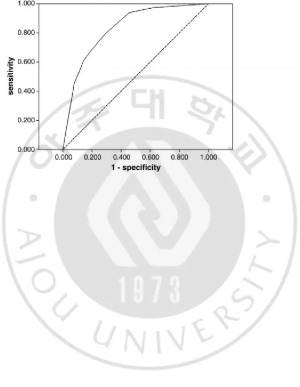
Figure 1. Receiver operating characteristic (ROC) curve of the SI score for prediction of significant inflammation. The area under the ROC curves was 0.831..……… 16
- 4 -
Table 1. Baseline characteristics of the patients………..…… 10 Table 2. Univariate and multivariate analysis for variables associated with
significant inflammation ………....12 Table 3. AUROCs for predicting significant inflammation, cut off value and new
variables……….... 13 Table 4.Predictors of significant inflammation according to multiple logistic regression
- 5 -
I. INTRODUCTION
Chronic hepatitis B (CHB) is one of the most common infections worldwide that can progress to cirrhosis, end stage liver disease and hepatocellular carcinoma (HCC)) [1-4]. Patients with significant hepatic inflammation and fibrosis are at the highest risk of developing these complications [5].
Liver biopsy is currently the gold standard in evaluating liver fibrosis and necroinflammation in patients with parenchymal liver diseases [6]. It provides important information regarding the severity of necroinflammatory activity and fibrosis, which are features predicting a treatment response and prognosis [7, 8]. However, the procedure is limited by sampling error, the risk of complications, patient discomfort, and poor inter-observer concordance [6, 9, 10].
Inflammatory activity grading is valuable information by which to guide management decisions for patients with chronic hepatitis. According to consensus recommendations, moderate or severe inflammatory activities have been considered for antiviral therapy in patients with chronic hepatitis [11, 12]. Serum alanine aminotransferase (ALT) and aspartate aminotransferase (AST) are most commonly used serologic tests for assessing hepatitis activity, but the inflammatory activity grades are not well-correlated with serum ALT or AST in patients with CHB. Hence, there is a need to develop an accurate non-invasive means to assess the severity of liver fibrosis and inflammation in chronic liver disease. Previous studies have focused on serum markers for the detection of liver fibrosis [13-17], and few studies have been performed to predict liver inflammation in patients with CHB [13, 18-20].
The aims of this study were to investigate the ability of serum markers to identify significant necroinflammation and to determine which combination of blood tests best predicts significant inflammation in patients with CHB.
- 6 -
II. PATIENTS AND METHODS
A. Patients Selection
Between October 2005 and June 2009, 384 consecutive patients with hepatitis B virus (HBV) infections who underwent liver biopsies in 6 centers (Ajou University Hospital, Hallym University Chuncheon, CHA University Hospital, Inje University Busan Paik Hospital, Pusan National University Hospital and Catholic University St. Vincent’s Hospital) in South Korea were recruited in this study. The inclusion criteria were CHB patients who underwent a liver biopsy and had not received antiviral treatment within 6 months prior this study’s commencement. Patients were not included if there were other causes of liver disease or decompensated cirrhosis.
Informed consent to participate in the study was obtained from all of the study subjects. The study protocols were approved by the Institutional Review Board of Human Research of Ajou University Hospital and the local Research Ethics Committees at all participating hospitals.
B. Histologic examination
All patients had liver biopsy specimens of at least 10 mm in length containing at least 6 complete portal tracts. The mean length of the liver biopsy sample was 12.6 mm (±3.3 mm) and the mean number of portal tracts was 13.2 (±6.3).
Necroinflammation was graded according to the Batts–Ludwig scoring system [21]. Based on the Batts–Ludwig scoring system, grade 1 denotes necroinflammatory activities largely confined to the portal areas with grades 2–3 denoting an extension beyond the portal areas, and grade 4 signifying confluent necrosis in the form of bridging necrosis. All biopsy specimens were analyzed by two hepatopathologists (Y.B. Kim and Y.N. Park)
- 7 -
who were blinded to patient clinical characteristics and serum measurements. At the end of this study, discrepancies between the two pathologists were resolved by joint discussion at the microscope
C. Biochemical Markers
Serum samples were obtained and routine blood tests were performed at the time of liver biopsy and processed immediately. The serum biochemical parameters included total bilirubin, alanine aminotransferase (ALT), aspartate transaminase (AST), γ-glutamyl transpeptidase (GGT), alkaline phosphatase (ALP), albumin, blood urea nitrogen (BUN), creatinine, a-fetoprotein (AFP), prothrombin time, blood glucose, triglycerides, and total cholesterol levels.
The serum a2-macroglobulin (a2MG) level was measured with an automatic nephelometer (Dadebehring, Marburg, Germany). The serum haptoglobin level was assessed using an immunoturbidimetric assay on a Roche COBAS Integra 800 automated chemistry analyzer (Roche Diagnostic). The serum apolipoprotein A1 level was determined with an ELISA kit (Roche, Basel, Swizerland). The serum hyaluronic acid (HA) concentration was measured with a Corgenix HA quantitative test kit (Corgenix, Inc., Westminster, CO, USA). The reference values were 1.75-4.2 g/L for a2MG, 30-200mg/dL for haptoglobin, 1.08-1.76 g/L for apolipoprotein A1, and 10-800 ng/mL for HA. The matrix metalloproteinase (MMP)-2 and tissue inhibitor of metalloproteinase (TIMP)-1 levels were determined with Quantikine® Immunassay kits (R & D Systems,
Inc., Minneapolis, MN, USA). The procollagen III N-terminal peptide (PIIINP) level was assessed by the UniQ PIIINP radioimmunoassay (Orion Diagnostica, Inc., Espoo, Finland); the reference range was 2.3 ~ 6.4 μg/L. The alpha-1 antitrypsin level was measured with the Cobas Integra immunoturbimetric assay (Roche, Basle, Switzerland).. D. Statistical analysis
- 8 -
The patients’ characteristics are given as the mean ± SD as appropriate. The variables that differed between the patients with significant inflammation and without significant inflammation were identified by chi-square tests and independent t test analyses. To identify the predictive factors, we used a multivariate logistic regression with forward selection for the significant variable on the univariate analysis (P < 0.05). Then we decided a cutoff value for each factor at the level of greater than 70% both for sensitivity and specificity on the area under receiver operating characteristic (AUROC) curve. To derive the binary or categorical predictors required for a clinically useful scoring system, we assigned a simplified score value to each factor. Each factor with a cutoff value or above gave score 1, and below cutoff value gave score 0. We performed a logistic regression analysis with a stepwise forward selection by the new scores, for construction of a new prognostic model of significant inflammation. The predictive value of this new prognostic model was assessed by AUROC curve and its optimal cutoff values were chosen at the point of maximizing the sum of the sensitivity and specificity. Diagnostic accuracies were evaluated by calculating the sensitivities, specificities, positive predictive values (PPVs), and negative predictive values (NPVs). All statistical tests were two-sided and performed with SPSS 12.0 (SPSS, Chicago, IL, USA).
- 9 -
III. RESULTS
A. Patient characteristics
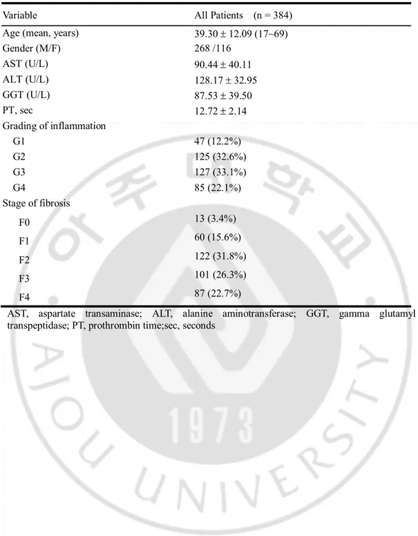
The baseline characteristics of the enrolled patients are depicted in Table 1. Three hundred eighty eight patients were included (268 men and 116 women) between 17 and 69 years of age (mean, 39.30 ± 12.09 years).
The grade of inflammation was converted into a binomial variable of significant inflammation (Batts–Ludwig inflammatory activity grades 3 or 4) vs. no significant inflammation (grades 0, 1, or 2). These thresholds were selected because they are generally considered indications for antiviral therapy [22]. Significant inflammation was present in 212 patients (55.2%). The distribution of the inflammatory activity grades was as follows: grade 1=47 (12.2%); grade 2=125 (32.6%); grade 3=127 (33.1%); and grade 4=85 (22.1%). Liver cirrhosis was present in 87 patients (22.7%) (Table 1).
- 10 -
Table 1. Baseline characteristics of the patients.
Variable All Patients (n = 384) Age (mean, years) 39.30 ± 12.09 (17~69) Gender (M/F) 268 /116 AST (U/L) 90.44 ± 40.11 ALT (U/L) 128.17 ± 32.95 GGT (U/L) 87.53 ± 39.50 PT, sec 12.72 ± 2.14 Grading of inflammation G1 47 (12.2%) G2 125 (32.6%) G3 127 (33.1%) G4 85 (22.1%) Stage of fibrosis F0 13 (3.4%) F1 60 (15.6%) F2 122 (31.8%) F3 101 (26.3%) F4 87 (22.7%)
AST, aspartate transaminase; ALT, alanine aminotransferase; GGT, gamma glutamyl transpeptidase; PT, prothrombin time;sec, seconds
- 11 -
B. Discriminatory variables in univariate and multivariate logistic regression analysis for predicting significant inflammation
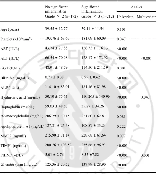
We identified the predictors of significant inflammation by univariate and multivariate analyses. Univariate logistic regression analysis of the variables revealed that AST, ALT, bilirubin, GGT, ALP, HA, TIMP1, PIIINP, a1-antitrypsin, platelet counts, haptoglobin and apolipoprotein A1 levels were significantly different between the patients with or without significant inflammation (Table 2).
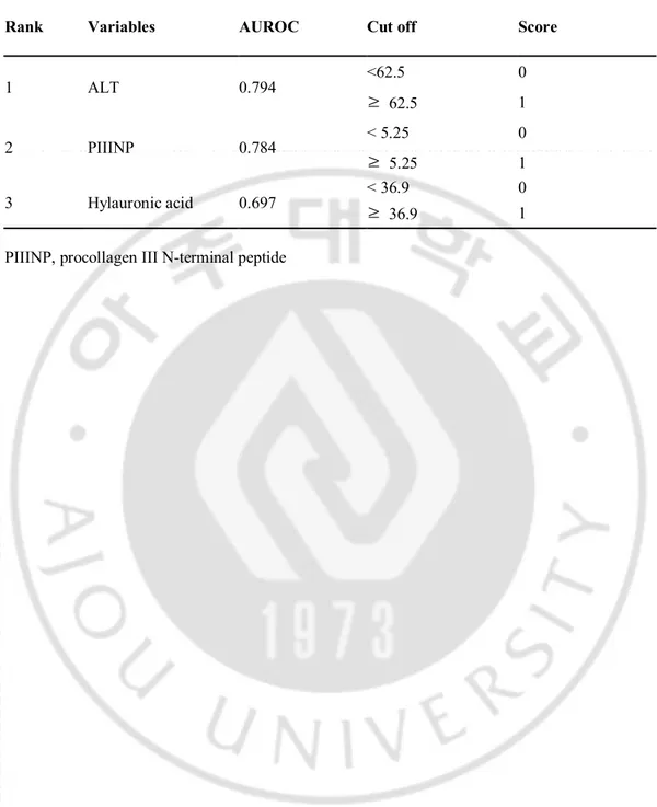
Given the association observed between many of the covariates, multiple logistic regression analyses were performed, simultaneously controlling for all covariates with statistical significance (P < 0.05). Multiple logistic regression analysis by stepwise forward selection identified the high ALT (p=0.000), high HA (p=0.043) and high PIIINP (p=0.001) as independent predictors of significant inflammation. Table 3 shows AUROCs of three independent predictors of significant inflammation (ALT: 0.794, HA: 0.784, PIIINP: 0.697).
- 12 -
Table 2. Univariate and multivariate analysis for variables associated with significant inflammation No significant inflammation Grade ≤ 2 (n=172) Significant inflammation Grade ≥ 3 (n=212) p value Univariate Multivariate Age (years) 39.55 ± 12.77 39.11 ± 11.54 0.101 Platelet (x103/mm3) 193.76 ± 63.67 181.09 ± 60.09 0.047 AST (IU/L) 43.74 ± 27.88 128.33 ± 116.73 <0.001 ALT (IU/L) 66.54 ± 70.98 178.17 ± 173.92 <0.001 <0.001 GGT (IU/L) 48.91 ± 48.79 114.50 ± 211.59 0.001 Bilirubin (mg/dL) 0.77 ± 0.38 0.99 ± 0.62 <0.001 ALP (IU/L) 114.10 ± 85.91 181.16 ± 81.98 <0.001 Hyaluronic acid (ng/mL) 50.10 ± 75.61 110.265 ± 140.96 <0.001 0.043 Haptoglobin (mg/dL) 59.03 ± 48.67 35.27 ± 34.26 <0.001 α2-macroglobulin (mg/dL) 206.29 ± 70.15 221.60 ± 62.87 0.081 Apolipoprotein A1 (mg/dL) 127.31 ± 26.58 108.57 ± 35.23 0.222 MMP2 (ng/mL) 215.90 ± 71.14 228.68 ± 61.64 0.072 TIMP1 (ng/mL) 200.76 ± 103.52 255.66 ± 96.93 <0.001 PIIINP (㎍/L) 5.01 ± 2.76 8.55 ±7.82 <0.001 0.001 α1-antitrypsin (mg/dL) 125.36 ± 20.52 137.99 ± 28.90 <0.001
MMP, matrix metalloproteinase; TIMP, tissue inhibitor of metalloproteinase; PIIINP, procollagen III N-terminal peptide
- 13 -
Table 3. AUROCs for predicting significant inflammation, cut off value and new variables
Rank Variables AUROC Cut off Score
1 ALT 0.794 <62.5 0 ≥ 62.5 1 2 PIIINP 0.784 < 5.25 0 ≥ 5.25 1 3 Hylauronic acid 0.697 < 36.9 ≥ 0 36.9 1 PIIINP, procollagen III N-terminal peptide
- 14 -
C. Construction of the new prediction formula for predicting significant inflammation
We assigned a new variable of 0 or 1 by making the cutoff value of each of the markers on the point giving sensitivity and specificity simultaneously greater than 70% on the ROC curve (ALT=62.5 IU/L, HA=36.9 ng/mL, PIIINP=5.25 μg/L, Table 3). Next, we performed logistic regression analyses by using new values (ALT score, HA score, PIIINP score), for the construction of a new predicting model of significant inflammation (Table 4). The derived formula for predicting significant inflammation (SI score) was as follows;
SI score = 1.773 × ALT score + 1.599 × PIIINP score + 0.677 × HA score – 1.962
The ROC curve of the new predictive model for significant inflammation is demonstrated in Figure 1. The AUROC of the SI score was 0.831 (CI: 0.788-0.875, P=0.000), which was higher than that of each of the single markers.
Next, the diagnostic performance of the SI score was analyzed. The sensitivity, specificity, positive predictive value and negative predictive value of the SI score for predicting significant inflammation were 79.5, 70.8, 76.8, and 74.3%, respectively, at a cutoff score of 0.0625.
- 15 -
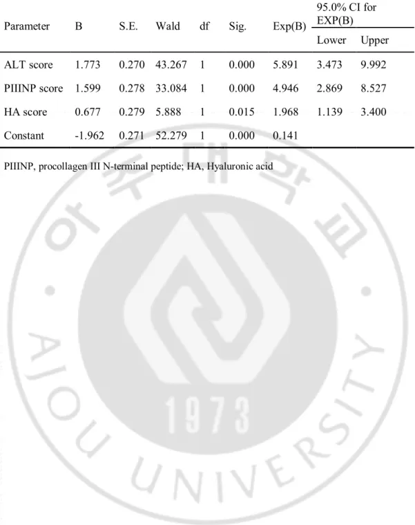
Table 4. Predictors of significant inflammation according to multiple logistic regression analysis
Parameter B S.E. Wald df Sig. Exp(B)
95.0% CI for EXP(B) Lower Upper ALT score 1.773 0.270 43.267 1 0.000 5.891 3.473 9.992 PIIINP score 1.599 0.278 33.084 1 0.000 4.946 2.869 8.527 HA score 0.677 0.279 5.888 1 0.015 1.968 1.139 3.400 Constant -1.962 0.271 52.279 1 0.000 0.141
- 16 -
Figure 1. Receiver operating characteristic (ROC) curve of the SI score for prediction of significant inflammation. The area under the ROC curves was 0.831
- 17 -
IV. DISCUSSION
We have investigated the relationship among biochemical variables and liver necroinflammation as assessed by histologic examination of liver biopsy specimens in patients with CHB. Based on a multivariate logistic regression analysis, three variables were identified as independent predictors for significant inflammation (ALT, procollagen III N-terminal peptide, and hyaluronic acid). These variables were used to generate the scoring system, which we call the “significant inflammation (SI) score”, that could predict significant inflammation with an AUROC of 0.831. The advantage of our scoring system is the use of a logistic regression, which has facilitated the appropriate weighting of the parameters that compose the SI score. By applying the appropriate cutoff score, the presence of significant inflammation could be diagnosed with a sensitivity of 79.5% and specificity of 70.8% in patients with CHB.
Most non-invasive markers have been assessed for liver fibrosis staging and reports on serum markers for liver inflammation and are relatively rare [13, 19, 23]. The Actitest has been validated as a serum marker for necroinflammatory activity in patients with chronic viral hepatitis [13, 24]. Myers et al. used the Fibrotest and Actitest to discriminate mild from extensive fibrosis and inflammation in patients with CHB [13]. The same study demonstrated that
- 18 -
Actitest had an AUROC of 0.82 for predicting significant necroinflammation. In our study, we tried to evaluate all possible markers affecting liver inflammation and/or fibrosis. We tested five variables of Fibrotest (total bilirubin, GGT, a2-macroglobulin, apolipoprotein A1 and haptoglobin), other direct/indirect markers of liver fibrosis and/or inflammation, and included a novel candidate marker (such as a1-antitrypsin). Of the serum markers evaluated in our study, the ALT was the most accurate predictor of necroinflammation and yielded the highest AUROC. However, serum ALT levels can fluctuate spontaneously with time and a single value is unlikely to be representative. It seems that the addition of other serum markers is required to overcome this limitation.
HA is a glycosaminoglycan, a high molecular weight polysaccharide synthesized by hepatic stellate cells and degraded by sinusoidal endothelial cells of the liver [25-27]. In the study by Oberti et al., the serum HA level was considered the most sensitive test for screening in viral hepatitis B and C [28]. A previous study also showed that the serum HA level was the best predictor of extensive liver fibrosis and inflammation for HBeAg-negative CHB [20].
PIIINP is one of the frequently reported direct fibrosis markers, and serum levels of PIIINP are known to reflect the degree of liver fibrosis in alcoholic liver disease, viral hepatitis, and primary biliary cirrhosis. The PIIINP level is correlated with aminotransferase levels in patients with chronic hepatitis [29],
- 19 -
however, the correlation between the PIIINP and aminotransferase levels may also reflect that PIIINP is a marker of the inflammatory response driven by HBV replication. Moreover, elevated levels of serum PIINP have been found in active liver disease such as chronic active hepatitis and active cirrhosis [30-32]. In patients with chronic hepatitis C, PIIINP had correlated with inflammatory activity as well as liver fibrosis, especially reflecting disease activity within and around portal tracts [33]. Considering the complex interrelation between hepatic fibrogenesis and the inflammatory process in vivo, it is impossible to separate pure inflammation from fibrosis in patients with chronic liver disease in the clinical setting. Our results suggest that PIIINP and HA should not be regarded as markers linked only with liver fibrosis, but are also influenced by necroinflammatory activity.
In our current study, we attempted to validate the usefulness of α1-antitrypsin, a novel liver inflammation marker from proteomics study results. Alpha1-antitrypsin, the major serine protease inhibitor in human circulation, is an acute phase reactant and its levels rise following tissue injury and inflammation [34, 35]. A previous serum proteomic study revealed that different fragments of α1-antitrypsin were expressed in patients with HBV infection [18]. In our study, the α1-antitrypsin level was significantly higher in patients with significant
- 20 -
inflammation in a univariate analysis, but it was not a statistically significant variable in a multivariate analysis. Because we did not analyze the different isoforms of α1-antitrypsin, further research will be needed to evaluate clinical usefulness of analysis of isoforms of α1-antitrypsin.
Our study had some limitations. First, our study lacked a validation group, thus our results need to be validated in the independent patient population. Second, our study did not separate HBeAg positive from HBeAg negative patients. The differences in disease behavior in these two groups had been clearly demonstrated in earlier studies [13, 36]. Third, the PIIINP and HA are not routinely measured in the clinical setting and the lack of availability of these serum markers in many centers makes it difficult to apply the proposed scoring system in a daily clinical practice.
In conclusion, we have developed a novel scoring system that is accurate in distinguishing patients with or without significant inflammation and the measurement of serum ALT, PIIINP and HA appears useful for predicting significant inflammation in patients with CHB. It has yet to be determined whether the addition of a novel serum marker of inflammation increases the diagnostic accuracy of the scoring system.
- 21 -
Acknowledgement
This work was supported by a grant from the Ministry of Health and Welfare, Republic of Korea (no. A050021)
- 22 -
References
[1]. Lai CL, Ratziu V, Yuen MF, Poynard T. Viral hepatitis b. Lancet. 2003;362:2089-94.
[2]. Kwon SY, Lee CH. Epidemiology and prevention of hepatitis b virus infection. Korean J Hepatol. 2011;17:87-95.
[3]. Suk KT, Baik SK, Yoon JH, Cheong JY, Paik YH, Lee CH, et al. Revision and update on clinical practice guideline for liver cirrhosis. Korean J Hepatol. 2012;18:1-21.
[4]. Lee HS, Kim JK, Cheong JY, Han EJ, An SY, Song JH, et al. Prediction of compensated liver cirrhosis by ultrasonography and routine blood tests in patients with chronic viral hepatitis. Korean J Hepatol. 2010;16:369-75.
[5]. Fattovich G, Brollo L, Giustina G, Noventa F, Pontisso P, Alberti A, et al. Natural history and prognostic factors for chronic hepatitis type b. Gut. 1991;32:294-8.
[6]. Poynard T, Ratziu V, Bedossa P. Appropriateness of liver biopsy. Can J Gastroenterol. 2000;14:543-8.
[7]. Liaw YF, Tai DI, Chu CM, Chen TJ. The development of cirrhosis in patients with chronic type b hepatitis: A prospective study. Hepatology. 1988;8:493-6. [8]. Chien RN, Liaw YF, Atkins M. Pretherapy alanine transaminase level as a determinant for hepatitis b e antigen seroconversion during lamivudine therapy in patients with chronic hepatitis b. Asian hepatitis lamivudine trial group. Hepatology. 1999;30:770-4.
[9]. Bedossa P, Dargere D, Paradis V. Sampling variability of liver fibrosis in chronic hepatitis c. Hepatology. 2003;38:1449-57.
- 23 -
et al. Sampling error and intraobserver variation in liver biopsy in patients with chronic hcv infection. Am J Gastroenterol. 2002;97:2614-8.
[11]. Easl international consensus conference on hepatitis b. 13-14 september, 2002: Geneva, switzerland. Consensus statement (short version). J Hepatol. 2003;38:533-40.
[12]. Conjeevaram HS, Lok AS. Management of chronic hepatitis b. J Hepatol. 2003;38 Suppl 1:S90-103.
[13]. Myers RP, Tainturier MH, Ratziu V, Piton A, Thibault V, Imbert-Bismut F, et al. Prediction of liver histological lesions with biochemical markers in patients with chronic hepatitis b. J Hepatol. 2003;39:222-30.
[14]. Imbert-Bismut F, Ratziu V, Pieroni L, Charlotte F, Benhamou Y, Poynard T. Biochemical markers of liver fibrosis in patients with hepatitis c virus infection: A prospective study. Lancet. 2001;357:1069-75.
[15]. Vallet-Pichard A, Mallet V, Nalpas B, Verkarre V, Nalpas A, Dhalluin-Venier V, et al. Fib-4: An inexpensive and accurate marker of fibrosis in hcv infection. Comparison with liver biopsy and fibrotest. Hepatology. 2007;46:32-6. [16]. Wai CT, Greenson JK, Fontana RJ, Kalbfleisch JD, Marrero JA, Conjeevaram HS, et al. A simple noninvasive index can predict both significant fibrosis and cirrhosis in patients with chronic hepatitis c. Hepatology. 2003;38:518-26.
[17]. Rosenberg WM, Voelker M, Thiel R, Becka M, Burt A, Schuppan D, et al. Serum markers detect the presence of liver fibrosis: A cohort study. Gastroenterology. 2004;127:1704-13.
[18]. He QY, Lau GK, Zhou Y, Yuen ST, Lin MC, Kung HF, et al. Serum biomarkers of hepatitis b virus infected liver inflammation: A proteomic study. Proteomics. 2003;3:666-74.
- 24 -
H, et al. Noninvasive markers of liver fibrosis and inflammation in chronic hepatitis b-virus related liver disease. Am J Gastroenterol. 2006;101:2537-45. [20]. Montazeri G, Estakhri A, Mohamadnejad M, Nouri N, Montazeri F, Mohammadkani A, et al. Serum hyaluronate as a non-invasive marker of hepatic fibrosis and inflammation in hbeag-negative chronic hepatitis b. BMC Gastroenterol. 2005;5:32.
[21]. BATTS K P LJ. Chronic hepatitis. An update on terminology and reporting. Am J Surg Pathol. 1995;19:1409-17.
[22]. Easl clinical practice guidelines: Management of chronic hepatitis b. J Hepatol. 2009;50:227-42.
[23]. Poynard T, McHutchison J, Manns M, Myers RP, Albrecht J. Biochemical surrogate markers of liver fibrosis and activity in a randomized trial of peginterferon alfa-2b and ribavirin. Hepatology. 2003;38:481-92.
[24]. Poynard T, Zoulim F, Ratziu V, Degos F, Imbert-Bismut F, Deny P, et al. Longitudinal assessment of histology surrogate markers (fibrotest-actitest) during lamivudine therapy in patients with chronic hepatitis b infection. Am J Gastroenterol. 2005;100:1970-80.
[25]. McGary CT, Raja RH, Weigel PH. Endocytosis of hyaluronic acid by rat liver endothelial cells. Evidence for receptor recycling. The Biochemical journal. 1989;257:875-84.
[26]. Guechot J, Laudat A, Loria A, Serfaty L, Poupon R, Giboudeau J. Diagnostic accuracy of hyaluronan and type iii procollagen amino-terminal peptide serum assays as markers of liver fibrosis in chronic viral hepatitis c evaluated by roc curve analysis. Clin Chem. 1996;42:558-63.
[27]. Gressner AM, Bachem MG. Cellular sources of noncollagenous matrix proteins: Role of fat-storing cells in fibrogenesis. Seminars in liver disease. 1990;10:30-46.
- 25 -
[28]. Oberti F, Valsesia E, Pilette C, Rousselet MC, Bedossa P, Aube C, et al. Noninvasive diagnosis of hepatic fibrosis or cirrhosis. Gastroenterology. 1997;113:1609-16.
[29]. Trinchet JC, Hartmann DJ, Pateron D, Laarif M, Callard P, Ville G, et al. Serum type i collagen and n-terminal peptide of type iii procollagen in chronic hepatitis. Relationship to liver histology and conventional liver tests. J Hepatol. 1991;12:139-44.
[30]. Chang TT, Lin HC, Lee SD, Tsai YT, Lee FY, Jeng FS, et al. Clinical significance of serum type-iii procollagen aminopropeptide in hepatitis b virus-related liver diseases. Scand J Gastroenterol. 1989;24:533-8.
[31]. Ramadori G, Zohrens G, Manns M, Rieder H, Dienes HP, Hess G, et al. Serum hyaluronate and type iii procollagen aminoterminal propeptide concentration in chronic liver disease. Relationship to cirrhosis and disease activity. Eur J Clin Invest. 1991;21:323-30.
[32]. Misaki M, Shima T, Yano Y, Sumita Y, Kano U, Murata T, et al. Basement membrane-related and type iii procollagen-related antigens in serum of patients with chronic viral liver disease. Clin Chem. 1990;36:522-4.
[33]. Gabrielli GB, Capra F, Casaril M, Squarzoni S, Tognella P, Dagradi R, et al. Serum laminin and type iii procollagen in chronic hepatitis c. Diagnostic value in the assessment of disease activity and fibrosis. Clin Chim Acta. 1997;265:21-31. [34]. Travis J, Salvesen GS. Human plasma proteinase inhibitors. Annu Rev Biochem. 1983;52:655-709.
[35]. Dickson I, Alper CA. Changes in serum proteinase inhibitor levels following bone surgery. Clin Chim Acta. 1974;54:381-5.
[36]. Hui AY, Chan HL, Wong VW, Liew CT, Chim AM, Chan FK, et al. Identification of chronic hepatitis b patients without significant liver fibrosis by a simple noninvasive predictive model. Am J Gastroenterol. 2005;100:616-23.
- 26 - - 국문요약 –
만성
B형 간염 환자에서
간의
염증 괴사 활동도 예측을 위한 혈청 마커의 개발
아주대학교대학원의학과 조효정 (지도교수: 정재연) 목적: 이 연구의 목적은 만성 B형간염 환자에서 중등도 이상의 염증 괴사 활동도를 예측하는 혈청 마커를 개발하는 것이다. 방법: 간 조직 검사를 시행한 만성 B형 간염 환자 총 384명의 환자가 연구 에 포함 되었다.결과: 혈청 ALT 및 Hyaluronic acid(HA), procollagen III N-terminal peptide(PIIINP)가 Batts-Lugwig inflammatory activity grade 3 이상의 염증 정도를 예측하는 독립 인자였으며, 이 항목들을 이용하여 중등도 이상의 염증괴사 활동도를 예측하기 위한 식을 개발하였다. Significant inflammation (SI) score = 1.773 × ALT score + 1.599 × PIIINP score + 0.677 × HA score - 1.962. SI score 의 ROC 곡선 아래 역역은 0.831 이었고, 민감도 및 특이도, 양성 예측도, 음성 예측도는 각각 79.5%, 70.8%, 76.8%, 74.3% 였다.
결론: 만성 B 형 간염 환자에서 ALT, hyaluronic acid (HA) and procollagen III N-terminal peptide (PIIINP)를 이용하여 비침습적이고 정확한 염증 모델을 개발하였다.
핵심어: 만성 B형 간염, 염증 예측 모델, hyaluronic acid, procollagen III N-terminal peptide