저작자표시-변경금지 2.0 대한민국 이용자는 아래의 조건을 따르는 경우에 한하여 자유롭게 l 이 저작물을 복제, 배포, 전송, 전시, 공연 및 방송할 수 있습니다. l 이 저작물을 영리 목적으로 이용할 수 있습니다. 다음과 같은 조건을 따라야 합니다: l 귀하는, 이 저작물의 재이용이나 배포의 경우, 이 저작물에 적용된 이용허락조건 을 명확하게 나타내어야 합니다. l 저작권자로부터 별도의 허가를 받으면 이러한 조건들은 적용되지 않습니다. 저작권법에 따른 이용자의 권리는 위의 내용에 의하여 영향을 받지 않습니다. 이것은 이용허락규약(Legal Code)을 이해하기 쉽게 요약한 것입니다. Disclaimer 저작자표시. 귀하는 원저작자를 표시하여야 합니다. 변경금지. 귀하는 이 저작물을 개작, 변형 또는 가공할 수 없습니다.
Production of Monoclonal Antibodies
for Toxoplasma gondii Lactate
Dehydrogenase
by
Minh Anh Dang Trinh
Major in Molecular Medicine
Department of Medical Sciences
The Graduate School, Ajou University
Production of Monoclonal Antibodies
for Toxoplasma gondii Lactate
Dehydrogenase
by
Minh Anh Dang Trinh
A Dissertation Submitted to The Graduate School of Ajou University
in Partial Fulfillment of the Requirements for the Degree of
Master of Biomedical Sciences
Supervised by
Ho-Joon Shin, Ph.D.,
Sun Park, M.D., Ph.D.
Major of Molecular Medicine
Department of Medical Sciences
The Graduate School, Ajou University
This certifies that the dissertation
of Minh Anh Dang Trinh is approved.
SUPERVISORY COMMITTEE
_____________________________
Sun Park
_____________________________
Ho-Joon Shin
_____________________________
Kyongmin Kim
- ABSTRACT-
Production of Monoclonal Antibodies for Toxoplasma gondii Lactate
Dehydrogenase
Background/Purpose: Toxoplasma gondii is one of the most common parasitic infections of human and other warm blooded animals. Because of the geographical distribution of toxoplasmosis, there is a need to develop a sensitive, rapid, simple and an inexpensive test for the early screening of patiens. In this study, to create the dipstick kit that has high sensitivity against LDH (lactate dehydrogenase) of T. gondii, at the first of all, hybridoma cell lines producing monoclonal antibodies with high affinity and specificity against T. gondii LDH (TgLDH) were established.
Methods: After 6-8 week old BALB/c mice were immunized with synthetic peptides of TgLDH, splenocytes were harvested. To get the monoclonal antibodies (McAbs), splenocytes were fused with myeloma cells using PEG by a cell fusion technique. Hybridoma cells were selected by ELISA and subcloned through a limiting dilution method. McAbs were characterized by isotyping.
Results: Eight McAb-producing hybridoma cells were selected. After three times of limiting dilution, eight clones, 1A10, 1B12, 3B2, 3B6, 1C12, 3C8, 3G6 and 3H1, were selected. OD values of eight monoclonal antibodies were ranged from 0.938 to 1.269 by ELISA. All eight McAbs isotypes were IgM class.
Conclusion: These results indicated that eight hybridoma clones had strong reactivity to TgLDH and may be useful for diagnosis to T. gondii infection.
Key Words: Toxoplasma gondii, lactate dehydrogenase, cell fusion technique, monoclonal antibodies.
TABLE OF CONTENTS
ABSTRACT...i
TABLE OF CONTENTS... iii
LIST OF FIGURES ...v
LIST OF TABLE ...vi
I. INTRODUCTION ...1
A. Morphology ...1
B. Life cycle ...4
C. Diagnosis ...7
D. Lactate dehydrogenase of parasites ...7
E. Purpose of this study...8
II. MATERIALS AND METHODS ...
9
A
.
Culture of myeloma cells ...9B. Antigen ...9
C. Immunization of mice with TgLDH ...9
D. Cell fusion ...10
E. ELISA screening ...11
F. Selection of hybridoma cells ...11
G. Large scale production of McAbs
...
12A. Antibody formation of immunized mice ...13
B. Cell fusion...15
C. Isolation of antibody-producing hybridoma cells ...16
D. Producing large scale of antibodies ...17
E. The specificity of McAbs with T. gondii lysate...18
F. Isotyping of monoclonal antibodies ...20
IV. DISCUSSION...21
V. CONCLUSION...23
REFERENCES ...24
LIST OF FIGURES
Fig. 1. Morphology of T. gondii ...3
Fig. 2. Life cycle of T. gondii . ...6
Fig. 3. Polyclonal antibody titers from the TgLDH-5-immunized mice. ...14
Fig. 4. Antibody titers of the the hybridoma cell culture supernatant after cell fusion ...15
Fig. 5. Antibody titers of the hybridoma cell culture supernatant after third limiting dilution ...16
Fig. 6. Antibody titers of McAb-containing ascitic fluids ...17
Fig. 7. Antibody titers of the hybridoma cell culture supernatant using T. gondii lysate as antigen ...18
Fig. 8. Antibody titers of McAb-containing ascitic fluids using T. gondii lysate as antigen...19
LIST OF TABLE
Table 1. Amino acids sequence of synthetic peptide for TgLDH-5 ...9
I. INTRODUCTION
Toxoplasma gondii is one of the most common pathogen in the world. A huge
portion of the world’s population (over 30 million in France, and over 80 million people in the USA) is infected with T. gondii. The parasites were first recovered in the North African rodent called Ctenodactylus gondi, hence the species was named gondii. It can infect virtually all mammals but the definitive host is the house cat and other members of the Felidae (Garcia and Bruckner, 1997a).
This organism is an obligate intracellular parasite and is found in two forms in humans. The actively proliferating trophozoites or tachyzoites (endozoites) are usually seen in the early, more acute phases of the infection. The resting forms or bradyzoites (cystozoites) are primarily found in muscle and the brain (Garcia and Bruckner, 1997b).
Although T. gondii infection is usually asymptomatic in individuals with normal immune systems, it can cause severe syndromes including congenital malformations such blindness, mental retardation, and hydrocephaly in fetuses exposed in uterus. Recently, more attention has been given to T. gondii because toxoplasmosis is the most common opportunistic infection in immunocompromised patients with AIDS or in transplant patients (Luft and Remington, 1992).
A. Morphology
Various strains of T. gondii differ in virulence and certain biological and genetic characteristics. The life cycle of T. gondii includes various stages:
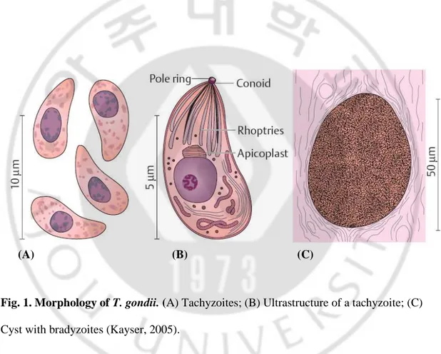
Tachyzoites (endozoites) are proliferative forms that reproduce rapidly within a parasitophorous vacuole in nucleate host cells by means of endodyogeny (endodyogeny: formation of two daughter cells from a mother cell by endogenous budding) (Fig. 1A, 1B). The tachyzoites are sickleshaped (toxon: bow) cells about 4~7 µm long and 2~4 µm wide. An apical complex is located at the anterior pole, consisting of serveal components, including the conoid (a conical structure of spirally arranged microtubulin), a pole ring complex, the rhoptries, and micronemes (Fig. 1B). The apical complex contributes to parasite penetration into host cells. The nucleus is located in the posterior half of the cell. More recent investigations have revealed that T. gondii and several other apicomplexan protozoa (e.g., Plasmodium, Sarcocystis) contain, in addition to the chromosomal and mitochondrial genomes, a further genome consisting of circular DNA (35 kb) localized in a special organelle called the apicoplast. This organelle, usually located anterior to the nucleus and close to the Golgi apparatus, is surrounded by several membranes and possesses outer and inner membrane complexes. There is evidence that the apicoplast has evolved from the plastids of endosymbiontic green or red algae. The function of the apicoplast remains uncertain but is indispensable for the host cell and may be a new and specific target for chemotherapy. Tachyzoites also multiply in experimental animals and cell cultures. This stage is highly labile outside of a host and usually does not survive the stomach passage following ingestion.
Bradyzoites (cystozoites) are stages produced by slow reproduction within the cysts (4~8 x 2~4 µm). The cysts develop intracellularly in various tissues, have a relatively resistant wall, grow as large as 150 µm, and can contain up to several thousand bradyzoites
(Fig. 1C). They have a long lifespan in the host. Humans and animals can be infected by peroral ingestion of meat containing cysts (Fig. 2).
Oocysts are rounded and encysted stages of the organism, surrounded by a resistant cyst wall, and are approximately 9 x 14 µm in size. They are the final product of a sexual reproductive cycle in the intestinal epithelia of Felidae. They contain a zygote and are shed in feces (Fig. 2). Sporulation takes place within two to four days, producing two sporocysts with four sporozoites each. Sporulated oocysts are infective for human and animals.
Fig. 1. Morphology of T. gondii. (A) Tachyzoites; (B) Ultrastructure of a tachyzoite; (C) Cyst with bradyzoites (Kayser, 2005).
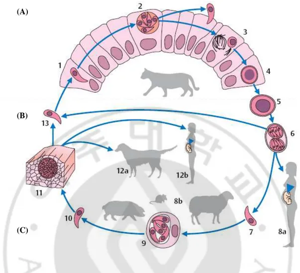
B. Life cycle
The developmental cycle of T. gondii involves three phases: intestinal, external, and extraintestinal (Fig. 2).
The intestinal phase: Production of sexual forms (gamogony) takes only place in enterocytes of definitive hosts. Only domestic cats, and several other felid species of little epidemiological significance, can function as definitive hosts for T. gondii. Only extraintestinal development is seen in intermediate hosts (pigs, sheep, and many other animal species) as well as in dead-end hosts (humans). Following primary infection of a cat with cysts in raw meat, asexual reproductive forms at first develop in the small intestine epithelium, with sexually differentiated stages and oocysts following later. The oocysts are shed in feces after a prepatent period of three to nine days. When cats are infected with sporulated oocysts, the prepatent period is extended to 20–35 days because in these cases the intestinal development of T. gondii is preceded by an extraintestinal asexual reproduction. Oocyst shedding lasts for only a few days to a maximum of three weeks, but can be highly intensive (up to 600 million oocysts per eat during the patent period).
External phase: Oocysts excreted in cat feces sporulate at room temperature within two to four days, rendering them infective. Kept moist, they remain infective for up to five years and are not killed by standard disinfectant agents. They die within a few minutes at
temperatures exceeding 55oC or by cooling to -13oC.
Extraintestinal phase: This phase follows a peroral infection with oocysts or cysts and is observed in intermediate hosts (humans, dogs, sheep, pigs, other vertebrates and birds),
or lymph to various organs and multiply in nucleate host cells, especially in the reticulohistiocytic system, in musculature, and in the CNS. Repeated endodyogenic cycles produce as many as 32 individual daughter cells in the expanding host cell before it bursts. The tachyzoites thus released attack neighboring cells. These processes result in focal necroses and inflammatory reactions in affected tissues. Generalization of the infection can lead to colonization of the placenta and, about three to four weeks later, infection of the fetus.
Bradyzoites that elicit no inflammatory reactions in the near vicinity are produced early in
the course of the infection. Such bradyzoites (cystozoites) are found above all in the CNS, in
the skeletal and heart muscles as well as in the retina, the uterine wall, and other organs. They can remain viable for years without causing noticeable damage to the host.
Fig. 2. Life cycle of T. gondii. (A) Development in the definitive host (cat): 1 Toxoplasma that has penetrated into a small intestine epithelial cell; 2 asexual reproductive stage with merozoites. 3 production of sexual forms (gamogony) and formation of the zygote; 4 oocyst. (B) External phase with sporogony: 5 unsporulated oocyst, shed in cat feces; 6 sporulated oocyst with two sporocysts each containing four sporozoites. (C) Development in an intermediate host (mammals, birds, humans): 7 sporozoite released in organism following oral ingestion of oocysts; 8a human infection; 8b infection of various animal species; 9 asexual reproductive stages (tachyzoites) in a somatic cell; 10 free tachyzoite; 11 cyst with bradyzoites in musculature; 12a infection of a dog with Toxoplasma cysts in animal meat;
(A)
(B)
C. Diagnosis
Diagnosis of toxoplasmosis can be aided by biologic, serologic, histologic or molecular methods or by combination of the above. Clinical signs of toxoplasmosis are nonspecific and cannot be depended on for a definite diagnosis; toxoplasmosis clinically mimics several other infectious diseases.
Many serologic tests have been used to detect antibodies to T. gondii; these include the Sabin-Feldman dye test, the indirect hemagglutination assay, the indirect fluorescent antibody assay (IFA), the direct agglutination test, the latex agglutination test (LAT), the enzyme-linked immunoabsorbent assay (ELISA) and the immunoabsorbent agglutination assay test (IAAT). The Sabin-Feldman dye test, which is still considered as the “gold standard” for detection T. gondii in human, is labour-intensive and has the disadvantage that it requires a continuous supply of live parasites. Therefore, most epidemiological studies on
T. gondii infection now use different tests for antibody detection (Dubey, 1988; Tenter,
2000). However, these tests are all time consuming and require a laboratory equipped with proper instruments and trained personnel. Therefore there is a need to develop a sensitive, rapid, simple and an inexpensive test for the early screening of patients.
D. Lactate dehydrogenase of parasites
It is well known that parasites, within hosts, derive much of their energy from anaerobic glycolysis. Glycolytic enzymes are essential for the survival of the parasites and appear to be potential targets for chemotherapeutic attacks. LDH is a terminal glycolytic
enzyme, which catalyzes the interconversion of puruvate and lactate in the presence of the nicotinamide adenine dinucleotide coenzyme. Compounds that inhibit LDH have also been shown to inhibit growth of Plasmodium falciparum in cultured erythrocytes (Royer et al., 1986) and T. gondii in cultured fibroblasts (Dando et al., 2001). It has been suggested that lactate dehydrogenase (LDH), therefore represents a judicious target for drug therapy against these organisms (Kavanagh et al., 2004).
E. Purpose of this study
To create the rapid diagnostic dipstick kit that has high sensitivity against lactate dehydrogenase (LDH) of Toxoplasma gondii, first of all, hybridoma cell lines producing monoclonal antibodies with high affinity and specificity against T. gondii LDH (TgLDH) were established.
II. MATERIALS AND METHODS
A. Culture of myeloma cells
F0 cells (a mouse myeloma cell line) were cultured in Roswell Park Memorial Institute (RPMI)-1640 medium (Welgene, Korea) containing 10% fetal bovine serum (FBS) (GIBCO, New York, USA) at 37℃ in 5% CO2 incubator.
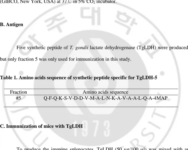
B. Antigen
Five synthetic peptide of T. gondii lactate dehydrogenase (TgLDH) were produced, but only fraction 5 was only used for immunization in this study.
Table 1. Amino acids sequence of synthetic peptide specific for TgLDH-5
Fraction Amino acids sequence
#5 Q-F-Q-K-S-V-D-D-V-M-A-L-N-K-A-V-A-A-L-Q-A-4MAP
C. Immunization of mice with TgLDH
To produce the immune splenocytes, TgLDH (50 µg/100 µl) was mixed with an equal volume of Freund’s complete adjuvant (Sigma laboratories, UAS), and mixture was injected intraperitoneally (i.p.) into 6~8 week old BALB/c mice. At second and third time, the mice were boosted biweekly for another 4 weeks with TgLDH (25 µg/100 µl) mixed
immunized mouse serum was assayed by indirect enzyme-linked immunosorbent assay (ELISA). For the final boost, the TgLDH (5 µg/100 µl) was injected intravenously without the adjuvant.
D. Cell fusion
To produce cell lines that secrete McAb, we practiced a cell-fusion technique by the method of previous paper (Kohler and Milstein, 1975; Lee et al., 2007). After blood was harvested, the spleen was extracted from an immunized mouse, and splenocytes were harvested. Then, myeloma cells were harvested from culture flask. After washing 3 times with incomplete RPMI-1640 by centrifuge at 1,500 rpm for 3 min, myeloma cells and spleen cells were suspended together in incomplete RPMI-1640. The cell suspension was centrifuged at 1,500 rpm for 3 min and the pellet was mixed with 50% polyethylene glycol (PEG) (Sigma laboratories, USA). Essentially, for effective cell fusion, the mixture was mixed with 1 ml of 50% PEG solution for 1 min at 37℃ in water bath by gently shacking, and then diluted with 19 ml incomplete RPMI-1640. The cell suspension was centrifuged at 1,000 rpm for 5 min, and the supernatant was removed. After cell fusion, the cells were distributed with 100 µl of RPMI-1640 medium containing 20% FBS in individual wells of 96 well culture plate and maintained at 37℃ in 5% CO2 incubator. At one day after cell
fusion, 100 µl of Hypoxanthine-Aminopterin-Thymidine (HAT) medium were added to each well (Littlefield, 1964). HAT medium was changed at second and third days after fusion. Then culture medium was replaced with 100 µl of Hypoxanthine-Thymidine (HT) medium.
E. ELISA screening
Antigen (synthetic TgLDH peptide and T. gondii lysate) was diluted (0.1 µg/100 µl) with 0.05 M carbonate-bicarbonate buffer (pH 9.6), then added to each well of an ELISA plate (Nunc, Denmark) and incubated for over night at 4℃. After washing three times with PBS containing 5% Tween 20 (PBST), it was blocked by 3% bovine serum albumin (BSA) for 2 h at 4℃. After washing three times with PBST, immune serum diluted at 1:500 ratio or hybridoma cell culture supernatant were added on each well (100 µl/well) and incubated for 2 h at 37℃. After 3 times washing with PBST, secondary antibody (100 µl/well) conjugated to alkaline phosphatase (Sigma laboratories) diluted at 1:5,000 ratio was added and incubated for 1 h at 37℃. After washing three times with PBST, substrate buffer (100 µl/well) containing р-NPP tablet (p-nitrophenyl phosphate 20 mg) was added. After incubating for 10~20 min at room temperature, absorbance was read at 405 nm.
F. Selection of hybridoma cells
Hybridoma cells producing monoclonal antibodies were selected by the limiting dilution method. Hybridoma cells were seeded on a 96 well culture plate with 0.2 cells per well. After incubation about 10 days, the hybridoma cells were checked with an inverted microscope and then screened by ELISA. Colonies showing high antibody titer were selected, and then transferred to 24 well culture plates. Selected hybridoma clonies were moved into 75-cm2 culture flask.
G. Large scale production of McAbs
6~8 week old BALB/c mice were injected intraperitoneally with 200 µl of Freund’s incomplete adjuvant. After four days, mice were injected intraperitoneally with hybridoma cells. For 1~2 weeks, ascites were collected from mice peritoneum by using syringe. After collection, ascites were stored for 30 min at 4℃, and centrifuged at 12,000 rpm for 5 min at 4℃. Collecting supernatant was filtered. The affinity of antibodies was checked by ELISA.
H. Monoclonal antibody isotyping
The McAb isotypes were determined by using the IsoStrip mouse monoclonal antibody isotyping kit (Roche, Indiana, USA) according to the manufacturer’s instructions. All isotypes of IgA, IgM, IgG2a and IgG2b, IgG3 were checked.
III. RESULTS
A. Antibody formation of immunized mice
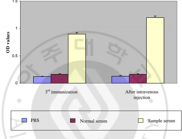
Sera obtained from immunized mice were assayed for antibody formation by indirect ELISA with synthetic peptide of TgLDH-5 (fraction 5) as the antigen (Table 1). Sera from unimmunized mice were used as the negative control. Fig. 3 showed the polyclonal antibody formation for TgLDH-5 antigen. OD values of sera from immunized mice were 0.903 after third immunization. After intravenous injection, the OD values of sera were increased to 1.204, otherwise OD values of normal mouse serum and PBS were 0.16 and 0.13, respectively (Fig. 3).
0 0.5 1 1.5 O D v a lu e s
Fig. 3. Polyclonal antibody titers from the TgLDH-5 immunized mice. OD405 values of
each serum were screened by indirect ELISA (n=3). Sample and normal serum were diluted in PBS at 1:500 ratio, respectively. PBS; control.
3rd immunization After intravenous
injection
PBS Normal serum
Sample serum
B. Cell fusion
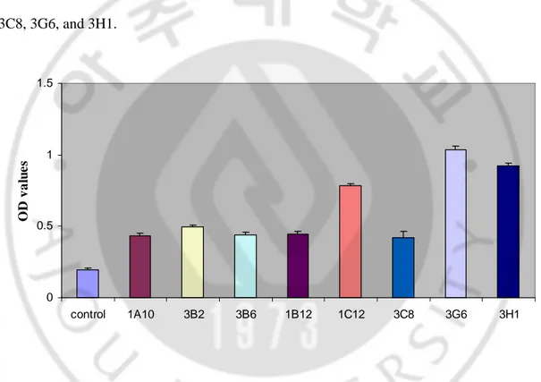
Microscopic examination of the 96 wells revealed the growth of hybridoma cells (Data is not shown). Approximately 60% fusion rate was accomplished in a plate. After final cell fusions, twelve hybridoma cell clones were produced, which the antibody formation for TgLDH-5 were detected by indirect ELISA. After three times of limiting dilution, eight hybridoma clones were selected. The OD values of them ranged from 0.435 to 1.035 and control was 0.192. Eight hybridoma cell clones were named as 1A10, 3B2, 3B6, 1B12, 1C12, 3C8, 3G6, and 3H1. 0 0.5 1 1.5 control 1A10 3B2 3B6 1B12 1C12 3C8 3G6 3H1 O D v a lu e s
Fig. 4. Antibody titers of the hybridoma cell culture supernatant after cell fusion. After hybridoma cells were checked by microscopy, OD405 values of each culture supernatant were
C. Isolation of antibody-producing hybridoma cells
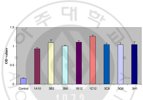
All of eight clones were selected by three times of limiting dilution. Using hybridoma cell culture supernatant after the third limiting dilution, the OD values of them ranged from 0.938 to 1.269 and control was 0.156 (Fig. 5). Among eight clones, 1C12 indicated the highest value.
0 0.5 1 1.5 Control 1A10 3B2 3B6 1B12 1C12 3C8 3G6 3H1 O D v a lu e s
Fig. 5. Antibody titers of the hybridoma cell culture supernatant after third limiting dilution. OD405 values of each culture supernatant were screened by indirect ELISA (n=3).
D. Producing large scale of antibodies
To produce large scale of antibodies, hybridoma cells were intraperitoneally injected to BLAB/c mice and ascitic fluids containing McAbs were harvested. The ascites were diluted at 1:500 ratio with PBS and screened by indirect ELISA. OD values ranged from 1.089 to 1.314, otherwise OD values of normal and PBS were 0.127 and 0.116, respectively (Fig. 6). 0 0.5 1 1.5 PBS Normal 1A10 3B2 3B6 1B12 1C12 3C8 3G6 3H1 O D v a lu e s
Fig. 6. Antibody titers of McAb-containing ascitic fluids. OD405 values of ascetic fluids
were screened by indirect ELISA (n=3). All ascites and normal serum were diluted in PBS at 1:500 ratio. TgLDH-5 peptide was used as antigen. Normal; serum from normal mouse. PBS; control.
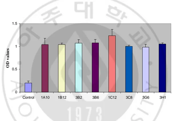
E. The specificity of McAbs with T. gondii lysate
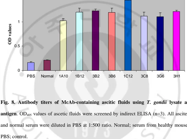
To investigate whether the McAbs were responsive to lysate of T. gondii, indirect ELISA was performed with T. gondii lysate used as antigen. The OD values of cell culture supernatants from eight hybridoma clones ranged from 0.986 to 1.239 and control was 0.203 (Fig. 7). In cases of eight ascites, the antibody titers ranged from 1.025 to 1.445, otherwise OD values of PBS and normal were 0.162 and 0.212, respectively (Fig. 8).
0 0.5 1 1.5 Control 1A10 1B12 3B2 3B6 1C12 3C8 3G6 3H1 O D v a lu e s
Fig. 7. Antibody titers of the hybridoma cell culture supernatant using T. gondii lysate as antigen. OD405 values of each culture supernatant were screened by indirect ELISA (n=3).
0 0.5 1 1.5 PBS Normal 1A10 1B12 3B2 3B6 1C12 3C8 3G6 3H1 O D v a lu e s
Fig. 8. Antibody titers of McAb-containing ascitic fluids using T. gondii lysate as antigen. OD405 values of ascetic fluids were screened by indirect ELISA (n=3). All ascites
and normal serum were diluted in PBS at 1:500 ratio. Normal; serum from healthy mouse. PBS; control.
F. Isotyping of monoclonal antibodies
The McAb isotypes were determined with a mouse monoclonal antibody isotyping kit (Roche, Indiana, USA). All of eight monoclonal antibodies were IgM class.
Table 2. Isotype profile of eight hybridoma clones producing monoclonal antibody
Clones Isotype 1A10 1B12 3B2 3B6 1C12 3C8 3G6 3H1 IgM IgM IgM IgM IgM IgM IgM IgM
IV. DISCUSSION
T. gondii, an intracellular protozoan parasite, is the etiological agent of the
worldwide human and animal toxoplasmosis. Until now, there is no vaccine to prevent toxoplasmosis in humans. Clinical signs of toxoplasmosis are non-specific and are not sufficiently characteristic for a definite diagnosis. In fact, toxoplasmosis mimics several other infectious diseases. Therefore, diagnosis of the infection is important not only for treatment but from the point of view epidemiology and prevention. But diagnosis of any infectious disease depends on isolation of the organism and demonstration of an antibody response to the infection. In the case of toxoplasmosis the organism is difficult to isolate because it is very difficult to culture. The number of organism seen in histological sections or isolated from enlarged lymph nodes is very small. Isolation of organisms from other tissues is also a difficult and time consuming procedure. Diagnosis therefore depends on serological tests (Fleck, 1989).
The most popular test has proved to be the toxoplasma dye test. The disadvantages of this test are that it requires the use of live organisms, is complicated, and needs fresh frozen human plasma. These difficulties have led to a search for specific and sensitive substitutes (Fleck, 1989). Many different antibody tests have been investigated such as the indirect hemagglutination assay, indirect fluorescent antibody assay, the direct agglutination test, the latex agglutination test, ELISA and the immunosorbent agglutination assay test. These tests are quite sensitive and specific and have been proved very useful in confirming the diagnosis and in seroepidemiological studies (Remington, 2000). However, these tests are all time consuming and require a laboratory equipped with proper instruments and
trained personnel. These distinct drawbacks make them unsuitable for direct use in the field and in laboratories where no refrigeration, incubator or expensive spectrophotometric equipment are available. Because of the geographical distribution of toxoplasmosis, there is a need to develop a sensitive, rapid, simple and an inexpensive test for the early screening of patients. With the development of monoclonal antibody in 1980s, assays for detecting T.
gondii antigens were developed (Hafid, 1995).
Recently, immunochromatographic tests, rapid diagnostic tests, have been developed.
Lactate dehydrogenase (LDH) are the most common antigens used in
Immunochromatographic tests (Makler and Hinrichs, 1993).
In the present study, McAbs against the TgLDH were produced and characterized.
Eight hybridoma cells with high ELISA titer were selected. After three times of limiting dilution, all eight clones had strong reactivity to TgLDH by indirect ELISA.
All eight McAbs generated in this study were IgM class. Serologic test for detection of IgM antibodies are commonly performed for the diagnosis of acute acquired toxoplasma infection in the immunocompetent patients (Remington, 1995). Because IgM antibodies may persist for many months or even years following the acute infection (Bobic, 1992; Bono, 1989; Handman, 1980).
In this study, the eight hybridoma cell lines producing TgLDH specific McAbs were established.Although the further studies to investigate the sensitivity against patient sera are needed, these McAbs may be used to create the rapid diagnostic dipstick kit.
V. CONCLUSION
To create the rapid diagnostic dipstick kit that has high sensitivity against LDH (lactate dehydrogenase) of T. gondiil, first of all, hybridoma cells producing monoclonal antibodies with high affinity and specificity must be generated. This study established hybridoma cells producing McAbs against T. gondii LDH (TgLDH).
After cell fusion, hybridoma clones secreting antibodies against TgLDH were screened by ELISA. After three times of limiting dilution, eight clones, 1A10, 1B12, 3B2, 3B6, 1C12, 3C8, 3G6 and 3H1, were selected. The OD values of cell culture supernatant and ascites from eight hybridoma clones ranged from 0.936 to 1.239 and 1.089 to 1.314, respectively. All eight McAbs generated in this study were IgM class.
These results indicated that eight hybridoma clones had strong reactivity to TgLDH and may be useful for diagnosis to T. gondii infection.
REFERENCES
1. Barbero S and Ponte PL: Infectious diseases in the fetus and newborn infant. Arch Sci Med 134: 413-435, 1977
2. Black MW and Boothroyd JC: Lytic cycle of Toxoplasma gondii. Microbiol Mol Biol Rev 64: 607-623, 2000
3. Bobic B, Sibalic D, and Djurkoviv-Djakovic O: High levels of IgM antibodies specific for
Toxoplasma gondii in pregnancy 12 years after primary toxoplasma infection. Gynecol Obstet Invest 31: 182-184, 1991
4. Bohne W, Heesemann J, et al: Reduced replication of Toxoplasma gondii is necessary for induction of bradyzoite-specific antigens: a possible role for nitric oxide in triggering stage conversion. Infect Immun 62: 1761-1767, 1994
5. Dando C, Schroeder ER, et al: The kinetic properties and sensitivities to inhibitors of lactate dehydrogenases (LDH1 and LDH2) from Toxoplasma gondii: comparisons with pLDH from Plasmodium falciparum. Mol Biochem Parasitol 118: 23-32, 2001
6. Del Bono V, Canessa A, Bruzzi P, Fiorelli MA, and Terragna A: Significance of specific immunoglobulin M in the chronological diagnosis of 38 cases of toxoplasmic lymphadenopathy. J Clin Microbiol 27: 2133-2135, 1989
8. Dubey JP: Advances in the life cycle of Toxoplasma gondii. Int J Parasitol 28: 1019-1024, 1998
9. Dubley JP, Lindsay DS, and Speer CD: Structures of Toxoplasma gondii tachyzoites, bradyzoites, and sporozoites and biology and development of tissue cysts. Clin
Microbiol. Rev 11: 267-299, 1998
10. Fleck DG: Annotation: Diagnosis for Toxoplasmosis. J Clin Pathol 42: 191-193, 1989
11. Galfre G and Milstein C: Preparation of monoclonal antibodies: strategies and procedures. Methods Enzymol 73: 3-46, 1981
12. Garcia SL and Bruckner AD: Tissue protozoa (Toxoplasma gondii). In Diagnostic Medical Parasitology (ed Garcia SL & Bruckner AD) Washington, DC: American Society for Microbiology, pp.111–121, 1997a
13. Garcia SL and Bruckner AD: Parasitic infections in the compromised host (Toxoplasma
gondii). In Diagnostic Medical Parasitology (ed. Garciam SL and Bruckner AD)
Washington, DC: American Society for Microbiology, pp.423–424, 1997b
14. Hafid J, Tran Manh Sung R, Raberin H, Akono ZY, Pozzetto B and Jana M: Detection of circulating antigens of Toxoplasma gondii in human infection. Am J Trop Med Hyg 52: 336-339, 1995
15. Handman E and Remington JS: Antibody responses to toxoplasma antigens in mice infected with strains of different virulence. Infect Immun 29: 215-220, 1980
16. Johnson AM: Speculation on possible life cycles for the clonal lineages in the genus toxoplasma. Parasitol Today 13: 393-397, 1997
17. Kayser FH, Bienz KA , Eckert J, Zinkernagel RM: Medical Microbiology. In Pathology: Protozoa, Stuttgart, Germany, Georg Thieme Verlag, pp.508-514, 2005
18. Kavanagh KL, Elling RA and Wilson DK: Structure of Toxoplasma gondii LDH1: active-site differences from human lactate dehydrogenases and the structural basis for efficient APAD+ use. Biochemistry 43: 879-889, 2004
19. Kikuchi T, Nagata T, Furuta T: Production and characterization of a monoclonal antibody against nucleoside triphosphate hydrolase from Toxoplasma gondii. J Eukaryot
Microbiol (Suppl): 195S-196S, 2001
20. Kohler G and Milstein C: Continuous cultures of fused cells secreting antibody of predefined specificity. Nature 256: 495-497, 1975
21. Lee YJ, Kim JH, Jeong SR, Song KJ Kim KM, Park S, Park MS, Shin HJ: Production of Nfa1-specific monoclonal antibodies that influences the in vitro cytotoxocity of
Naegleria fowleri trophozoites on microglial cells. Parasitol Res 101: 1191-1196, 2007
22. Littlefield JW: The use of drug-resistant markers to study the hybridization of mouse fibroblasts. Exp Cell Res 41: 190-196, 1966
23. Luft BJ and Remington JS: Toxoplasmic encephalitis in AIDS. Clin Inf Dis 15: 211-222, 1992
24. Makler MT and Hinrichs DJ: Measurement of the lactate dehydrogenase activity of
Plasmodium falciparum as an assessment of parasitemia. Am J Trop Me Hyg 48:
205-210, 1993
25. Montoya JG: Laboratory diagnosis of Toxoplasma gondii infection and toxoplasmosis. J
Infect Dis 185 (Suppl 1): 73-82, 2002
26. Remington JS and Desmonts G: Toxoplasmosis. In infectious of the fetus and newborn infant (ed. Remington JS and Kllein JO) Philadelphia,WB Saunders, pp.140-243, 1995
27. Remington JS, McLeod R, Thulliez P and Desmonts G: Toxoplasmosis. In Infectious diseases of the fetus and newborn infant (ed. Remington JS and Klein J) Philadelphia, WB Saunders, pp.205–346, 2000
28. Sethi KK, Endo T, Brandis H: Hybridomas secreting monoclonal antibody with specificity for Toxoplasma gondii. J Parasitol 66: 192-6, 1980
29. Strannegard O: The formation of Toxoplasma antibodies in rabbits. Act Pathol Microbiol
Scand 71: 439-449, 1967
30. Tenter AM, Heckeroth AR, et al: Toxoplasma gondii: from animals to humans. Int J
Parasitol 30: 1217-1258, 2000
31. Vander Jagt DL, Hunsaker LA and Heidrich JE: Partial purification and characterization of lactate dehydrogenase from Plasmodium falciparum. Mol Biochem Parasitol 4: 255-264, 1981
32. Weiss LM, Udem SA, et al: Western blot analysis of the antibody response of patients with AIDS and toxoplasma encephalitis: antigenic diversity among Toxoplasma strains.
J Infect Dis 157: 7-13, 1988
33. Yang S and Parmley SF: Toxoplasma gondii expresses two distinct lactate dehydrogenase homologous genes during its life cycle in intermediate hosts. Gene 184: 1-12, 1997
-국문요약-
톡소포자충의
톡소포자충의
톡소포자충의
톡소포자충의 lactate dehydrogenase
lactate dehydrogenase
lactate dehydrogenase
lactate dehydrogenase를
를
를
를 이용한
이용한
이용한
이용한
단클론
단클론
단클론
단클론 항체의
항체의
항체의
항체의 생산
생산
생산
생산
아주대학교 대학원 의학과 당 트린 민안 (지도교수: 신 호 준, 박 선) 목적 목적목적 목적: 톡소포자충의 lactate dehydrogenase에 대하여 높은 민감성과 정확도를 가지는 진단 키트를 만들기 위해서 톡소포자충의 lactate dehydrogenase에 맞 는 펩타이드를 합성한 후 세포 융합 기술을 이용하여 단클론 항체를 만들었다. 방법 방법 방법 방법 : 6-8주령의 BALB/c 마우스에 TgLDH5를 면역시킨 후 비장을 적출하여 PEG를 사용하여 세포 융합을 시켰다. 그리고, 세포 융합 된 세포들의 isotyping 을 확인하였다. 결과 결과 결과 결과: 8개의 융합된 세포들을 얻을 수 있었는데 그 이름을 1A10, 1B12, 3B2, 3B6, 1C12, 3C8, 3G6 그리고 3H1이라 명명하였다. 또한, 8개의 단클론 항체들 의 O.D값은 0.938에서 1.269를 보였고 isotyping을 확인한 결과 모두 IgM이었 다.결론 결론결론 결론: 8개의 단클론 항체들 모두 TgLDH와 강하게 반응하였다. 핵심어 핵심어핵심어 핵심어: 톡소포자충, lactate dehydrogenase, 세포 융합 기술, 단클론항체