저작자표시-비영리-변경금지 2.0 대한민국 이용자는 아래의 조건을 따르는 경우에 한하여 자유롭게 l 이 저작물을 복제, 배포, 전송, 전시, 공연 및 방송할 수 있습니다. 다음과 같은 조건을 따라야 합니다: l 귀하는, 이 저작물의 재이용이나 배포의 경우, 이 저작물에 적용된 이용허락조건 을 명확하게 나타내어야 합니다. l 저작권자로부터 별도의 허가를 받으면 이러한 조건들은 적용되지 않습니다. 저작권법에 따른 이용자의 권리는 위의 내용에 의하여 영향을 받지 않습니다. 이것은 이용허락규약(Legal Code)을 이해하기 쉽게 요약한 것입니다. Disclaimer 저작자표시. 귀하는 원저작자를 표시하여야 합니다. 비영리. 귀하는 이 저작물을 영리 목적으로 이용할 수 없습니다. 변경금지. 귀하는 이 저작물을 개작, 변형 또는 가공할 수 없습니다.
碩士學位論文
넙치(
Paralichtys olivaceus
)의
점액포자충성 여윔증에 대한
톨트라주릴을 이용한 치료법연구
Therapeutic study of myxosporean emaciation disease in olive flounder (Paralichtys
olivaceus) using toltrazuril
濟州大學校 大學院
海洋生命科學科
姜 味 來
Therapeutic study of myxosporean
emaciation disease in olive flounder
(Paralichtys olivaceus) using
toltrazuril
Mi-Rae Kang
(Supervised by professor Joon Bum Jeong)
A thesis submitted in partial fulfillment of the
requirement for the degree of Master of Science
Department of Marine Biomedical Science
GRADUATE SCHOOL
JEJU NATIONAL UNIVERSITY
목 차
목 차 ··· ⅰ ABSTRACT ··· ⅲ LIST OF FIGURES ··· ⅳ LIST OF TABLES ··· ⅶ I. 서론 ··· 1 II. 재료 및 방법 ··· 4 1. 실험어 ··· 4 2. 시험제제 ··· 5 3. In vitro 상에서의 점액포자충 배양조건 설정 ··· 6 3-1. 점액포자충 배양을 위한 cell lin 설정 ··· 6 3-2. 점액포자충 배양을 위한 온도설정 ··· 7 4. In vitro 상에서의 시험제제 투여 ··· 8 4-1. 세포에 대한 시험제제의 독성 실험 ··· 8 4-2. 점액포자충에 대한 시험제제의 치료효과 실험 ··· 9 5. In vivo 상에서의 시험제제 투여 ··· 10 6. DNA 분리 ··· 11 7. PCR ··· 12 III. 결과 ··· 14 1. In vitro 상에서의 점액포자충 배양조건 설정 ··· 142. In vitro 상에서의 시험제제 효과 ··· 17 2-1. 세포에 대한 시험제제의 반응 ··· 17 2-2. 점액포자충에 대한 시험제제의 치료효과 ··· 22 3. 약욕실험에서의 시험제제 효과 ··· 28 3-1. 약욕실험에서의 누적폐사율 관찰 ··· 28 3-2. PCR ··· 29 IV. 고 찰 ··· 32 V. 요약 ··· 35 VI. 참고문헌 ··· 36 VII. 감사의글 ··· 42
ABSTRACT
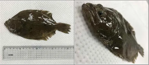
Since 2007, at Jeju olive flounder farm, emaciated fishs with a size of about 20 cm has been showing symptoms such as body color blackening, decreased fish body and death. In 2015-2017, the average cumulative mortality rate caused by emaciation disease of olive flounder in jeju accounted for 10.3%. Various studies have been conducting on the causative agents that cause these emaciation disease. Parvicapsula sp. and Enteromyxum leei were confirmed at the emaciated symptoms olive flounder.
In order to search candidates for the treatment of myxosporean emaciation disease of olive flounder, Paralichthys olivaceus, in vitro
and in vivo experiments were conducted using products such as Coxi-stop, Coxiclin, and BLEAN80. In the case of Coxi-stop, whose major component is toltrazuril, in vitro experiment using BF-2 cells showed a tendency of reducing the activity of myxosporea and did not exhibit cytotoxicity such as cell lysis. In the in vivo activity measurements, the experimental group immersed with Coxi-stop showed a lower cumulative mortality rate than the control group. Toltrazuril is effective in the treatment of parasitic diseases such as cattle, pigs, and poultry. which has been reported to be effective as a treatment for parasitic diseases of fish. This study result is similar to the previous report that toltrazuril has a therapeutic effect on parasitic fish disease.
This study suggests that toltrazuril is a potential candidate for the treatment of emaciation disease of olive flounder.
LIST OF FIGURES
Fig. 1. The external sign of the emaciated symptoms olive flounder. ··· 4 Fig. 2. Immersion treatment tank with olive flounder with emaciation
disease. ··· 10 Fig. 3. Observation on 3 day after inoculation of myxosporea in HeLa
cell, BF-2 cell and CHSE-214 cell. (A), Normal HeLa cell; (B), HeLa cell inoculated by myxosporea; (C), Normal BF-2 cell; (D), BF-2 cell inoculated by myxosporea; (E), Normal CHSE-214 cell; (F), CHSE-214 cell inoculated by myxosporea; Bar arrow, myxosporea. ···15 Fig. 4. Observation of changes in BF-2 cell on 3 day after inoculation
of myxosporea cultured at 20℃ and 25℃. (A), Normal BF-2 cell; (B), BF-2 cell inoculated by myxosporea at 20℃; (C), BF-2 cell inoculated by myxosporea at 25℃; Bar arrow, myxosporea. ···16 Fig. 5. Observation of BF-2 cells change in the cytotoxicity test of
the test formulation at 2, 4, 24 h after inoculation of Coxi-stop, Coxiclin, BLEAN80. Control, Normal BF-2 cell. ····18 Fig. 6. Observation of BF-2 cells change in the cytotoxicity test of
the test formulation at 2, 4, 24 h after inoculation of Coxi-stop, Coxiclin, BLEAN80. Experimental group 1, BF-2 cell inoculated by Coxi-stop. ···19 Fig. 7. Observation of BF-2 cells change in the cytotoxicity test of
the test formulation at 2, 4, 24 h after inoculation of Coxi-stop, Coxiclin, BLEAN80. Experimental group 2, BF-2
cell inoculated by Coxiclin. ··· 20 Fig. 8. Observation of BF-2 cells change in the cytotoxicity test of
the test formulation at 2, 4, 24 h after inoculation of Coxi-stop, Coxiclin, BLEAN80. Experimental group 3, BF-2 cell inoculated by BLEAN80. ··· 21 Fig. 9. Observation of the therapeutic effect of the test formulation
against myxosporea in BF-2 cell inoculated by myxosporea isolated from emaciated olive flounder at 2, 4, 24 h after inoculation of Coxi-stop, Coxiclin or BLEAN80. N.C, negative control, Normal BF-2 cell. ··· 23 Fig. 10. Observation of the therapeutic effect of the test formulation
against myxosporea in BF-2 cell inoculated by myxosporea isolated from emaciated olive flounder at 2, 4, 24 h after inoculation of Coxi-stop, Coxiclin or BLEAN80. P.C, positive control, BF-2 cell inoculated by myxosporea; Bar arrow, myxosporea. ··· 24 Fig. 11. Observation of the therapeutic effect of the test formulation
against myxosporea in BF-2 cell inoculated by myxosporea isolated from emaciated olive flounder at 2, 4, 24 h after inoculation of Coxi-stop, Coxiclin or BLEAN80. Experimental group 1, BF-2 cell inoculated by myxosporea and Coxi-stop; Bar arrow, myxosporea. ··· 25 Fig. 12. Observation of the therapeutic effect of the test formulation
against myxosporea in BF-2 cell inoculated by myxosporea isolated from emaciated olive flounder at 2, 4, 24 h after inoculation of Coxi-stop, Coxiclin or BLEAN80. Experimental group 2, BF-2 cell inoculated by myxosporea and Coxiclin; Bar arrow, myxosporea. ··· 26
Fig. 13. Observation of the therapeutic effect of the test formulation against myxosporea in BF-2 cell inoculated by myxosporea isolated from emaciated olive flounder at 2, 4, 24 h after inoculation of Coxi-stop, Coxiclin or BLEAN80. Experimental group 3, BF-2 cell inoculated by myxosporea and BLEAN80; Bar arrow, myxosporea. ··· 27 Fig. 14. PCR amplification using EM-F/R primer set for detection of
Parvicapsula sp. (Lane 1-8) and EL-F/R primer set for detection of Enteromyxum leei (Lane 9-16) from total nucleic acids of kidney (Lane 1, 3, 5, 7, 9, 11, 13 and 15) and intestine (Lane 2, 4, 6, 8, 10, 12, 14 and 16) of emaciated olive flounder. Lane 1, 2, 9 and 10, Immersion treatment of Coxiclin; Lane 3, 4, 11 and 12, Immersion treatment of Coxi-stop; ; Lane 5, 6, 13 and 14, Immersion treatment of BLEAN80; Lane 7, 8, 15 and 16, Non-treatment (Control); M, 100 bp DNA ladder. ··· 31
LIST OF TABLES
Table 1. Test formulation used in the experiment ··· 5 Table 2. PCR primers used in this study ··· 13 Table 3. Comparison of the numbers of survived fish and PCR results
in emaciated olive flounder after immersion treatment of Coxi-stop, Coxiclin and BLEAN80 ··· 30
Ⅰ. 서 론
우리나라 양식 산업에 있어 넙치는 대표적인 양식어류중 하나로 2019년부터 2020년 상반기까지를 기준으로 국내 양식어류 생산량 중 약 51%의 높은 비 중을 차지하고 있다(Korean statistical information service, KOSIS, 2020). 그러나 넙치 양식 과정 중 발생하는 다양한 바이러스나 기생충, 세균 성질병으로 인하여 폐사가 지속적으로 발생하고 있으며, 이로 인해 국내 넙치 양식 산업에 큰 피해를 입히고 있다. 또한, 이전에 발생한 사례가 없었던 새로 운 질병으로 인한 피해가 가중되는 경우 새로운 질병이 발생한 후에는 예방대 책이 부족한 실정이므로 대량폐사로 이어질 수 있어(Cho et al., 2019) 이에 대한 대책이 필요한 실정이다. 2015년부터 2017년까지를 기준으로 국내 넙치생산량의 약 61%를 차지하 는 제주넙치의 경우(KOSIS, 2020), 제주넙치의 폐사피해를 조사 한 결과 2015년에는 28.69%, 2016년에는 32.65%, 2017년에는 26%의 폐사를 보 였으며 제주넙치의 폐사원인을 분석해보았을 때, 여윔증에 의한 폐사가 2016 년에는 8.82%, 2017년에는 16.2%로 점차 증가하는 것이 보고되었다(Shim et al., 2019). 제주 넙치 양식장에서는 2007년부터 20㎝ 전후 크기의 넙치에서 체색흑화, 어체중 감소, 여위는 등의 증상이 1-3주간에 걸쳐 나타나며 폐사에 이르는 여윔증이 발생하고 있으며, 국내에서 넙치에 여윔증을 일으키는 원인체에 대 한 정확한 원인은 밝혀지지 않고 있다. 이에 따라, 여윔증을 일으키는 원인체 에 관한 연구가 다양하게 진행되고 있으며(Kim et al., 2011; Choi et al., 2012; Sekiya et al., 2016; Kim et al., 2015a, 2015b, 2017; Kim et al., 2018; Kim and Jeong, 2018; Shin et al., 2018a, 2018b), Kim et al. (2015a)에 따르면 국내에서 발생하는 여윔증 원인체를 분석한 결과
할 수 있는 PCR 방법을 개발하였고, Kim et al. (2018)은 2014년부터 2015년까지 제주도 60개소의 넙치양식장을 대상으로 정기적인 여윔증 모니 터링을 실시하여 여윔증상 넙치로부터 Parvicapsula sp.가 공통적으로 검출된 다는 것을 보고하였고, Sekiya et al. (2016)에 의하여 Enteromyxum leei
(E.leei) 도 여윔증상 넙치에서 다수 검출되는 것을 확인하였다. 어류 여윔증 은 대부분 점액포자충이 그 원인인 것으로 알려져 있으며, Shin et al. (2018b)은 여윔증상을 보이는 넙치에서 점액포자충인 Parvicapsula anisocaudata와 E.leei가 검출되는 것을 확인하였다. 이에 Kim et al.
(2018)은 우리나라 넙치 여윔증의 원인은 점액포자충이 두가지 이상 인 것으 로 보고하였다.
현재 어류의 기생충성질병에 대한 치료제에 대한 연구들이 다양하게 진행되 고 있지만(Ahn et al., 2017; Athanassopoulou et al., 2009; Iglesias et al., 2002; Kang and Kim, 2015; Monlnar K. and Ostoros G., 2007) 여 윔증상을 유발하는 점액포자충에 대한 예방대책 및 치료제 개발에 대한 연구 는 미비한 실정이다. 이에 본 연구에서는 1980년대에 처음 개발되어 현재까 지 소, 돼지, 가금류, 양 등의 기생충성 질병 치료에 효과가 있으며, 특히 축 산분야에서 점액포자충에 의한 콕시듐증의 치료 및 예방 목적으로 이용되고 있는 톨트라주릴(Toltrazuril) (EMEA, 1998; Greif G, 2000; Joachim et al., 2019; Kim et al., 2010; Maes et al., 2007; Olsen et al., 2012; Vanparijs et al., 1989)을 사용하여 점액포자충에 의한 넙치 여윔증 치료에 도 효과가 있는지 알아보기 위해 실험을 진행하였다. 톨트라주릴은 점액포자충의 한 종류인 콕시듐 원충의 세포 내 기생단계에서 세망내피계와 골지체의 종대와 비정상적인 핵 주위공간을 유발하여 원충의 구 조변화를 일으켜 원충을 사멸하게 하며, 원충의 호흡기관에서 효소의 감소를 유도하는 작용을 하는 것으로 알려져 있다(Poultry Research, 2014). 또한, 톨트라주릴은 독성이 강한 제제로서, 사람이 직접적으로 섭취할 경우 부작용 을 일으킬 수도 있어(Ai et al., 2011), EU (European union)에서는 동물에 대하여 최대허용기준(Maximum residue limits, MRLs)을 설정하여 사용하
고 있다(Hong et al., 2019). 그러나, 국내 동물용의약품 공정서 에는 톨트라 주릴에 대한 분석 자료가 없어 외국의 분석 자료를 참고하여 동물용의약품을 분석하고 있으며(Jeong et al., 2017), 수산물에 대한 잔류허용기준도 없었 다. 그러나 최근 국내에서 톨트라주릴을 분석하는 방법이 개발되었고(Jeong et al., 2017), 양식 넙치에 발생하는 기생충성 질병의 치료제로서 평가연구 를 할 수 있도록 톨트라주릴 분석법이 개발되었다(Hong et al., 2019). 톨트 라주릴은 어류의 기생충성 질병 치료제로써 연구되고 있으며(Ahn et al., 2017; Athanassopoulou et al., 2009; Iglesias et al., 2002; K. Monlnar K. and Ostoros G., 2007; Mehlhorn et al., 1988; Schmahl and Mehlhorn, 1988; Schmahl et al., 1989a, 1989b, 1991) 이전의 연구들에 서 어류의 기생충성 질병 치료제로써 효과를 보인다고 보고되었다 (Athanassopoulou et al., 2009; Iglesias et al., 2002; Mehlhorn et al., 1988; Schmahl and Mehlhorn, 1988; Schmahl et al., 1989a, 1989b, 1991). 이에 톨트라주릴을 주성분으로 하여 생산되는 Coxi-stop, Coxiclin 제품을 시험제제로 사용하여 여윔증상 넙치에 치료 효과가 있는지 확인하고자 하였다.
Ⅱ. 재료 및 방법
1. 실험어 여윔증상을 보이며 대량 폐사가 발생하고 있는 제주도 소재 넙치양식장으로 부터 넙치(평균 체장 23.8±0.2 cm, 평균 체중 104 ± 14.1 g) 45마리를 운반 용기에 10마리 이하가 되도록 나누어 밀봉한 뒤, 제주대학교 해양과학대 학에 위치한 실험수조에 옮겼다. 수조는 80 L의 플라스틱 수조에 해수 60 L 를 유지하며 매일 환수하였고, 공기를 주입해 주었으며 수온은 15±0.5℃로 유지하였다. 모든 실험군은 Coxi-stop, Coxiclin, BLEAN80 및 대조군 (control group)으로 나누어, 각 그룹당 총 10마리를 5마리씩 2개의 수조로 나누어 실험을 진행하였다. 여윔증상이 있는 넙치 중 5마리를 임의로 선택한 후 신장과 장 조직을 적출하여, 여윔증 원인체로 알려진 두 종류의 점액포자 충 Parvicapsula sp. 및 E. leei에 의한 감염유무를 PCR (Polymerase chain reaction) 방법으로 확인하기 위해 DNA분리에 사용하였다.2. 시험제제
본 연구에서는 톨트라주릴 성분이 함유된 Coxi-stop (WooGene B&G. Co., Ltd), Coxiclin (WooGene B&G. Co., Ltd) 제품과 양식현장에서 혼합 성 단미사료로 사용되고 있는 BLEAN80 (㈜대명산업) 제품을 시험제제로 사 용하였다. 각 시험제제들에 대한 성분 함량 표는 다음과 같다(Table 1).
Table 1. Test formulation used in the experiment
Test formulation Ingredient Content (1 L)
Coxi-stop Toltrazuril 50g
Sodium docusate 2.5g
Sodium propionate 1.8g
Sodium benzoate 1.8g
Simethicone 0.5g
Citric acid anhyous 4g
Coxiclin Toltrazuril 25g
Triethanolamine 300g
Polyetylene glycol suitable amount
BLEAN80 Sodium chloride 0.79% ≥
Magnesium sulfate 0.54% ≥ Potassium sulfate 0.88% ≥ Copper sulfate 0.11% ≥ Zinc sulfate 0.64% ≥ Manganese(II) sulfate 1.80ppm ≥ Ferrous sulfate 33.92ppm ≥
3. In vitro 상에서의 점액포자충의 배양조건 설정
3-1. 점액포자충 배양을 위한 cell line 설정
본 연구에서는 in vitro상에서 점액포자충이 배양될 수 있도록 cell line을 사용하여 배양조건에 대한 연구를 진행하였다. 사람의 자궁경부암 조직에서 유래되어 인체과학 실험에서 가장 많이 이용되고 있으며 최근 스쿠치카충의 배양을 시도하였다고 알려진 HeLa cell을 분양받아 CO2가 공급되는 37℃에 배양하며 실험을 준비하였고, 어류에서 유래되어 어류의 바이러스 실험에 많 이 이용되고 있는 bluegill fin (BF-2) cell과 chinook salmon embryo (CHSE-214) cell을 각각 25℃와 20℃에 배양하며 80%의 cell frequency 를 유지하도록 하였다. 이후, 여윔증 감염이 확인된 넙치의 신장을 적출하여 1% antibiotic-antimycotic (Gibco, BRL)을 첨가한 Dulbecco’s Modified Eagle Medium (DMEM, Gibco, USA) 으로 세척한 후 5% fetal bovine serum (FBS)과 1% antibiotic-antimycotic을 첨가한 DMEM 이 들어 있는 petri dish에 두고 30분간 반응시키며 점액포자충이 유리되도록 하였다. 80% cell frequency로 배양 중인 3종의 cell은 세포배양액을 모두 버려준 뒤 5% FBS과 1% antibiotic-antimycotic을 첨가한 DMEM 5 mL을 넣어준 후, 점 액포자충이 부유된 부유액 1 mL씩 3종의 cell에 각각 접종하였다. 점액포자 충이 감염된 각각의 cell은 각 cell의 배양적정온도에서 3일간 배양 후 광학현 미경을 통하여 관찰하였으며 실험은 2반복 진행하였다.
3-2. 점액포자충 배양을 위한 온도설정
in vitro상에서 점액포자충이 배양될 수 있는 적정온도를 설정하기 위하여 점액포자충이 감염된 BF-2 cell을 배양온도를 다르게 설정하여 연구를 진행 하였다. 먼저는 여윔증 감염이 확인된 넙치의 신장을 적출하여 1% antibiotic-antimycotic을 첨가한 DMEM 으로 세척한 후 5% FBS와 1% antibiotic-antimycotic을 첨가한 DMEM 5 mL이 들어 있는 petri dish에 두고 30분간 반응시키며 점액포자충이 유리되도록 하였다. 또한, 80% cell frequency로 배양 중인 BF-2 cell의 세포배양액을 모두 버려준 뒤 5% FBS 과 1% antibiotic-antimycotic을 첨가한 DMEM 5 mL을 넣어준 후, 점액포 자충이 부유된 부유액 1 mL씩 cell에 각각 접종하였다. 점액포자충을 접종한 각각의 BF-2 cell은 20℃, 25℃로 나누어 3일간 배양 후 광학현미경을 통하 여 관찰 하였다.
4. In vitro 상에서의 시험제제 투여
4-1. 세포에 대한 시험제제의 독성 실험
본 연구에서는 시험제제의 안전성을 확인하기 위해 세포를 이용한 세포독성 실험을 진행 하였다. 먼저, 80% cell frequency로 배양 중인 BF-2 cell의 세포배양액을 모두 버려준 뒤 5% FBS와 1% antibiotic-antimycotic을 첨가 한 DMEM 5 mL을 넣어준 후, Coxi-stop, Coxiclin은 원액으로 100 μL, BLEAN80은 원액으로 200 μL를 넣고 25℃에서 반응시켰으며 대조군에는 어떤 시험제제도 접종하지 않았다. 시험제제 접종 후 2시간, 4시간, 24시간째 에 시험제제 투여에 따른 세포 반응을 광학현미경을 통하여 관찰하였다. Coxi-stop의 경우, 시험제제가 불투명한 하얀색을 띄고있어 세포관찰이 잘 되지 않아 접종 후 2시간, 4시간, 24시간째에 5% FBS와 1% antibiotic-antimycotic을 첨가한 DMEM 6 mL으로 세척한 후 광학현미경을 통하여 관찰하였다.
4-2. 점액포자충에 대한 시험제제의 치료효과 실험
세포에 점액포자충을 감염시킨 후 시험제제를 접종하였을 때, 점액포자충의 변화를 관찰하고자 하였다. 먼저, 여윔증 감염이 확인된 넙치의 신장을 적출하 여 1% antibiotic-antimycotic을 첨가한 DMEM 으로 세척한 후 5% FBS와 1% antibiotic-antimycotic을 첨가한 DMEM 5 mL이 들어 있는 petri dish 에 두고 30분간 반응시키며 점액포자충이 유리되도록 하였다. 또한, 80% cell frequency로 배양 중인 BF-2 cell의 세포배양액을 모두 버려준 뒤 5% FBS과 1% antibiotic-antimycotic을 첨가한 DMEM 5 mL을 넣어준 후, 점 액포자충이 유리된 부유액 1 mL씩 BF-2 cell에 각각 접종하였다. 점액포자 충을 접종한 각각의 BF-2 cell에 Coxi-stop, Coxiclin은 원액으로 100 μ L, BLEAN80은 원액으로 200 μL를 넣고 25℃에서 반응시켰고, 대조군에는 어떤 시험제제도 접종하지 않았다. 시험제제 접종 후 2시간, 4시간, 24시간째 에 시험제제 투여에 따른 세포 및 점액포자충의 반응을 광학현미경을 통하여 관찰하였다.
5. In vivo 상에서의 시험제제 투여
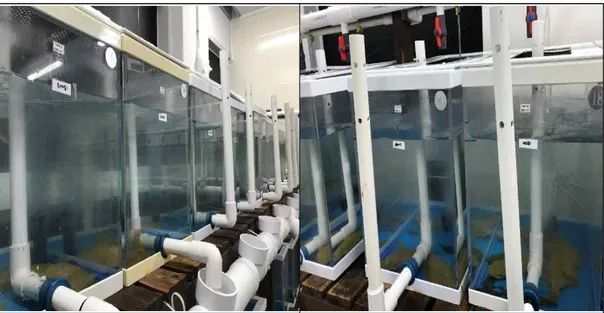
In vivo 상에서 여윔증상 넙치에 대한 Coxi-stop, Coxiclin 및 BLEAN80 의 치료 효과를 조사하고자 약욕실험을 진행하였다. 그 방법은 다음과 같다.
Parvicapsula sp. 및 E. leei에 의하여 복합적으로 감염된 것이 확인된 넙치 를 8개의 80 L 수조에 각각 5마리씩 되도록 넣어주었고, 2개의 수조가 한 group이 되도록 하여 시험제제를 투여하였다. Coxi-stop, Coxiclin은 각각 0.3 mL/L씩, BLEAN80은 0.6 mL/L의 농도가 되도록 약욕 처리하였다. 대 조군에는 어떤 시험제제도 처리하지 않았다. 약욕은 16일간 매일 1시간 동안 진행하였고, 이후 모든 실험군과 대조군의 해수는 모두 환수해 주었다. 약욕이 진행되는 16일 동안 매일 폐사를 확인하였고 실험기간 동안의 누적폐사율을 분석하였다. 또한, 실험기간 중 폐사한 넙치와 실험 종료 후 살아남은 넙치는 모두 신장과 장을 적출하여 DNA 분리 후 PCR 분석에 사용하였다.
Fig. 2. Immersion treatment tank with Olive flounder with emaciation disease.
6. DNA 분리
폐사한 넙치와 살아남은 넙치로부터 신장과 장 조직을 적출하여 DNeasy Blood & Tissue Kit (Qiagen, Hilden, Germany)를 사용하여 total DNA 를 분리하였다. 먼저 각 조직 30 mg에 ATL buffer 180 μL와 proteinase K 20 μL를 첨가하여 56℃에서 조직이 녹을 때까지 반응시켰다. 반응 후, AL buffer 200 μL를 섞은 다음 ethanol 200 μL를 더하여 spin column에 옮겨 6,000 x g (gravity)로 1분간 원심 분리하였다. Column을 새로운 tube로 옮긴 후 AW1 buffer와 AW2 buffer 500 μL를 이용하여 세척과정 을 거친 후, AE buffer 200 μL를 첨가하여 total DNA를 분리하였다. 분리 된 DNA는 실험 전까지 -20℃에 보관하였다.
7. PCR
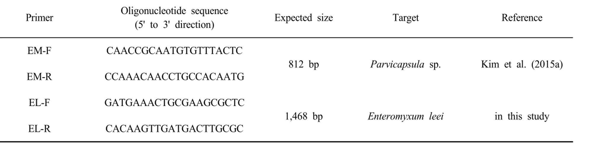
두 종류의 점액포자충이 복합 감염된 넙치로부터 각 병원체를 검출하기 위 하여, Parvicapsula sp.의 검출을 위한 EM-F/R primer set (812 bp) 및
E. leei의 검출을 위한 EL-F/R primer set (1,468 bp)를 각각 사용하여 PCR을 실시하였으며(Table 2), PCR은 microtube에 1 μM의 각 primer, 2.5 mM의 각 dNTP, 10 x G-Taq Buffer, 2.5 U G-Taq DNA polymerase (Gene Pro Themal Cycler Cosmo, Korea) 및 template DNA로서 추출된 핵산을 첨가한 후 distilled water로 PCR 혼합물의 최종 volume이 20 μL가 되게 하였다. Parvicapsula sp.를 검출하기 위한 PCR 조건은 95℃에서 3분간 pre-denaturation시킨 후, 95℃에서 30초 denaturation, 55℃에서 30초 annealing, 72℃에서 30초 extension의 반응 을 1회로 하여, 35회 반복하여 반응시켰다. 그리고 72℃에서 7분간 post-extension시켰다. Enteromyxum leei를 검출하기 위한 PCR 조건은 93℃에서 3분간 pre-denaturation시킨 후, 93℃에서 1분 denaturation, 5 2℃에서 1분 annealing, 72℃에서 1분 extension의 반응을 1회로 하여, 35 회 반복하여 반응시켰다. 그리고 72℃에서 5분간 post-extension시켰다. PCR 후 증폭 산물은 1 × TAE buffer를 전기영동을 위한 완충액으로 하여, 0.1 μL/mL SYBR ® Safe DNA gel stain이 첨가된 1% agarose gel 상에 서 전기영동한 후, UV 검출기를 이용하여, ultraviolet 상에서 검출되는 산물 의 크기를 관찰하였다.
Table 2. PCR primers used in this study
Primer Oligonucleotide sequence
(5' to 3' direction) Expected size Target Reference
EM-F CAACCGCAATGTGTTTACTC
812 bp Parvicapsula sp. Kim et al. (2015a)
EM-R CCAAACAACCTGCCACAATG
EL-F GATGAAACTGCGAAGCGCTC
1,468 bp Enteromyxum leei in this study
Ⅲ. 결 과
1. In vitro 상에서 점액포자충 배양조건 설정
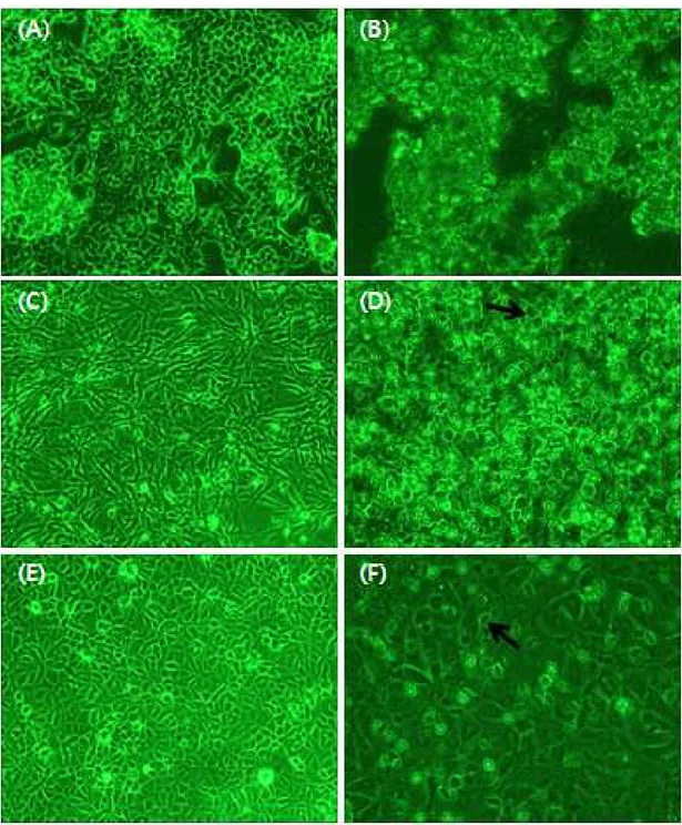
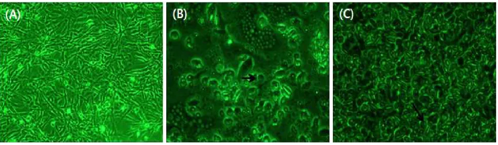
여윔증을 일으키는 원인체로 알려진 점액포자충에 대한 치료연구를 진행하기 앞서 점액포자충이 배양되는 조건을 설정하여 실험을 진행하고자 하였다. 이에 따라 인체에서 유래된 HeLa cell과 어류에서 유래된 BF-2 cell, CHSE-214 cell을 이용하여 점액포자충이 배양되는 cell line과 온도를 설정하였다. 점액포 자충이 감염된 HeLa cell의 경우, 적정배양온도 37℃에서 배양되며 확인되었던 포자가 3일째부터는 관찰되지 않았고, cell의 lysis만 확인되었다(Fig. 3A,B). 반면에 BF-2 cell과 CHSE-214 cell에서는 포자의 형태가 증가하는 것을 확인 할 수 있었다. 또한, CHSE-214 cell보다 BF-2 cell에서 보다 더 포자의 형태 가 증가하는 것을 확인하였으며, 두 번의 반복실험에서 동일한 결과를 확인할 수 있었다(Fig. 3C,D,E,F). 감염된 cell의 상등액을 이용하여 PCR을 실시한 결과, cell이 점액포자충에 감염된 것을 확인할 수 있었다. 점액포자충의 배양이 잘되 는 것으로 확인된 BF-2 cell에서 점액포자충 배양온도를 설정하기 위해 점액포 자충이 감염된 BF-2 cell을 온도를 다르게 하여 20℃, 25℃에서 배양하였다. 그 결과, 25℃에서 포자의 형태가 20℃보다 더 많이 관찰되었고(Fig. 4), 감염 된 cell의 상등액을 이용하여 PCR을 실시한 결과 20℃의 감염상등액 보다 25℃ 의 감염 상등액에서 점액포자충의 농도가 높은 결과를 확인하였다.
Fig. 3. Observation on 3 day after inoculation of myxosporea in HeLa cell, BF-2 cell and CHSE-214 cell. (A), Normal HeLa cell; (B), HeLa cell inoculated by myxosporea; (C), Normal BF-2 cell; (D), BF-2 cell inoculated by myxosporea; (E), Normal CHSE-214 cell; (F), CHSE-214 cell inoculated by myxosporea; Bar arrow, myxosporea.
Fig. 4. Observation of changes in BF-2 cell on 3 day after inoculation of myxosporea cultured at 20℃ and 25℃. (A), Normal BF-2 cell; (B), BF-2 cell inoculated by myxosporea at 20℃; (C), BF-2 cell inoculated by myxosporea at 25℃; Bar arrow, myxosporea.
2. In vitro 상에서 시험제제의 효과
2-1. 세포에 대한 시험제제의 반응
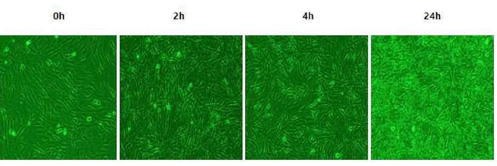
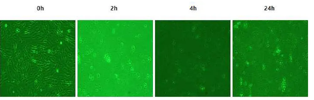
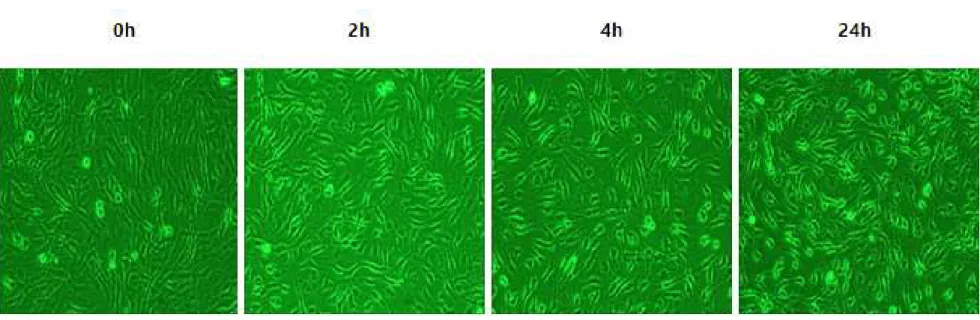
본연구에서 사용되는 시험제제인 Coxi-stop, Coxiclin 및 BLEAN80에 대하 여 안전성을 확인하기 위하여 세포독성 실험을 진행하였다. 실험군과 대조군으로 나누어 각각의 시험제제를 접종한 후, 세포의 변화를 관찰하였다. 시험제제를 첨 가하지 않은 대조군에서는 BF-2 cell의 어떤 변화도 관찰되지 않았다(Fig. 5). Coxi-stop을 첨가한 실험군의 경우 시간의 경과에 따라 시험제제가 세포에 흡 착되었으나 Cell lysis현상이 관찰되지 않는 것으로 보아 세포독성이 없는 것으 로 확인되었다(Fig. 6). Coxiclin을 첨가한 실험군의 경우, 시험제제 접종 2시간 이후부터 cell lysis 현상이 관찰되기 시작하여 대조군에 비하여 높은 세포독성 이 확인되었다(Fig. 7). BLEAN80을 첨가한 실험군의 경우 시간의 경과에 따라 세포의 모양이 일부 변성되는 것이 관찰되어 일부 세포독성이 있는 것으로 관찰 되었다(Fig. 8).
Fig. 5. Observation of BF-2 cells change in the cytotoxicity test of the test formulation at 2, 4, 24 h after inoculation of Coxi-stop, Coxiclin, BLEAN80. Control, Normal BF-2 cell.
Fig. 6. Observation of BF-2 cells change in the cytotoxicity test of the test formulation at 2, 4, 24 h after inoculation of Coxi-stop, Coxiclin, BLEAN80. Experimental group 1, BF-2 cell inoculated by Coxi-stop.
Fig. 7. Observation of BF-2 cells change in the cytotoxicity test of the test formulation at 2, 4, 24 h after inoculation of Coxi-stop, Coxiclin, BLEAN80. Experimental group 2, BF-2 cell inoculated by Coxiclin.
Fig. 8. Observation of BF-2 cells change in the cytotoxicity test of the test formulation at 2, 4, 24 h after inoculation of Coxi-stop, Coxiclin, BLEAN80. Experimental group 3, BF-2 cell inoculated by BLEAN80.
2-2. 점액포자충에 대한 시험제제의 치료효과
감염 넙치의 신장으로부터 유리시킨 점액포자충을 BF-2 cell에 넣어주고, Coxi-stop, Coxiclin 및 BLEAN80과 대조군으로 나누어 각각의 시험제제를 접 종한 후, 세포와 점액포자충의 변화를 관찰하였다. 시험제제와 점액포자충을 첨 가하지 않은 음성대조군에서는 BF-2 cell의 어떤 변화도 관찰되지 않았다(Fig. 9). 시험제제를 첨가하지 않고 점액포자충만 접종시킨 양성대조군에서는 점액포 자충의 움직임이 매우 활발하게 나타났고, 접종 후 4시간째부터는 BF-2 cell의 모양이 조금씩 변화하기 시작하였으며, 접종 후 24시간째까지도 점액포자충이 잘 배양되고 있는 것을 확인하였다(Fig. 10). Coxi-stop을 첨가한 실험군의 경 우, 시험제제 접종 2시간째부터 점액포자충의 움직임과 수가 감소하기 시작하여 반응 4시간째에 확연하게 감소하는 것이 관찰되었고(Fig. 11) 양성대조군(Fig. 10)과 비교해보아도 점액포자충의 수가 감소하는 것을 확인할 수 있었다. 또한, 시험제제의 세포독성 반응을 관찰한 결과(Fig. 6)와 동일하게 접종 후 24시간째 까지도 cell lysis 현상이 관찰되지 않았고, 음성대조군(Fig. 9)과 비교해 보았을 때에도 cell lysis 현상이 관찰되지 않아 실험에 사용한 농도에서의 세포독성은 거의 없는 것으로 확인되었다. Coxiclin을 첨가한 실험군의 경우, 시험제제 접종 2시간 이후부터 cell lysis 현상이 관찰되기 시작하여 세포독성 반응을 관찰한 결과(Fig. 7)와 동일하게 세포독성이 강한 것으로 확인되었고, cell lysis 현상과 함께 점액포자충의 움직임이 감소하는 것이 관찰되었다(Fig. 12). BLEAN80을 첨가한 실험군의 경우, 접종 후 2시간째까지도 양성대조군(Fig. 10)과 비교해 보았을 때 점액포자충의 움직임에는 변화가 없었고, 접종 후 4시간 이후부터 점 액포자충의 움직임이 조금씩 감소하였으며, 시간의 경과에 따라 cell lysis 현상 이 일부 관찰되었다(Fig. 13).
Fig. 9. Observation of the therapeutic effect of the test formulation against myxosporea in BF-2 cell inoculated by myxosporea isolated from emaciated olive flounder at 2, 4, 24 h after inoculation of Coxi-stop, Coxiclin or BLEAN80. NC, negative control, Normal BF-2 cell.
Fig. 10. Observation of the therapeutic effect of the test formulation against myxosporea in BF-2 cell inoculated by myxosporea isolated from emaciated olive flounder at 2, 4, 24 h after inoculation of Coxi-stop, Coxiclin or BLEAN80. PC, positive control, BF-2 cell inoculated by myxosporea; Bar arrow, myxosporea.
Fig. 11. Observation of the therapeutic effect of the test formulation against myxosporea in BF-2 cell inoculated by myxosporea isolated from emaciated olive flounder at 2, 4, 24 h after inoculation of Coxi-stop, Coxiclin or BLEAN80. Experimental group 1, BF-2 cell inoculated by myxosporea and Coxi-stop; Bar arrow, myxosporea.
Fig. 12. Observation of the therapeutic effect of the test formulation against myxosporea in BF-2 cell inoculated by myxosporea isolated from emaciated olive flounder at 2, 4, 24 h after inoculation of Coxi-stop, Coxiclin or BLEAN80. Experimental group 2, BF-2 cell inoculated by myxosporea and Coxiclin; Bar arrow, myxosporea.
Fig. 13. Observation of the therapeutic effect of the test formulation against myxosporea in BF-2 cell inoculated by myxosporea isolated from emaciated olive flounder at 2, 4, 24 h after inoculation of Coxi-stop, Coxiclin or BLEAN80. Experimental group 3, BF-2 cell inoculated by myxosporea and BLEAN80; Bar arrow, myxosporea.
3. 약욕실험에서의 시험제제의 효과 3-1. 약욕실험에서의 누적폐사율 관찰 두 종류의 점액포자충이 복합 감염된 여윔증상 넙치에 각 시험제제들을 처리 하였을 때 넙치의 누적폐사율에 어떠한 영향을 주는지 알아보기 위하여, 시험제 제를 16일간 매일 약욕하며 발생하는 누적폐사율을 조사하였다(Table 3). Coxi-stop을 사용하여 약욕한 실험군의 경우, 실험이 진행되는 동안 약욕 9일 째에 한 마리만 폐사한 것으로 나타났다. 또한, BLEAN80을 사용하여 약욕한 실험군에서는 약욕 3, 4일째에 3마리가 폐사하였고 그 이후에는 폐사가 일어나 지 않았다. 시험제제를 처리하지 않은 대조군에서는 약욕실험을 시작한지 2일째 부터 폐사가 나타나기 시작하여 실험기간 동안 10마리 중 6마리가 폐사하였다. In vitro 실험에서 세포독성을 보였던 Coxiclin을 사용한 실험군에서는 2일째부 터 폐사가 나타나기 시작하였고, 실험 기간 동안 8마리의 누적폐사가 발생하였 다. 각 시험제제를 사용한 약욕실험이 종료된 후, 각 그룹에 따라 누적폐사율을 보았을 때 대조군에서는 60%의 누적폐사율을 보였고, Coxi-stop, BLEAN80을 사용한 실험군에서는 각각 10%, 30%로 대조군에 비해 낮은 누적폐사율이 나타 났다. Coxiclin을 사용한 실험군에서는 대조군에 비해 더 높은 80%의 누적폐사 율을 나타내었다. 실험기간이 종료된 후, 각 시험제제를 투여한 실험군의 살아있 는 넙치들을 육안으로 관찰하였을 때, 대조군의 넙치에 비하여 여윔증상이 일부 완화되었으며 좀 더 활동적인 행동양상을 나타내었다.
3-2. PCR
약욕실험기간 동안 폐사한 넙치와 약욕실험 종료 후 살아있는 넙치를 대상으로 점액포자충이 검출되는지 확인하기 위하여 PCR 검사를 실시하였다. 약욕실험이 진행되는 동안 폐사한 넙치와 실험이 종료된 후 살아있는 넙치 대부분의 검체에 서 Parvicapsula sp.와 E. leei 점액포자충 모두 검출되었다. Parvicapsula sp. 는 신장에서 97.5%, 장에서 62.5%가 검출되었고 E. leei는 신장에서 47.5%, 장에서 87.5% 검출되었다(Table 3, Fig. 14). 이를 통해, Parvicapsula sp.는 주로 신장, E. leei는 장이 주요한 감염 표적장기라는 것을 확인할 수 있었다.
Table 3. Comparison of the numbers of survived fish and PCR results in emaciated olive flounder after immersion treatment of Coxi-stop, Coxiclin and BLEAN80
Group Total no.
No. of dead fish No. of Survived fish PCR result
Parvicapsula sp. Enteromyxum leei
Kidney (positive/total) Intestine (positive/total) Kidney (positive/total) Intestine (positive/total) Coxi-stop 10 1 9 9/10 7/10 8/10 9/10 Coxiclin 10 8 2 10/10 5/10 3/10 10/10 BLEAN80 10 3 7 10/10 6/10 6/10 8/10 Control 10 6 4 10/10 7/10 2/10 8/10
Fig. 14. PCR amplification using EM-F/R primer set for detection of
Parvicapsula sp. (Lane 1-8) and EL-F/R primer set for detection of
Enteromyxum leei (Lane 9-16) from total nucleic acids of kidney (Lane 1, 3, 5, 7, 9, 11, 13 and 15) and intestine (Lane 2, 4, 6, 8, 10, 12, 14 and 16) of emaciated olive flounder. Lane 1, 2, 9 and 10, Immersion treatment of Coxiclin; Lane 3, 4, 11 and 12, Immersion treatment of Coxi-stop; ; Lane 5, 6, 13 and 14, Immersion treatment of BLEAN80; Lane 7, 8, 15 and 16, Non-treatment (Control); M, 100 bp DNA ladder.
Ⅳ. 고 찰
최근 국내의 수산양식 현장에서 발생하는 수산생물 질병으로 인한 폐사피해가 매년 거듭되고 있어 산업적으로 피해가크다(shim et al., 2019). 양식장에서 발 생하는 수산생물 질병으로 인한 감염성 질병은 경제적 손실의 원인이 되고 있으 며(Walker and Winton, 2010), 이전에 발생한 사례가 없었던 새로운 질병으로 인한 피해가 가중되는 경우 새로운 질병이 발생한 후에는 예방대책이 부족한 실 정이므로 대량폐사로 이어질 수 있어(Cho et al., 2019) 이에 대한 연구가 필요 하다. 양식넙치의 경우 2000년 중반 이후부터 여윔증이 발생하기 시작하였고, 2017 년부터 2019년 상반기까지를 기준으로 우리나라의 넙치생산량 중 58.8% (11,660톤)를 차지하고있는(KOSIS, 2019) 제주넙치의 경우, 제주넙치가 폐사 한 원인중 여윔증에 의한 폐사가 2015년에는 6.71%, 2016년에는 8.82%, 2017년에는 16.2%로 점차 증가하고 있다(Shim et al., 2019). 이에 여윔증상 을 보이는 넙치를 대상으로 한 원인체 연구를 통하여 Parvicapsula sp., E. leei 등의 점액포자충이 검출되는 것을 확인하였다(Kim et al., 2015a, 2018; Sekiya et al., 2016; Shin et al., 2018b). 그러나 아직 점액포자충에 의한 여 윔증 발병에 대한 예방대책 및 치료에 대한 연구는 부족하며 이에 대한 연구가 필요한 상황이다.
이에 주로 축산분야에서 점액포자충성 질병의 치료제로 사용되고 있으며, 어류 의 기생충성 질병 치료제로써 연구가 진행되어(Athanassopoulou et al., 2009; Hong et al., 2019; Iglesias et al. 2002; Mehlhorn et al., 1988; Schmahl and Mehlhorn, 1988; Schmahl et al., 1989a, 1989b, 1991) 효과가 있는 것 으로 보고되어진(Athanassopoulou et al., 2009; Iglesias et al., 2002; Schmahl and Mehlhorn, 1988; Schmahl et al., 1989a, 1989b) 톨트라주릴 성분이 함유된 기생충 구제제를 사용하여 점액포자충 감염에 의한 여윔증상 넙 치에 대한 치료효과 연구를 진행하였다.
본 연구에서는 톨트라주릴 성분이 함유된 Coxi-stop, Coxiclin 및 양식현장에 서 혼합단미사료로 사용중인 BLEAN80 등의 제품이 점액포자충에 의한 여윔증 에 대한 치료 후보 제제로서 가능성이 있는지를 in vitro 및 in vivo 실험을 통 하여 조사하였다. 심각한 여윔증상 보이며, Parvicapsula sp. 및 E. leei의 점 액포자충의 복합감염이 확인된 넙치를 대상으로 각 시험제제에 대한 약욕실험을 실시하였고, Coxi-stop과 BLEAN80을 사용하여 약욕한 실험군은 실험 기간 동안의 누적폐사율이 각각 10%, 30%로 조사되었고, 누적폐사율이 60%로 나타 난 대조군에 비해 낮은 수치를 보였다. 특히, Coxi-stop의 경우, in vivo 실험에서 약욕 시의 누적폐사율이 대조군에 비해 매우 낮게 나타났고, in vitro 실험중 세포독성을 관찰한 결과 Coxi-stop 을 첨가한 실험군에서 cell lysis 현상이 나타나지 않아 자체 세포 독성도 낮은 것으로 확인되었으며(Fig. 6), 시간이 경과함에 따라 점액포자충의 움직임과 충 의 수가 감소하는 것이 관찰되었다. 이는 어류에 톨트라주릴을 사용하여 약욕하 였을 때 기생충이 감소하는 효과를 보인다는(Schmahl and Mehlhorn, 1988; Schmahl et al., 1989b) 보고와 유사한 결과이며, 점액포자충성 여윔증에 대한 치료효과의 가능성을 보여준다.
Coxiclin의 경우, Coxi-stop과 마찬가지로 주성분은 톨트라주릴로 동일하지만, 다른 용매를 사용하여 생산된 제품으로 이러한 용매의 차이에 의하여 in vitro
실험에서 cell lysis와 같은 세포 독성 현상과 in vivo 실험에서 대조군보다 높 은 누적폐사율 결과를 보인 것으로 사료된다.
BLEAN80의 경우, in vitro 및 in vivo 실험에서 대조군에 비하여 점액포자충 활성 억제와 누적폐사율이 감소하는 차이가 나타나긴 했지만, 그 차이가 크지 않 고 in vitro 실험에서 약한 cell lysis 현상이 관찰되어 향후 치료 효과를 입증하 기 위해서는 추가적인 실험이 뒤따라야 할 것이다. 또한, 약욕실험이 진행되는 동안 폐사한 넙치와 실험이 종료된 후 살아있는 넙 치를 대상으로 점액포자충이 검출되는지 확인 하기위해 진행한 pcr 검사에서 실 험군, 대조군 모두에서 대부분 점액포자충이 검출되었다(Table 3, Fig. 14). 이 는 점액포자충에 의한 여윔증에 치료가능성을 보인 본 연구결과와 종합하여 보 았을 때, 점액포자충을 어류의 체외로 배출하는 작용이 아닌 어류의 체내에서 활
성을 억제하는 작용을 하여 약욕실험 중 폐사한 넙치와 약욕실험이 종료된 후 살아있는 넙치에서도 대부분 점액포자충이 검출되는 것으로 사료되어진다.
톨트라주릴(Toltrazuril)은 Myxobolus sp., Henneguya sp., Ichthyophthirius multifillis, Trichodina spp., Apiosoma spp.와 같은 어류의 기생충성 질병에 치료효과가 있는 것으로 보고되었으며(From et al., 1992; Mehlhorn et al., 1988; Schmahl and Mehlhorn, 1988; Schmahl et al., 1989a, 1989b, 1991), 본 연구에서 점액포자충에 의한 넙치의 여윔증 치료에도 유사한 효과가 있는 것으로 확인되었다. Ahn et al. (2017)에따르면 in vitro 실험을통해 톨트 라주릴이 어류의 기생충성 질병에 대한 치료효과가 거의 없다고 보고하였으나, Schmahl et al. (1989b)은 먹이와 함께 공급하는 방법보다 약욕실험에서 효과 가 있다고 보고한 것으로 보아 치료방법, 치료농도 등에 대한 추가적인 실험이 뒤따라야 할 것이다. 본 연구에서는 점액포자충에 의해 발생한 넙치의 여윔증 치료에 대한 후보 물 질로서 톨트라주릴 성분의 사용 가능성을 제시하였고, 본 연구결과는 향후 여윔 증 치료제 개발에 기초자료로 활용 가능할 것이다.
Ⅴ. 요 약
2007년부터 제주넙치 양식장에서 20cm전후 크기의 넙치에서 체색흑화, 어체 중 감소, 여위는증상이 1-3주간 지속되며 폐사에 이르는 여윔증이 발생하기 시 작하였다. 2015-2017년에는 제주넙치가 폐사한 원인 중 여윔증에 의한 평균 누적 폐사율이 10.3%를 차지했다. 이러한 여윔증을 일으키는 원인체에 대한 연 구가 다양하게 진행되어 여윔증상을 보이는 넙치로부터 Parvicapsula sp.와 E.leei가 다수 검출되는 것을 확인하였다. 그러나 여윔증상을 유발하는 점액포자 충에 대한 예방대책 및 치료제에 대한 연구는 미비한 실정이다. 이에 본 연구에서는 소, 돼지, 가금류등의 기생충성 질병 치료에 효과가 있으며, 어류의 기생충성 질병 치료제로써 효과를 보인다고 보고되어진 톨트라주릴 성분 이 함유된 Coxi-stop, Coxiclin과 양식현장에서 혼합성 단미사료로 사용되고 있 는 BLEAN80 (㈜대명산업) 제품을 in vitro, in vivo 실험을 통하여 여윔증상 넙치에 치료효과가 있는지 확인하고자 하였다. Coxi-stop의 경우 BF-2 cell을 이용한 in vitro 실험에서 시간의경과에 따라 강한 흡착성을 보이나 세포독성은 관찰되지 않았으며, 점액포자충의 움직임과 충의 수가 감소하는 것이 관찰 되었 다. 또한, 약욕실험에서 대조군의 누적폐사율은 60% Coxi-stop의 경우는 10% 로 낮게 나타났다. Coxi-stop과 주성분은 톨트라주릴로 동일하지만, 다른 용매 를 사용한 Coxiclin의 경우 BF-2 cell 이용한 실험에서는 시간의 경과에따라 강한 cell lysis 현상이 나타나며 점액포자충의 움직임과 충의 수가 감소하였고, 약욕실험에서 대조군보다 높은 누적폐사율을 보였다. 이는 점액포자충에 대한 효 과가 있으나, Coxi-stop과 다른 용매로 인하여 높은 세포독성, 누적폐사율을 보 인 것으로 사료된다. BLEAN80의 경우 BF-2 cell 이용한 실험에서는 시간의 경과에 따라 약한 cell lysis 현상이 나타나며 점액포자충의 움직임과 충의 수가 감소하였고, 약욕실험에서 대조군보다 낮은 30%의 누적폐사율을 보였다. 이에 본 연구에서는 톨트라주릴 성분이 점액포자충에 의한 여윔증 넙치의 치료물질 후보로서의 가능성을 제시하였다.Ⅵ.참고문헌
Ahn, M.J., Won, S.H., Kang, B.J., Gong, P., Yoo, E.H., Dharaneedharan, S. and Jang, Y.H.: In vitro effect of two commercial anti-coccidial drugs against myxospores of Kudoa septempunctata genotype ST3 (Myxozoa, Multivalvulida). Parasite., 24, 11, 2017. https://doi.org/10.1051/parasite/2017012
Ai, L., Sun, H., Wang, F., Chen, R. and Guo, C.: Determination of diclazuril, toltrazuril and its two metabolites in poultry tissues and eggs by gel permeation chromatography-liquid chromatography-tandem mass spectrometry. J. Chromato. B., 879:1757-1763, 2011. https://doi.org/10.1016/j. jchromb.2011.04.021.
Athanassopoulou, F., Bitchava, K. and Pappas, I.S.: An overview of the treatments of parasitic disease in Mediterranean aquaculture. Options Méditerr Ser A., 86:65–83, 2009.
Cho, M.Y., Kim, K.I., Min, E.Y. and Jung, S.H.: Global outbreaks and strategies to control the emerging diseases in aquaculture farms in Korea. Ocean Policy Research., 34(1):67-88, 2019.
Choi, H.S., Jun, L.J., Kim, S.M., Jeong, H.D., Kim, Y.K., Lim, H.Y., Yeo, I.K. and Jeong, J.B.: Clinical features of fish with parhogens isolated from emaciatd olive flounder Paralichthys olivaceus. J. Fish pathol., 25(2): 67-76, 2012. https://doi.org/10.7847/jfp.2012.25.2.067
EMEA (The European Agency for the Evaluation of Medicinal Products Veterinary Medicines Evaluation Unit). Committee for veterinary medicinal products, toltrazuril, Summary report (1). EMEA/MRL/314/97-FINAL., London, U.K., 1988.
From, J., Karas, N. and Vorderrncicr, T.: Trials with toltrazuril against Ichthyaphthirius multifiliis Fouquet, 1876. Bull. Eur. Ass. Fish Pathol.,
12(4):137-138, 1992.
Grief, G.: Immunity to coccidiosis after treatment with toltrazuril. Parasitol Res., 86:787-790, 2000. https://doi.org/10.1007/s004360000218
Hong, D.H., Kim, A.H., Lee, K.J., Yoon, M.C., Son, K.T., Kim, M.S., Kim, N.Y., Jung, S.H. and Jo, M.R.: Determinations of Toltrazuril and Toltrazuril Sulfone Levels in Olive Flounder Paralichthys olivaceus Samples Using Liquid Chromatography Electrospray Ionization Tandem Mass Spectrometry. J. Fish Aquat Sci., 52(5):461-467, 2019. https://doi.org/10.5657/KFAS.2019.0461
Iglesias, R., Parama, A., Alvarez, M.F., Leiro, J. and Sanmartin, M.L.: Antiprotozoals effective in vitro against the scuticociliate fish pathogen Philasterides dicentrarchi. Diseases of Aquatic Organisms., 49:191-197, 2002.
Jeong, K.H., Jeong, M.Y., Park, H.C., Md Akil Hossain, Kim, D.G., Lee, K.J. and Kang, J.W.: Development of high-performance liquid chromatography methods for the anticoccidials - toltrazuril and diclazuril. Korean J. Vet Res., 57(4):223-226, 2017. https://doi.org/10.14405/kjvr.2017.57.4.223
Joachim, A., Guerra, N., Hinney, B., Hodžić, A., Karembe, H., Shrestha, A and Sperling, D.: Efficacy of injectable toltrazuril-iron combination product and oral toltrazuril against early experimental infection of suckling piglets with Cystoisospora suis. Parasites & Vectors., 12:272, 2019. https://doi.org/10.1186/s13071-019-3527-3
Kang, Y.J. and Kim, K.H.: Immunochemotherapy with doxycycline and CpG-ODN 1668 for treatment of scuticociliatosis in olive flounder (Paralichthys olivaceus).
Journal of Aquaculture., 435:143-145, 2015.
https://doi.org/10.1016/j.aquaculture.2014.09.024
Kim, K.R., Kim, Y.M., Lee, E.W., Lee, D.S. and Lee, M.S.: Screening of Antiviral Activity from Natural Plants against Feline Calicivirus. Journal of Life Science.,
19(7):928-933, 2009. https://doi.org/10.5352/jls.2009.19.7.928
Kim, M.S., Lim, J.H., Hwang, Y.H., Park, B.K., Song, I.B. and Yun, H.I.: Plasma disposition of toltrazuril and its metabolites, toltrazuril sulfoxide and toltrazuril sulfone, in rabbits after oral administration. Vet Parasitol., 169:51-56, 2010. https://doi. org/10.1016/j.vetpar.2009.12.011.
Kim, N.E., Kim, A.R., Roh, H.J., Gang, K.S. and Kim, D.H.: A case report of farmed olive flounder Paralichthys olivaceus infected with Myxosporean Parvicapsula anisocaudata. J. Fish Pathol., 31(2):123-129, 2018.
Kim, S.M., Jun, L.J., Park, M.A., Jeong, H.D. and Jeong, J.B.: Characterization of the myxosporean parasite isolated from emaciated olive flounders Paralichthys olivaceus on Jeju island. J. Fish Aquat Sci., 48(3):337-345, 2015a. https://doi.org/10.5657/kfas.2015.0337
Kim, S.M., Jun, L.J., Park, M.A., Jung, S.H., Jeong, H.D. and Jeong, J.B.: Monitoring of emaciation disease in cultured olive flounder Paralichthys olivaceus in Jeju (2010-2013), Korea. J. Fish Aquat Sci., 48(5):719-724, 2015b. https://doi.org/10.5657/kfas.2015.0719
Kim, S.M., Jun, L.J., Lee, D.W., Park, H.K., Kim, J.S. and Jeong, J.B.: Hematological Analysis and Non-specific Immune Responses of Emaciated Olive Flounder, Paralichthys olivaceus in Korea. JFMSE., 29(6):1758-1767, 2017. https://doi.org/10.13000/JFMSE.2017.29.6.1758
Kim, S.M. and Jeong, J.B.: Quantitative analysis of a myxosporean parasite, parvicapsula sp. detected from emaciated olive flounder, paralichthys olivaceus in Korea. J. Fish Pathol., 31(2):101-107, 2018. https://doi.org/10.7847/jfp.2018.31.2.101 Kim, S.M., Jun, L.J., Lee, D.W., Park, H.K., Jeong, H.D., Kim, J.S. and Jeong, J.B.:
Monitoring of emaciation disease in culture Paralichthys olicaceus of Jeju island during 2014-2015. Fish Aquat Sci., 21(17):1-7, 2018.
https://doi.org/10.1186/s41240-018-0094-z
Kim, Y.K., Jeong, J.B., Lee, M.K., Park, S.I., Park, M.A., Choe, M.K. and Yeo, I.K.: Pathophysiology for olive flounder Paralichthys olivaceus suffering from emaciation. J. Fish Pathol., 24(1):11-18, 2011. https://doi.org/10.7847/jfp.2011.24.1.011
KOSIS (Korean statistical information service, 국가통계포털). 2020.
Maes, D., Vyt, P., Rabaeys, P. and Gevaert, D.: Effects of toltrazuril on the growth of piglets in herds without clinical isosporosis. Vet. J., 173:197-199, 2007. https://doi.org/10.1016/j. tvjl.2005.07.002.
Mehlhorn, H., Schmahl, G. and Haberkorn, A.: Toltrazuril effective against a broad-spectrum of protozoan parasites. Parasitol. Res., 75:64-66, 1988. https://doi.org/10.1007/bf00931192
Monlnar K. and Ostoros G.: Efficacy of some anticoccidial drugs for treating coccidial enteritis of the common carp caused by Goussia carpelli (Apicomplexa: Eimeriidae). Acta Veterinaria Hungarica, 55: 67–76, 2007. https://doi.org/10.1556/avet.55.2007.1.7
Olsen, J., Björklund, E., Krogh, K.A. and Hansen, M.: Development of an analytical methodology for the determination of the antiparasitic drug toltrazuril and its two metabolites in surface water, soil and animal manure. Analytica Chimica Acta., 755:69-76, 2012. https://doi.org/10.1016/j.aca.2012.10.015.
Poultry Research. It is necessary to improve productivity through the management of coccidiosis. Prevention of spawning MG infection. Retrieved from http://cavac.co.kr/info/info02_view.asp?no=922#. 2014.
Schmahl, G. and Mehlhorn, H.: Treatment of fish parasites. 4. Effects of sym. triazinone (toltrazuril) on monogenea. Parasitol. Res., 75:132-143, 1988. https://doi.org/10.1007/bf00932713
Schmahl, G., Mehlhorn, H. and Taraschewski, H.: Treatment of fish parasites. 5. The effects of sym. triazinone (toltrazuril) on skin parasitic Ciliophora (Ichthyophthirius multifiliis Fouquet 1876, Glossatella amoeba Grenfell, 1884, and Trichodina sp. Ehrenberg, 1831. Eur. J. Protistol., 24:152-161, 1989a. https://doi.org/10.1016/s0932-4739(89)80044-6
Schmahl, G., Mehlhorn, H. and Taraschewski, H.: Treatment of fish parasites. 7. Effects of sym. triazinone (toltrazuril) on developmental stages of Myxobolus sp. Butschli, 1882 (Myxosporea myxozoa): A light and electron microscopic study. Eur. J. Protistol., 25:26-32, 1989b. https://doi.org/10.1016/s0932-4739(89)80075-6 Schmahl, G., Senaud, J. and Mehlhorn, H.: Treatment of fish parasites. 8. Effects of
sym triazinone (toltrazuril) on developmental stages of Henneguya sp. (Myxosporea, Myxozoa). light and electronmicroscopic study. Arch. Protistenkd., 140:83-94, 1991. https://doi.org/10.1016/s0003-9365(11)80175-2
Sekiya, M., Setsuda, A., Sato, H., Song, K.C., Han, J.K., Kim, G.J. and Yeo, I.K.: Enteromyxum leei (Myxosporea: Bivalvulida) as the cause of myxosporean emaciation disease of farmed olive flounders(paralichthys olivaceus) and a turbot(Scophthalmus maximus) on Jeju Island, Korea. Parasitol. Res., 115:4229-4237, 2016. https://doi.org/10.1007/s00436-016-5200-5
Shim, J.D., Hwang, S.D., Jang, S.Y., Kim, T.W. and Jeong, J.M.: Monitoring of the mortalities in oliver flounder (Paralichthys olivaceus) farms of Korea. J. Fish Pathol., 32(1):29-35, 2019. http://dx.doi.org/10.7847/jfp.2019.32.1.029
Shin, S.P., Jin, C.N., Sohn, H.C. and Lee, J.H.: Parvicapsula curvatura n. sp. in cultured olive flounder paralichthys olivaceus and phylogenetic characteristics of the genus Parvicapsula. Diseases Of Aquatic Organisms Dis Auat Org., 130:199-207, 2018a. https://doi.org/10.3354/dao03276
verifying an etiological agent of emaciation disease in cultured olive flounder Paralichthys olivaceus in Korea. Aquaculture, 493, 18-25, 2018b. https://doi.org/10.1016/j.aquaculture.2018.04.041
Vanparijs, O., Marsboom, R. and Desplenter, L.: Diclazuril, a new broad spectrum anticoccidial drug in chichens.: 1. Dose titration studies and pilot floor pen trials. Poult. Sci, 68:489-495, 1989. https://doi.org/10.3382/ps.0680489
Walker, P.J. and J.R. Winton.: Emerging viral diseases of fish and shrimp. Veterinary Research., 41(6):51, 2010. https://doi.org/10.1051/vetres/2010022
Ⅶ. 감사의 글
뒤늦게 다시 학교로 돌아와 시작했던 석사생활을 마무리 하면서 졸업논문을 쓰 게 되었습니다. 짧다면 짧고 길다면 길었던 석사생활을 돌아보면 저 스스로는 아 직도 부족하고 뭔가 아쉬운 부분들이 많았지만, 생각해보면 많은분들의 도움 덕 분에 좋은 경험들을 할 수 있었습니다. 가장 먼저 뒤늦게 학교로 돌아왔을 때 흔쾌히 받아주셨던 나의 지도교수님 정 준범 교수님. 처음해보는 랩미팅, 실험들을 하면서 내 생각과는 전혀 다른 관점 으로 실험결과들을 볼 수 있다는 것도 알려주셨고, 기계적으로 실험을 하는 기술 적인 측면이 아니라 실험을 하면서 왜 이렇게 실험을 해야 하는지, 실험결과를 통해서 다음 실험에서는 어떠한 방법으로 실험을 해야하는지 스스로 끊임없이 생각하고 알 수 있도록 지도해 주셨어요. 저는 여전히 부족하지만 그 가르침 잊 지않고 노력하면서 더 성장해 나갈께요. 존경하고 감사 드립니다. 그리고 실험하는 과정가운데 아무리해도 자료도 찾지 못하고 막막할 때 마다 가 장 가까이에서 ‘이렇게 접근해서 찾아 보면 어떨까?’하며 꿀팁처럼 세세하게 도와주시고, 논문이나 보고서 같은 것을 처음 쓰게되어 너무 서툰 표현에도 ‘이 런표현은 어떨까?’ 하시면서 알려주시며 함께 해주시고 그 뿐 아니라 힘들어 보일때면 ‘커피한잔 하러갈까?’하시며 산책도 시켜주시면서 이런저런 이야기 들을 많이 해주셨던 전려진 박사님. 뻔한 표현이지만 그래도 정말 감사해요. 또 무엇보다 동기이지만 나이많은 언니라 마음 속시원히 표현하지 못하고 힘들 었을 텐데도 수업이나 과제 여러 가지들을 먼저 챙겨주던 예지. 지금은 함께 없 지만, 가장 힘들었던 첫 공격실험을 함께하며 고생했던 솔이, 현정이. 가장 긴시 간동안 함께하며 모자랐던 언니이자 누나였지만 묵묵히 도와주면서 잘 따라와 줬던 영하, 연수. 다시 함께하게 된 영준이. 열심히 노력하면서 잘 따라와 주는 지은이. 제일 일찍 실험실에 와서 준비하고 제일 부지런한 막내 응준이. 부족한 언니이자 누나였지만 참아주고 배려해주면서 함께해줘서 정말 고마워. 동기는 아 니지만 나의 찡찡을 들어주고 공감해주던 진짜 내가 멋지다고 생각하는 윤혜, 오랜만에 보아도 오히려 언니처럼 따뜻한 말 한마디와 격려로 챙겨주던 영수, 찰랑 찰랑 노란머리 휘날리며 긍정웃음 전파해주던 원선이도 고마워. 그리고 무엇보다 늘 언제나 위로와 격려와 내편이라는 인식을 주며 내 기둥이 되어준 엄마,아빠. 말은 틱틱대지만 내편 들어주던 우리언니, 형부. 이제 곧 태어 날 첫 조카 콩딱이까지 부족한 딸이자 가족이지만 늘 함께해줘서 너무 감사해요. 또, 대학원 생활을 잘 모르고 전공분야도 다르지만 늘 내 찡찡을 함께 들어주고 속시원한 팩트폭격으로 정신차리게 해주던 나의 영원한 고삼팸 살토 현주, 흰둥 이 며니, 문똥 소희. 대학교 친구는 진짜 친구가 아니라고 했지만 늘 함께하는 영원한 나의 쏘울메이트 밴똘. 대학원생활 모르던 나를 힘든 순간마다 함께해주 고 위로해주던 든든한 상히. 다시 학교갈 때 응원해주던 주연이 까지 너무 고마 워. 마지막으로 내 석사생활을 모두 함께하면서 누구보다 나 때문에 답답하기도 하고 그러면서도 위로해주던 든든한 우리오빠 석범오빠 고마워. 작은 논문 하나일 수 있지만, 이 논문 한편에는 짧은 석사생활의 모든 순간들과 함께해주셨던 많은 분들과의 기억이 있어서 저에게는 가장 소중한 논문이 될 것 같습니다. 부족한 제 옆에서 모든 시간들을 함께해주셔서 감사합니다.