Received 18 Sep 2015
|
Accepted 19 Apr 2016
|
Published 25 May 2016
Stepwise phosphorylation of p65 promotes NF-kB
activation and NK cell responses during target cell
recognition
Hyung-Joon Kwon
1
, Go-Eun Choi
1,2
, Sangryeol Ryu
3
, Soon Jae Kwon
1
, Sun Chang Kim
4
, Claire Booth
5
,
Kim E. Nichols
6
& Hun Sik Kim
1,7,8
NF-kB is a key transcription factor that dictates the outcome of diverse immune responses.
How NF-kB is regulated by multiple activating receptors that are engaged during natural killer
(NK)-target cell contact remains undefined. Here we show that sole engagement of NKG2D,
2B4 or DNAM-1 is insufficient for NF-kB activation. Rather, cooperation between these
receptors is required at the level of Vav1 for synergistic NF-kB activation. Vav1-dependent
synergistic signalling requires a separate PI3K-Akt signal, primarily mediated by NKG2D or
DNAM-1, for optimal p65 phosphorylation and NF-kB activation. Vav1 controls downstream
p65 phosphorylation and NF-kB activation. Synergistic signalling is defective in X-linked
lymphoproliferative disease (XLP1) NK cells entailing 2B4 dysfunction and required for
p65 phosphorylation by PI3K-Akt signal, suggesting stepwise signalling checkpoint for
NF-kB activation. Thus, our study provides a framework explaining how signals from
different activating receptors are coordinated to determine specificity and magnitude of
NF-kB activation and NK cell responses.
DOI: 10.1038/ncomms11686
OPEN
1Department of Biomedical Sciences, University of Ulsan College of Medicine, 86 Asanbyeongwon-Gil, Seoul 138-735, Korea.2Institute of Convergence
Bio-Health, Dong-A University, Busan, Korea.3Department of Food and Animal Biotechnology, Department of Agricultural Biotechnology, Research Institute for Agriculture and Life Sciences, Seoul National University, Seoul 151-921, Korea.4Department of Biological Sciences, Korea Advanced Institute of Science and Technology, Daejeon 305-701, Korea.5Molecular Immunology Unit, Institute of Child Health, University College London, London WC1N 1EH, UK. 6Department of Oncology, Division of Cancer Predisposition, St Jude Children’s Research Hospital, Memphis, Tennessee 38105-3678, USA.7Department of
Microbiology, University of Ulsan College of Medicine, Seoul 138-735, Korea.8Cellular Dysfunction Research Center, University of Ulsan College of Medicine,
N
atural killer (NK) cells serve pivotal roles in the early
defence against transformed and virus-infected cells and
also help shape adaptive immune responses by regulating
antigen-presenting cells and T-cell responses
1,2. These effector
functions involve the secretion of cytokines such as Interferon-g
(IFN-g) and tumor-necrosis factor-a (TNF-a) and the
contact-dependent cytolysis of target cells
3. NK cells can mount
selective responses against diseased cells via integration of
signals delivered by an array of germ line-encoded receptors
1.
To avoid inappropriate NK cell reactivity towards healthy
cells, signals from multiple activating receptors are kept in check
by inhibitory receptors such as killer cell Ig-like receptors and
CD94-NKG2A heterodimer specific for MHC class I molecules on
target cells. Even in the absence of such inhibition, engagement
of a single activating receptor is generally insufficient to
activate resting human NK cells because of a cell-intrinsic
inhibition
mechanism
4.
Efficient
activation
of
resting
NK cells requires combined stimulation by particular pairs of
coactivation receptors, which function in combination (hereafter
referred to as ‘synergistic’ signalling). This differs from the
activation of cytokine-stimulated NK cells, which no longer
require coactivation
5,6.
Receptor combinations that function synergistically include
2B4 (CD244) paired with NKG2D (CD314) or DNAM-1
(CD226), each with its unique signalling properties. 2B4 carries
an ITSM motif in its cytoplasmic tail and transmits activation
signals through recruitment of the small adaptor SAP and
SAP-associated tyrosine kinase Fyn
7,8. 2B4 signalling leads to
Vav1, p38 MAPK, Erk and PLC-g2 activation
9. Notably, in NK
cells from patients with the inherited immunodeficiency
X-linked
lymphoproliferative
disease
(XLP1),
which
lack
functional SAP expression, 2B4 fails to activate and may
instead deliver inhibitory signals
10. NKG2D associates with the
adaptor DAP10, which carries a YINM motif and signals through
recruitment
of
phosphatidylinositol-3-kinase
(PI3K)
or
Grb2-Vav1 complex
11. NKG2D signalling involves Akt and
MAPK Erk and Jnk. DNAM-1 signalling in NK cells remains
unclear. DNAM-1 is associated with Fyn and phosphorylated by
protein kinase C
12, which is required for optimal differentiation
of memory NK cells during cytomegalovirus infection
13.
NK cell activation through receptors for ligands present on
target cells can stimulate early cytokine and chemokine
production, as well as target cell killing. A recent study on
distinct NK subsets revealed CD56
dimNK cells, which are
regarded as being specialized in cytotoxicity, to be a prominent
source of cytokines upon contact with target cells
14. Such
cytokine responses, together with cytolytic activity, may
constitute
an
important
component
of
early
immune
surveillance. Although NK cell responses to soluble factors
have been extensively studied (for example, IFN-g production
by interleukin (IL)-12 and IL-18) (ref. 15), the molecular
mechanisms that control cytokine and chemokine production
during NK-target cell contact remain largely undefined.
Signalling by various surface receptors modulates the activity of
diverse transcription factors, which in turn induce the
reprogramming of gene transcription for cytokine and chemokine
production. A key transcription factor for such regulation is
nuclear factor-kB (NF-kB)
16,17. NK cells from patients deficient
for NF-kB components, such as NF-kB essential modulator
(NEMO) and inhibitor of kB (IkB) kinase b (IKKb), demonstrate
severe defects in IFN-g production and cytotoxic function upon
target cell recognition
18,19, thus revealing the pivotal role of
NF-kB in NK cell effector functions via receptor stimulation. The
signalling pathways leading to NF-kB activation in NK cells have
been characterized to some extent, but such studies are mostly
confined to a few NK cell-activating receptors associated with
immunoreceptor tyrosine-based activation motif (ITAM)-bearing
adaptor molecules such as DAP12, FcRg and CD3z (refs 20,21).
These include NKp30 in humans and NK1.1, Ly49D, Ly49H,
CD16 and NKG2D in mice. Unlike human NKG2D, murine
NKG2D can associate with both DAP12 and DAP10 (refs 22,23).
The signalling pathways downstream of ITAM-coupled receptors
in NK cells are considered similar to those triggered by the
antigen-specific receptors on B and T cells
9. In contrast,
it remains unclear how the signalling cascades induced by
non-ITAM-associated receptors (for example, NKG2D, 2B4,
DNAM-1) are coupled to NF-kB activation. Furthermore,
because of multiple receptor–ligand interactions that occur
during NK-target cell contact, it is important to understand
how signals from different NK cell receptors are coordinated to
control NF-kB activation.
Based on the requirement for cooperation among coactivation
receptors to trigger effective cytokine production by resting NK
cells, signalling by a single coactivation receptor may not suffice
to activate NF-kB. Instead, it may require integration of disparate
signals from coactivation receptors for proper NF-kB activation.
In this study, we reveal that cooperative engagement of 2B4 with
NKG2D or DNAM-1 is required to achieve signalling
competence for full NF-kB activation. Furthermore, we uncover
an unexpected checkpoint in NF-kB activation by revealing
a requirement for complementary signals to converge at the
level of Vav1 and downstream NF-kB p65 subunit. The
pathophysiological relevance of this finding is supported by
our identification of defective synergistic NF-kB activation and
NK cell responses centred on Vav1 in XLP1 NK cells, which lack
functional SAP, a signalling molecule required for 2B4-mediated
signalling.
Results
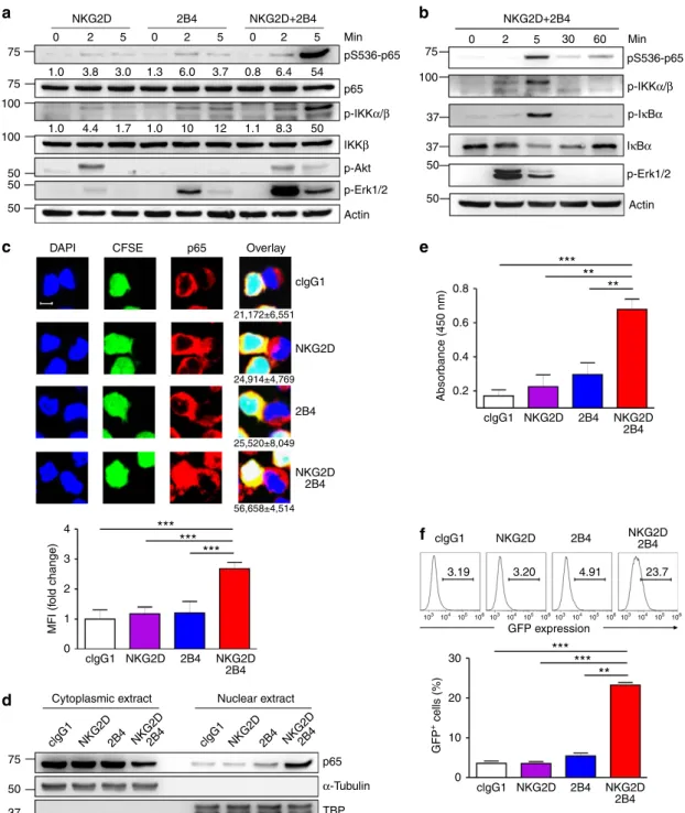
Synergistic activation of NF-jB by NKG2D and 2B4. Various
stimulatory receptors trigger distinct signalling pathways that
lead to NF-kB activation
24. Such diverse NF-kB-activating
stimuli, however, frequently converge on the IKK complex,
and signalling cascade downstream of IKK appears to be
well-conserved among most NF-kB activation pathways. This
includes the phosphorylation and degradation of the IkB family,
which releases bound NF-kB dimers for nuclear translocation and
target gene transcription. So far, little is known about the
mechanisms that couple non-ITAM-associated receptors to
NF-kB activation during the triggering of natural cytotoxicity
by NK cells. Thus, we first assessed NF-kB activation after
stimulating NK cells via NKG2D, 2B4 or both. Stimulation of
human NK cell line NKL with NKG2D or 2B4 alone resulted in
detectable but weak phosphorylation of IKK and Erk, whereas
co-engagement led to their synergistic phosphorylation (Fig. 1a).
Such an increase was distinct from Akt phosphorylation, which is
downstream of PI3K activation via NKG2D but not 2B4 (Fig. 1a).
Likewise, phosphorylation of the downstream NF-kB p65
subunit (pS536-p65) resembled that of IKK. Such synergistic
phosphorylation was evident at 5 min and decreased thereafter,
which coincided with the phosphorylation and degradation of
IkBa that links IKK activation and p65 nuclear translocation
(Fig. 1b). We next performed microscopy experiments to
assess nuclear translocation of p65 in individual NKL-target cell
conjugates. To facilitate NKL cell identification, they were labelled
with carboxyfluorescein succinimidyl ester (CFSE) and
conju-gated with unlabelled P815 target cells. In conjuconju-gated NKL cells,
little or weak nuclear translocation of p65 was observed after
stimulation with NKG2D or 2B4 alone (Fig. 1c). In contrast,
significant nuclear translocation of p65 was detected in
conju-gated NKL cells following NKG2D and 2B4 co-engagement
NKG2D DAPI MFI (f old change) GFP + cells (%) Absorbance (450 nm)
***
***
***
***
***
**
**
**
***
CFSE p65 Overlay 21,172±6,551 24,914±4,769 25,520±8,049 56,658±4,514 clgG1 NKG2D NKG2D 2B4 NKG2D2B4 NKG2D2B4 Nuclear extract Cytoplasmic extract 2B4 clgG1 NKG2D GFP expression 3.19 104 105 106 103 104 105 106 103 104 105 106 103 104 105 106 103 3.20 4.91 23.7 NKG2D 2B4 2B4 clgG1 clgG1 NKG2D NKG2D NKG2D 2B4 NKG2D 2B4 2B4 2B4 clgG1 p65 TBP α-Tubulin NKG2D 2B4 NKG2D 2B4 0 1.0 1.0 1.7 1.0 10 12 1.1 8.3 3.8 4.4 3.0 1.3 6.0 3.7 0.8 6.4 54 50 75a
c
e
f
d
b
75 100 100 50 50 75 37 37 100 50 50 50 4 30 0.2 0.4 0.6 0.8 20 10 0 3 2 1 0 75 clgG1 NKG2D2B4 clgG1 NKG2D2B4 50 37 2 5 0 2 5 0 2 5 0 2 5 30 60 2B4 NKG2D+2B4 NKG2D+2B4 Min Min p65 p-IKKα/β p-IKKα/β p-IκBα IκBα p-Akt Actin Actin p-Erk1/2 p-Erk1/2 IKKβ pS536-p65 pS536-p65Figure 1 | Synergistic activation of NF-jB by NKG2D and 2B4 co-engagement. (a) NKL cells rested in the absence of IL-2 for 24 h were stimulated with NKG2D and/or 2B4 by receptor crosslinking for the indicated time. Cell lysates were immunoblotted with Abs to phospho-p65 at serine 536 (pS536), p65, phospho-IKKa/b at serine 176/180 (pS176/180), IKKb, phospho-Akt at serine 473 (pS473), phospho-Erk1 and 2, or actin. The normalized intensities of the phosphorylated p65 and IKKa/b relative to their total forms are presented. (b) Rested NKL cells were treated as in a to stimulate NKG2D and 2B4 for the indicated time. Lysates were immunoblotted for phospho-p65, phospho-IKKa/b, phospho-IkBa at serine 32/36 (pS32/36), IkBa, phospho-Erk1/2 or actin. (c) Representative confocal images (top) of conjugates between rested NKL cells loaded with CFSE (green) and P815 target cells as indicated. Conjugates were fixed, permeabilized and stained with 4,6-diamidino-2-phenylindole (DAPI; blue) and mAb to p65, anti-mouse IgG-Biotin followed with Alexa Fluor 647 (red)-Streptavidin. The number beneath the overlay image is the mean nuclear fluorescence intensity (MFI)±s.d. of p65 from Z50 NKL-target cell conjugates. Statistical bar charts (bottom) for MFI of p65 in the nucleus are represented as fold change. Values represent mean±s.d. Scale bar, 5 mm. (d) Rested NKL cells were stimulated with plate-immobilized mAbs to NKG2D and/or 2B4 for 1 h. Equal amounts of protein from cytoplasmic and nuclear extracts were immunoblotted with mAb to p65. (e) Nuclear extracts collected as in d were added into a 96-well plate immobilized with double-stranded oligonucleotide containing the consensus NF-kB-binding sequence. The amount of p65 bound to the oligonucleotide was measured by colorimetric assay. Values represent mean±s.d. (f) Rested NKL cells transduced with a kB-GFP reporter construct were stimulated with plate-immobilized mAbs to NKG2D and/or 2B4 for 6 h. GFP expression in NKL-kB-GFP cells was analysed by flow cytometry, and representative result (top) and statistical bar charts (bottom) are shown. Values represent mean±s.d. Po0.05; **Po0.01; ***Po0.001 (two-sided Student’s t-test). Data are representative of at least three independent experiments.
(Fig. 1c). Supporting this, analysis of subcellular fractions revealed
a substantial increase in nuclear p65 and concomitant decrease in
cytoplasmic p65 in NKL cells stimulated with NKG2D and 2B4
but neither receptor alone (Fig. 1d). Further, the amount of p65
bound to oligonucleotide containing consensus NF-kB-binding
site was significantly increased in the nuclear extracts of NKL cells
following NKG2D and 2B4 co-engagement (Fig. 1e). Finally, to
evaluate the transcriptional activity of NF-kB, we generated NKL
reporter (NKL-kB-GFP) cells that express GFP under the control
of NF-kB transcription response elements. Proper functioning of
the reporter cells was demonstrated by increased GFP expression
following TNF-a treatment and its abrogation by prior treatment
with BAY11-7082 that inhibits NF-kB activation (Supplementary
Fig. 1). Compared with either receptor alone, NKG2D and 2B4
co-engagement induced an apparent and synergistic increase
in the proportion of cells expressing GFP (Fig. 1f). Thus, via
comprehensive analyses of conserved steps in the NF-kB
activation pathway, we conclude that robust NF-kB activation
is not achieved by stimulation with NKG2D or 2B4 alone, but
instead relies on the integration of distinct signals from NKG2D
and 2B4.
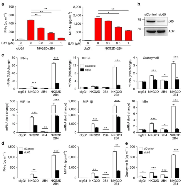
NF-jB is required for NK cell functions by NKG2D and 2B4.
Given NF-kB as a transcription factor important for gene
regulation, we assessed gene expression after stimulating NK
cells with NKG2D, 2B4 or both. Examination of a profile of 20
genes encompassing cytokines, chemokines, cytolytic pathway,
death receptor pathway, NF-kB pathway, apoptosis, IL-2
receptor and cytotoxic granule exocytosis revealed a synergistic
induction of diverse genes related to NK cell effector functions
(Supplementary Fig. 2), correlating with the increase in NF-kB
activation. Chemokine expression appears to be preferentially
triggered by engagement of a single receptor (especially NKG2D),
corroborating a recent study of the requirement for less
stimulation to induce chemokine production compared with
cytokine production or degranulation
14. Nonetheless, NKG2D
and 2B4 co-engagement still induced higher levels of chemokine
and cytokine gene expression.
Among the major genes induced by NF-kB are those encoding
cytokines and chemokines. The synergy-dependent activation of
NF-kB (Fig. 1) and mRNA expression of IFN-g and MIP-1a
(Supplementary Fig. 2) prompted us to test whether NF-kB is
required for NK cells to produce cytokines and chemokines
following NKG2D and 2B4 engagement. The synergistic
production of IFN-g and MIP-1a by NKL cells was diminished
in a dose-dependent manner following BAY11-7082 treatment
(Fig. 2a). To directly probe the role of NF-kB, we silenced the
expression of NF-kB p65 subunit, using small interfering RNA
(siRNA) (Fig. 2b). p65 knockdown caused marked reductions in
mRNA expression of IFN-g, TNF-a, MIP-1a/b, granzyme B and
IkBa induced by NKG2D and 2B4 co-engagement (Fig. 2c),
confirming the dependence of their transcription on NF-kB.
Similarly, the synergistic production of IFN-g and MIP-1a was
significantly reduced by p65 knockdown (Fig. 2d). To confirm
this finding in the context of physiological receptor–ligand
interactions, we stimulated NKL cells with P815 cells expressing
ULBP1 (a ligand for NKG2D) and/or CD48 (a ligand for 2B4).
Consistently, synergistic production of IFN-g and MIP-1a
following physiological stimulation was significantly diminished
by p65 knockdown (Supplementary Fig. 3).
Given the reports showing defective cytotoxicity of NK cells
from patients with NF-kB deficiency
18,19, we next assessed
whether NF-kB is also required for cytotoxic degranulation of
NK cells. The synergistic increase in degranulation, as assessed
by granzyme B release, was significantly decreased by p65
knockdown (Fig. 2e). Collectively, NF-kB could play an
indispensable
role in the production of cytokines and
chemokines, as well as the release of granzyme B by NK cells
via non-ITAM-associated receptors NKG2D and 2B4.
We next determined whether the findings obtained from NKL
cells were applicable to primary NK cells. NF-kB activation was
assessed using purified NK cells on a per-cell basis by flow
cytometry-based analysis of p65 phosphorylation. Similar to
experiments using NKL cells (Supplementary Fig. 4), the
proportion of responding cells synergistically increased following
combined stimulation of primary NK cells with NKG2D and
2B4 (Supplementary Fig. 5a). NKG2D and 2B4 co-engagement
also led to synergistic increase in the proportion of NK
cells expressing IFN-g or TNF-a (Supplementary Fig. 5b), which
was diminished after BAY11-7082 treatment (Supplementary
Fig. 5c). BAY11-7082 did not significantly affect the viability of
NKL cells and primary NK cells, as assessed by annexin-V/PI
staining (Supplementary Fig. 6). Collectively, these results suggest
that NKG2D and 2B4 coactivation is required to overcome a
threshold for NF-kB activation, leading to synergistic cytokine
production by NK cells.
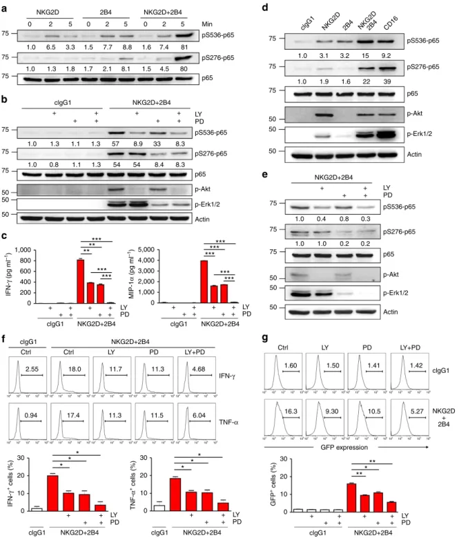
Disparate signals converge on the NF-jB p65 subunit. NF-kB
activation occurs primarily via IKK-dependent phosphorylation
and
degradation
of
IkB
proteins,
followed
by
nuclear
translocation of the released NF-kB dimers. In addition, optimal
NF-kB activation relies on post-translational modifications of
NF-kB subunits, such as p65 phosphorylation, by specific protein
kinases
25,26. These emerging and additional layers of NF-kB
regulation represent an important means for crosstalk between
different signalling pathways and determining context-specific
transcriptional responses
27. NKG2D and 2B4 co-engagement
induces two distinct pathways that lead to the activation of
PI3K-Akt and Vav1-dependent synergistic signalling involving
PLC-g2 and Erk
4. Given the site-selective phosphorylation of p65
through Akt and Erk pathways
26, Vav1-dependent synergy
achieved
by
a
proximal
convergence
of
signals
from
coactivation receptors may not suffice to activate NF-kB.
Although stimulation of NKL cells with NKG2D or 2B4 alone
induced weak p65 phosphorylation at serine 276 and 536,
NKG2D and 2B4 co-engagement functioned synergistically to
increase their phosphorylation (Fig. 3a). In support, synergistic
phosphorylation of p65 was observed after stimulation with P815
cells expressing ULBP1 and CD48 (Supplementary Fig. 7).
To assess the dependence of such phosphorylation on Akt
and Erk pathways, NKL cells were treated with an inhibitor of
PI3K (LY294002) or MEK (PD98059), which block Akt or Erk
activation, respectively. Notably, p65 phosphorylation at serine
276 fully depended on NKG2D and 2B4 co-engagement-induced
Erk activation (Fig. 3b). In contrast, p65 phosphorylation at
serine 536 largely depended on NKG2D-induced PI3K-Akt.
The PI3K-Akt and synergistic Erk pathway appear to be mutually
independent, given the insensitivity of synergistic Erk activation
to PI3K inhibitors and Akt activation to MEK inhibitors
4.
The functional significance of these pathways was supported by
significant impairment of IFN-g and MIP-1a production by an
inhibitor of each pathway and further by the combined inhibition
of both pathways (Fig. 3c). To probe the direct role of these
kinase pathways, we performed knockdown of Akt1 and/or Erk2
in NKL cells. Both Akt1 and Erk2 were required for optimal p65
phosphorylation and IFN-g and MIP-1a production following
coactivation, complementing the results with small-molecule
inhibitors (Supplementary Fig. 8).
We next assessed these findings using primary rested human
NK cells. Similar to the results seen in NKL cells, NKG2D and 2B4
co-engagement led to synergistic phosphorylation of p65 at serine
276 and 536 (Fig. 3d). Moreover, we observed that
phosphoryla-tion of S276-p65 was selectively dependent on Erk pathway,
whereas
phosphorylation
of
S536-p65
largely
relied
on
Akt(Fig. 3e). As expected, synergistic expression of IFN-g and
TNF-a was diminished by inhibiting Akt, Erk or both pathways
(Fig. 3f), correlating with reduced NF-kB activation, as determined
using reporter cells (NKL-kB-GFP; Fig. 3g). The combined
inhibition of Akt and Erk pathways demonstrated an additive
effect on dampening NF-kB activation, suggesting separate
involvement of Akt and Erk pathways in NF-kB activation.
To determine whether p65 phosphorylation is similarly
induced by a different combination of coactivation receptors,
primary NK cells were examined after engaging DNAM-1, 2B4 or
both. DNAM-1 synergizes with 2B4 to trigger effector
functions
28. Notably, crosslinking DNAM-1 induced Akt
phosphorylation, as did NKG2D, and 2B4 co-engagement
induced a synergistic phosphorylation of Erk, an apparent dual
phosphorylation of p65, and synergistic NF-kB activation
(Supplementary Fig. 9a,b). These suggest that the basis for
NF-kB activation is shared between DNAM-1 and NKG2D. In
contrast, crosslinking CD16, which mediates antibody-dependent
cellular cytotoxicity and is sufficient for cytokine production,
induced dual phosphorylation of p65 in addition to Akt and Erk
phosphorylation (Fig. 3d). These suggest distinct regulation of
p65 phosphorylation by coactivation receptors for natural
cytotoxicity that are incapable of activating alone. Thus,
co-engagement of 2B4 with NKG2D, or DNAM-1, was required
800
a
c
d
e
b
3,200 siControl GranzymeB siControl sip65 siControl sip65 siControl sip65 IκBα MIP-1β MIP-1α IFN-γ TNF-α sip65 p65 Actin 2,400 75 50 4 4 6 8 10 3 2 2 1 0 0 0 300 600 900 9,000 6,000 3,000 1,000 2,000 7,000 9,000 1,000 500 500 300 80 80 60 40 20 40 1,500 0 0 0 4 8 12 16 0 0 0 1,600 800 0**
**
***
***
***
***
***
***
***
***
***
***
***
**
**
***
***
***
***
**
**
**
**
*
*
*
*
**
**
600 400 IFN-γ (pg ml –1 ) MIP-1 α (pg ml –1) Gr anzyme B (pg ml –1) MIP-1 α (pg ml –1 ) IFN-γ (pg ml –1 ) mRNA (f old change) mRNA (f old change) mRNA (f old change) mRNA (f old change) mRNA (f old change) mRNA (f old change) 200 0 0 0 clgG1 clgG1 clgG1 NKG2D+2B4 NKG2D+2B4 NKG2D NKG2D 2B4 2B4 clgG1 NKG2D NKG2D 2B4 2B4 clgG1 NKG2D NKG2D 2B4 2B4 clgG1 NKG2D NKG2D 2B4 2B4 clgG1 NKG2D NKG2D 2B4 2B4 clgG1 NKG2D NKG2D 2B4 2B4 clgG1 NKG2D NKG2D 2B4 2B4 clgG1 NKG2D NKG2D 2B4 2B4 clgG1 NKG2D NKG2D 2B4 2B4 0 0.2 0.5 1 0 0.2 0.5 1 BAY (μM) BAY (μM)Figure 2 | NF-jB is required for cytokine and chemokine gene expression by NKG2D and 2B4 coactivation. (a) Rested NKL cells were pretreated with a NF-kB inhibitor BAY11-7082 at the indicated dose for 1 h and then stimulated with both NKG2D and 2B4 for 8 h. Thereafter, IFN-g and MIP-1a in the supernatants were measured by ELISA. Values represent mean±s.d. (b) NKL cells were transfected with 300 pmol of control siRNA or siRNA specific for p65. After 24 h, the cells were rested for another 24 h, and lysates were immunoblotted for p65 and actin. (c) Rested NKL cells transfected with control siRNA or p65-specific siRNA were stimulated with NKG2D and/or 2B4 for 3 h. Thereafter, total RNA was prepared from cells, reverse transcribed and the relative mRNA levels of IFN-g, TNF-a, granzyme B, MIP-1a, MIP-1b and IkBa were determined by real-time PCR and normalized to b-actin mRNA. Values represent mean±s.d. (d) Rested NKL cells transfected with control siRNA or p65-specific siRNA were stimulated as in c for 8 h. IFN-g and MIP-1a in the supernatants were measured by ELISA. Values represent mean±s.d. (e) Rested NKL cells that were transfected with control siRNA or p65-specific siRNA and stimulated with NKG2D and/or 2B4 for 2 h were used in a granzyme B release assay. Granzyme B in the supernatants was measured by ELISA. Error bars represent the s.d. *Po0.05; **Po0.01; ***Po0.001 (two-sided Student’s t-test). Data are representative of at least three independent experiments.
NKG2D
a
d
b
c
e
f
g
clgG1 clgG1 clgG1 clgG1 clgG1 clgG1 IFN-γ TNF-α Ctrl Ctrl 2.55 102 103 104 105 106102 103 104 105 106102 103 104 105 106102 103 104 105 106102 103 104 105 106 102 103 104 105 106 102 103 104 105 106 102 103 104 105 106 102 103 104 105 106 102 103 104 105 106 102 103 104 105 106102 103 104 105 106102 103 104 105 106102 103 104 105 106 102 103 104 105 106 102 103 104 105 106 102 103 104 105 106102 103 104 105 106 18.0 11.7 11.3 4.68 1.60 1.50 1.41 1.42 5.27 10.5 9.30 16.3 6.04 11.5 11.3 17.4 0.94 Ctrl LY PD LY+PD LY PD LY+PD clgG1 NKG2D 2B4 NKG2D2B4 CD16 + + + + + + + + + + + + + + + + + + + + + + + + + + + + + + + + + + + + + + + + + + + + 75 75 75 75 75 5,000 4,000 3,000 2,000 1,000 1,000 800 600 400 200 MIP-1 α (pg ml –1 ) IFN-γ (pg ml –1 ) IFN-γ + cells (%) TNF-α + cells (%) GFP + cells (%) 0 0 0 10 20 30 0 10 20 30 0 10 20 30 75 75 0 2 5 0 2 5 0 2 5 81 7.4 1.6 8.8 7.7 1.5 3.3 6.5 1.3 1.3 0.8 1.1 1.3 54 54 8.4 8.3 8.3 33 8.9 57 1.3 1.1 1.8 1.7 2.1 8.1 1.5 4.5 80 1.0 1.0 3.1 3.2 15 9.2 39 22 1.6 1.9 1.0 1.0 0.4 0.8 0.3 0.2 0.2 1.0 1.0 1.0 1.0 1.0 75 75 75 75 50 50 50 50 50 50 50 50 50 75 2B4 NKG2D+2B4 NKG2D+2B4 NKG2D+2B4 NKG2D+2B4 NKG2D+2B4 clgG1 NKG2D+2B4 clgG1 NKG2D+2B4 NKG2D+2B4 NKG2D + 2B4 NKG2D+2B4 Min LY PD pS536-p65 pS536-p65 pS536-p65 pS276-p65 pS276-p65 pS276-p65 p65 p65 p65 p-Akt p-Akt Actin *** *** ** ** * * * ** *** * * * *** *** *** *** *** *** Actin p-Erk1/2 LY PD LY PD LY PD LY PD LYPD LY PD pS536-p65 pS276-p65 p65 p-Akt Actin GFP expression p-Erk1/2 p-Erk1/2Figure 3 | NKG2D and 2B4 coactivation induces disparate and cooperative signals for NF-jB activation. (a) Rested NKL cells were stimulated with NKG2D and/or 2B4 by receptor crosslinking for the indicated time. Cell lysates were immunoblotted for phospho-p65 at serine 536 (pS536), phospho-p65 at serine 276 (pS276) or p65. The normalized intensities of the phosphorylated p65 relative to total p65 are presented. (b) Rested NKL cells were stimulated with NKG2D and 2B4 for 5 min after pretreatment with PI3K inhibitor (LY294002; 20 mM) and/or MEK inhibitor (PD98059; 20 mM) for 30 min. Lysates were analysed by immunoblotting for the indicated phosphorylations of p65. (c) Cytokine release assays with rested NKL cells after pretreatment with 20 mM LY294002 and/or 20 mM PD98059 for 30 min and then stimulation with NKG2D and 2B4 for 12 h in the presence of the inhibitor. Thereafter, IFN-g (left) and MIP-1a (right) in the supernatants were measured by ELISA. Values represent mean±s.d. (d) Primary rested NK cells after expansion were stimulated with the indicated receptors by receptor crosslinking for 5 min. Lysates were analysed by immunoblotting as inb. (e) Primary rested NK cells after expansion were stimulated and analysed by immunoblotting as inb. (f) Frequency of NK cells that displayed IFN-g or TNF-a expression after pretreatment of PBMCs with 20 mM LY294002 and/or 20 mM PD98059 for 1 h and then stimulation with P815 target cells as indicated in the presence of the inhibitor. After incubation for 6 h, cells were stained and analysed by flow cytometry. Representative result (top) and statistical bar charts (bottom) from three experiments are shown. Values represent mean±s.e.m. (g) Rested NKL-kB-GFP cells were pretreated with 20 mM LY294002 and/or 20 mM PD98059 for 1 h and then stimulated with plate-immobilized NKG2D and 2B4 for 6 h. GFP expression in the reporter NKL cells was analysed by flow cytometry, and representative result (top) and statistical bar charts (bottom) are shown. Values represent mean±s.d. *Po0.05; **Po0.01; ***Po0.001 (two-sided Student’s t-test). Data are representative of at least three independent experiments.
to achieve signalling competence for p65 phosphorylation and
NF-kB activation.
Signal amplification is insufficient for NF-jB activation. The
dependence of NK cell activation on synergistic signals is, in
part, relieved after c-Cbl knockdown
4. Thus, we examined
whether depleting c-Cbl enables NKG2D or 2B4 to bypass the
requirement for synergistic signals to induce p65 phosphorylation
and NF-kB activation. c-Cbl knockdown slightly enhanced p65
phosphorylation at serine 536 and 276 in response to NKG2D or
2B4 alone (Fig. 4a). However, it remained lower than p65
phosphorylation by their synergistic coactivation. In contrast,
c-Cbl knockdown substantially enhanced p65 phosphorylation at
both serines following synergistic coactivation.
Given the role of Akt and Erk in the phosphorylation of
S536-p65 and S276-S536-p65, we next assessed the effects of c-Cbl
knockdown on Akt and Erk phosphorylation. Although Akt
phosphorylation induced by NKG2D was markedly augmented
by c-Cbl knockdown, it was undetectable following 2B4
stimulation even after c-Cbl knockdown (Fig. 4a). In comparison,
c-Cbl knockdown caused a small increase in Erk phosphorylation
by NKG2D or 2B4 alone, but an apparent increase by their
synergistic coactivation. These results suggest that c-Cbl
knock-down may amplify signal input, but not trigger disparate signals
for NF-kB activation.
The extent of pS276-p65 induced by NKG2D, 2B4 or both
correlated with the level of Erk phosphorylation (Fig. 4a). In
contrast, the correlation between pS536-p65 and Akt
phosphor-ylation was observed following NKG2D and 2B4 coactivation, but
not NKG2D alone, although there was comparable Akt
phosphor-ylation. This suggests a checkpoint that restrains pS536-p65 by Akt
pathway, which could be overcome by synergistic coactivation. In
support, NF-kB activation and production of IFN-g and MIP-1a
by NKG2D and 2B4 coactivation but not by a single receptor was
markedly enhanced after c-Cbl knockdown (Fig. 4b,c). Together,
signal
amplification
from
individual
receptor
could
not
compensate for the lack of complementary signals from its partner
receptor for NF-kB activation.
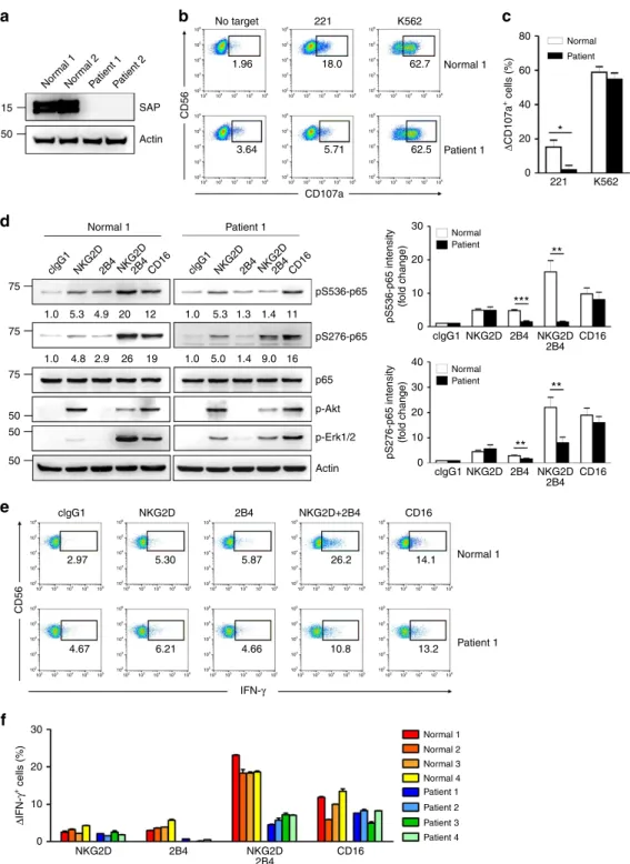
Defective NF-jB activation in XLP1 NK cells by NKG2D and 2B4.
We next assessed whether NF-kB activation requires cooperation
between coactivation receptors in pathophysiological contexts.
XLP1 is characterized by severe immunodeficiency resulting from
mutations in the SH2D1A gene encoding the SAP protein
29–31.
siControl
a
c
b
siControl siControl pS536-p65 pS276-p65 p65 p-Akt p-Erk1/2 c-Cbl Actin clgG1 2.03 103 104 105 106 103 104 105 106 103 104 105 106 103 104 105 106 103 104 105 106 103 104 105 106 103 104 105 106 103 104 105 106 2.15 3.88 22.7 39.2 9.07*
*
*
*
*
***
***
**
**
3.16 2.07 clgG1 NKG2D NKG2D 2B4 2B4 clgG1 NKG2D NKG2D 2B4 2B4 clgG1 NKG2D NKG2D 2B4 2B4 NKG2D GFP expression 2B4 NKG2D 2B4 75 1.3 1.8 17 1.1 1.5 4.4 44 1.0 1.0 2.0 2.1 16 2.0 3.0 5.6 63 clgG1 NKG2D2B4 NKG2D2B4 clgG1 NKG2D2B4 NKG2D2B4 75 50 40 30 20 GFP + cells (%) MIP-1 α (pg ml –1 ) IFN-γ (pg ml –1) 10 0 0 2,000 4,000 6,000 8,000 10,000 2,000 1,600 1,200 800 400 0 75 50 50 50 100 sic-Cbl sic-Cbl sic-Cbl siControl sic-Cbl siControl sic-CblFigure 4 | c-Cbl depletion augments but is not sufficient for NF-jB activation. (a) Rested NKL cells transfected with control siRNA or c-Cbl-specific siRNA were stimulated through the indicated receptors for 5 min. Cell lysates were immunoblotted for phospho-p65 at serine 536 (pS536), phospho-p65 at serine 276 (pS276), p65, phospho-Akt at serine 473 (pS473), phospho-Erk1 and 2, c-Cbl or actin The normalized intensities of the phosphorylated p65 relative to p65 are presented. (b) Rested NKL-kB-GFP cells transfected with control siRNA or c-Cbl-specific siRNA were stimulated with plate-immobilized mAbs specific for NKG2D and/or 2B4 for 6 h. GFP expression in NKL-kB-GFP cells was analysed by flow cytometry, and representative result (top) and statistical bar charts (down) are shown. Values represent mean±s.d. (c) Cytokine release assays with rested NKL cells transfected with control siRNA or c-Cbl-specific siRNA and stimulated with NKG2D and/or 2B4 for 8 h. IFN-g and MIP-1a in the supernatants were measured by ELISA. Values represent mean±s.d. *Po0.05; **Po0.01; ***Po0.001 (two-sided Student’s t-test). Data are representative of at least three independent experiments.
Given the requirement of SAP for 2B4-dependent NK cell
activation
32,33, we hypothesized that NK cells from XLP1 patients
would demonstrate defects in NF-kB activation and NK cell
responses following coactivation. We first tested the requirement
of SAP for NK cell activation during NKG2D and 2B4 synergy by
performing siRNA-mediated knockdown of SAP in NKL cells
(Supplementary Fig. 10a). The synergistic increases in Ca
2 þmobilization, NF-kB activation and IFN-g and MIP-1a release
were markedly diminished by SAP knockdown (Supplementary
Fig. 10b,c,d). Likewise, SAP knockdown markedly reduced the
synergistic increase in p65 phosphorylation and the proportion of
NK cells expressing IFN-g in primary NK cells following NKG2D
and 2B4 coactivation (Supplementary Fig. 11). The small increase
in IFN-g expression by ligating 2B4 but not NKG2D alone was
also selectively decreased by SAP knockdown.
Given these promising results, NK cells from XLP1 patients
were used to study the dependence of synergistic coactivation on
SAP expression. Among the four XLP1 patients examined, three
patients harboured macrodeletions in the SH2D1A gene that
resulted in complete loss of SAP expression, and one patient
harboured a missense mutation that reduced SAP expression, as
assessed by western blot analysis (Fig. 5a and Supplementary
Fig. 12). To probe the functional defects in XLP1 NK cells, we
measured target cell-induced degranulation, as determined by cell
surface expression of CD107a (ref. 28). Stimulation with K562 cell
line induced strong degranulation of both normal and XLP1 NK
cells (Fig. 5b,c). In contrast, XLP1 NK cells were severely
impaired in their ability to degranulate against Epstein–Barr virus
(EBV)-immortalized B-lymphoblastoid cell line 721.221 (referred
to as 221), an observation compatible with the defective killing of
EBV-infected B cells by XLP1 NK cells
32.
Next, we tested whether SAP is required for NF-kB activation
and NK cell responses in XLP1 NK cells. Notably, the apparent
dual phosphorylation of p65 at serine 536 and 276 following
NKG2D and 2B4 coactivation seen in normal NK cells
was significantly diminished in XLP1 NK cells (Fig. 5d).
The synergistic phosphorylation of Erk following coactivation
was also diminished in XLP1 NK cells, whereas Akt
phos-phorylation induced by NKG2D appeared to be unaffected.
In contrast, dual phosphorylation of p65 by CD16 ligation was
comparable in normal and XLP1 NK cells (Fig. 5d). Moreover,
NF-kB activation in XLP1 NK cells was measured by nuclear
translocation of p65 in individual NK-target cell conjugates.
A significant defect in nuclear translocation of p65 following
coactivation was detected in XLP1 NK cells relative to normal NK
cells (Supplementary Fig. 13). Corroborating these findings, the
proportion of NK cells expressing IFN-g was severely decreased
in XLP1 NK cells following NKG2D and 2B4 coactivation, but
not CD16 (Fig. 5e,f). Most strikingly, the small increase in IFN-g
expression by 2B4 ligation was selectively defective in XLP1 NK
cells, consistent with a critical role of SAP in 2B4-mediated
signalling. A similar but less severe defect was observed
when degranulation was induced through 2B4 alone and in
combination with NKG2D, but not through CD16 (Fig. 6a,b).
These results did not appear to be associated with defective
expression of coactivation receptors in XLP1 NK cells, given
comparable expression of various activating receptors, including
NKG2D, 2B4 and CD16 (Fig. 6c). Instead, our results suggest that
the functional deficiencies in XLP1 NK cells by NKG2D and
2B4 coactivation are most likely due to selective defects in
2B4-associated synergistic signalling to Erk, which is important
for optimal NF-kB activation and effector functions.
Enhanced
Vav1
signalling
during
NKG2D
and
2B4
coactivation is required to overcome inhibition by c-Cbl and
deliver synergistic signals for NK cell activation
4. Thus, we tested
whether aberrant Vav1 regulation is associated with such defects
seen in XLP1 NK cells. As reported, Vav1 phosphorylation was
induced in normal NK cells by engaging NKG2D or 2B4, and
was additive after co-engagement (Fig. 6d). Notably, Vav1
phosphorylation was impaired in XLP1 NK cells following 2B4
stimulation, but normal after NKG2D stimulation. Furthermore,
Vav1 phosphorylation after co-engaging NKG2D and 2B4 was
defective in XLP1 NK cells, similar to the level of phosphorylation
induced by NKG2D alone (Fig. 6d). Collectively, SAP deficiency
could cause a defect in 2B4, but not NKG2D, signalling at the
level of Vav1 and thereby impair the synergistic activation of Erk
and NF-kB following coactivation.
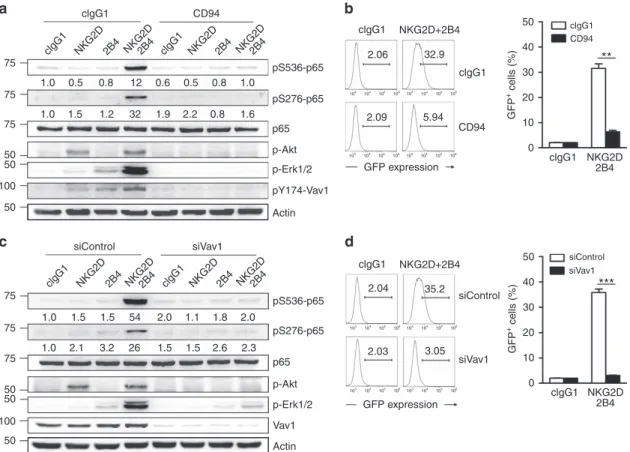
Vav1 is required for the synergistic activation of NF-jB. To
ensure that Vav1 is required for synergistic NF-kB activation,
we tested whether NF-kB activation following coactivation is
susceptible to Vav1 inhibition. Inhibitory signalling through
CD94-NKG2A can override Vav1-dependent activation of NK
cells
4. The synergistic phosphorylation of p65 and Erk and the
combined phosphorylation of Vav1 induced by NKG2D and
2B4 coactivation were all abrogated by co-crosslinking
CD94-NKG2A on NKL cells (Fig. 7a). Moreover, NKG2D-dependent
phosphorylation of Akt, required for pS536-p65, was also
abrogated by the same inhibition. Accordingly, the synergistic
increase in transcriptional activity of NF-kB was markedly
impaired by co-crosslinking CD94-NKG2A (Fig. 7b). These
results suggest the suppression of NF-kB activation as a
mechanism underlying the inhibitory function of
CD94-NKG2A. To ascertain the direct involvement of Vav1 in NF-kB
activation, we performed siRNA-mediated knockdown of Vav1.
Similarly, Vav1 knockdown abrogated the phosphorylation of
p65, along with that of Akt and Erk, and in turn transcriptional
activity of NF-kB following coactivation (Fig. 7c,d). Collectively,
our results suggest that NF-kB activation in NK cells by NKG2D
and 2B4 coactivation is under the control of Vav1 and
dominantly inhibited by CD94-NKG2A.
Discussion
Here we offer a new perspective on the regulation of NF-kB
activation in NK cells during target cell recognition. NF-kB
activation via non-ITAM-associated receptors (for example,
NKG2D, 2B4, DNAM-1) relies on coordinated engagement of
coactivation receptors, which together provide complementary
and independent signals leading to optimal Vav1 and p65
phosphorylation (Fig. 8). The importance of such regulation
between Vav1 and p65 in NF-kB activation was supported by
signalling and functional defects centred on Vav1 in XLP1 NK
cells following coactivation.
A prevailing view of the mechanism underlying synergistic
coactivation involves signal integration by receptor-proximal
adaptor proteins that promote downstream signalling events for
cytokine production and target cell killing
1. Supporting this, it
was shown that signals from synergizing receptors converge on
the
adaptor
protein
SH2
domain-containing
leukocyte
phosphoprotein of 76 kDa (SLP-76) through site-selective
phosphorylation of two tyrosines in SLP-76 (ref. 34). These two
phosphotyrosines enable simultaneous binding of Vav1-Nck
protein complex to both tyrosines on SLP-76, which leads to
Vav1-dependent synergistic signals. Vav1 is an essential
component for synergistic coactivation among combinations
of NKG2D, 2B4 and DNAM-1 (ref. 4). Thus, the dependence
of synergistic coactivation on the regulated interaction between
SLP-76 and Vav1 may represent a checkpoint in NK cell
activation that ensures proper specificity of NK cell responses.
Supporting this notion, the defective phosphorylation of Vav1 in
XLP1 NK cells, and the inhibition of Vav1 by inhibitory receptor
15
a
d
e
f
b
c
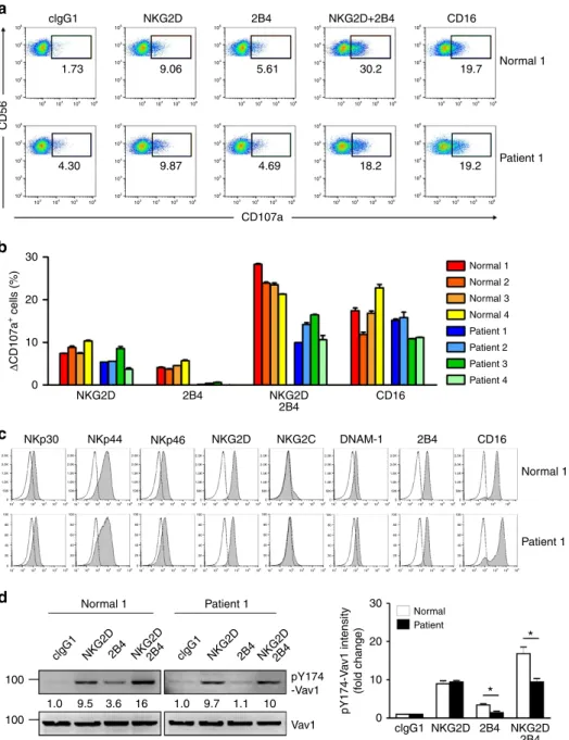
80 * ** ** ** *** 60 40 20 0 0 0 clgG1 NKG2D NKG2D 2B4 2B4 CD16 clgG1 clgG1 2.97 5.30 5.87 26.2 14.1 13.2 10.8 4.66 4.67 6.21 NKG2D NKG2D NKG2D 2B4 NKG2D+2B4 2B4 2B4 IFN-γ CD16 NKG2D NKG2D 2B4 2B4 CD16 CD16 10 10 20 20 30 30 40 221 K562 CD56 CD56 Δ IFN-γ + cells (%) Δ CD107a + cells (%) pS276-p65 intensity (f old change) pS536-p65 intensity (f old change) 75 1.0 1.0 1.0 5.3 5.3 1.3 1.4 11 16 9.0 1.4 5.0 1.0 4.9 2.9 26 19 4.8 20 12 75 75 50 50 50 30 20 10 0 SAP pS536-p65 pS276-p65 p65 p-Akt p-Erk1/2 Actin Normal 1 Normal 1 Normal Patient Normal Patient Normal Patient Patient 1 No target 103 103 104 104 105 105 106 106 102 102 103 104 105 106 102 103 104 105 106 103 104 105 106 102 103 104 105 106 102 103 104 105 106 102 103 104 105 106 102 102 103 104 105 106 102 103 104 105 106 102 103 104 105 106 102 103 104 105 106 103 104 105 106 102 103 104 105 106 102 103 104 105 106 102 103 104 105 106 102 103 104 105 106 102 102 103 104 105 106 102 103 104 105 106 102 103 104 105 106 102 103 104 105 106 102 103 104 105 106 102 103 104 105 106 102 103 104 105 106 102 103 104 105 106 102 103 104 105 106 102 103 104 105 106 102 103 104 105 106 102 103 104 105 106 102 103 104 105 106 102 103 104 105 106 102 103 104 105 106 102 1.96 3.64 5.71 CD107a 62.5 18.0 62.7 221 K562 Actin Nor mal 1 clgG1 NKG2D2B4NKG2D2B4CD16 clgG1 NKG2D2B4NKG2D2B4CD16 Patient 1Patient 2 Nor mal 2 50 Patient 1 Normal 1 Normal 1 Normal 2 Normal 3 Normal 4 Patient 1 Patient 1 Patient 2 Patient 3 Patient 4Figure 5 | XLP1 NK cells have defects in NF-jB activation and IFN-c expression in response to coactivation by NKG2D and 2B4. (a) Total lysates of primary expanded NK cells from representative normal or XLP1 patient donors were immunoblotted for SAP and actin. (b,c) Primary rested NK cells after expansion from normal or XLP1 patient donors were mixed with 221 or K562 cells in the presence of fluorochrome-conjugated anti-CD107a mAb for degranulation assay. After incubation for 2 h, cells were stained with fluorochrome-conjugated mAb to CD56, and the level of CD56þCD107aþNK cells was
measured using flow cytometry. (b) Representative result is shown. (c) Percent increase of CD107aþ NK cells obtained from normal or XLP1 donors after stimulation with target cells relative to CD107aþNK cells without target cells (DCD107aþcells). Values represent the mean±s.e.m. (d) Primary rested NK cells after expansion from normal or XLP1 patient donors were stimulated with the indicated receptors for 5 min. Lysates were immunoblotted for the indicated phosphorylations of p65. The normalized intensities of the phosphorylated p65 relative to p65 are presented. Representative result (left) and statistical bar charts (right) are shown. Values represent mean±s.e.m. (e,f) Primary rested NK cells after expansion from normal or XLP1 patient donors were mixed with P815 target cells as indicated. After incubation for 6 h, cells were stained with fluorochrome-conjugated mAb to CD56 and analysed by flow cytometry after intracellular staining of IFN-g. (e) Representative result is shown. (f) Percent increase of IFN-gþNK cells from individual normal or XLP1 patient donors after stimulation with the indicated receptors relative to IFN-gþNK cells without stimulation (DIFN-gþcells) is presented. Values represent mean±s.d. *Po0.05; **Po0.01; ***Po0.001 (two-sided Student’s t-test). Statistical bar charts in d show pooled data from three different donors.
or Vav1 knockdown resulted in abrogation of NF-kB activation
during coactivation.
In addition, our study revealed that Vav1-dependent
synergistic signalling was crucial but insufficient for full NF-kB
activation. The outcome of NF-kB activation also relied on
signal integration by NF-kB p65 subunit via specific
phos-phorylation at regulatory serine residues. Post-translational
modification of NF-kB subunit has long been appreciated as an
important regulatory mechanism, dictating its transcriptional
activity
and
target
gene
specificity
25.
Among
others,
phosphorylation of the p65 subunit plays a key role in
determining the specificity, strength and duration of
NF-kB-dependent gene programmes
24,27. Thus, such modification likely
serves to fine-tune NF-kB transcriptional activity, rather than
functions as a simple on-off switch
27. Although the possibility of
signal integration by other proteins cannot be excluded, our
results reveal that synergistic NF-kB activation is kept in check at
the level of p65 by the requirement for a PI3K-Akt signal, in
addition to the Vav1-Erk signal. In a T-cell study, Akt was shown
to fine-tune NF-kB signalling and transcription during CD3 and
clgG1
a
b
c
d
106 106 105 105 104 104 103 103 106 105 104 103 106 105 104 103 106 105 104 103 106 105 104 103 106 105 104 103 106 105 104 103 106 105 104 103 106 105 104 103 106 105 104 103 102 106 105 104 103 102 106 105 104 103 102 106 105 104 103 102 106 105 104 103 102 106 105 104 103 102 106 105 104 103 102 106 105 104 103 102 106 105 104 103 102 106 105 104 103 102 CD56 Δ CD107a + cells (%) pY174-V a v1 intensity (f old change) 1.73 30 100 30*
*
20 10 0 clgG1 NKG2D2B4 NKG2D2B4 clgG1 NKG2D2B4 NKG2D2B4 100 20 10 0 2.5K 2.0K 1.5K 1.0K 500 100 80 60 40 20 0 100 80 60 40 20 0 100 80 60 40 20 0 100 80 60 40 20 0 100 80 60 40 20 0 100 80 60 40 20 0 100 80 60 40 20 0 100 80 60 40 20 0 101102103104105 106 101 102103104105106 101 102 103 104 105106 101 102 103 104 105106101 102103104105106 101 102103104105 106 101102 103 104 105106 101 102 103104105106101102 103 104 105 106 101102 103 104 105106100 102 104 106 101 102103104105 106 101102 103 104 105 106 101102103104 105 106 101102 103 104 105 106 101 102 103 104 105106 0 2.5K 2.0K 1.5K 1.0K 500 0 2.5K 2.0K 1.5K 1.0K 500 0 2.5K 2.0K 1.5K 1.0K 500 0 2.5K 2.0K 1.5K 1.0K 500 0 2.5K 2.0K 1.5K 1.0K 500 0 2.5K 2.0K 1.5K 1.0K 500 0 2.5K 2.0K 1.5K 1.0K 500 0 4.30 9.06 9.87 4.69 18.2 19.2 5.61 30.2 19.7 NKG2D CD107a NKG2D NKG2D clgG1 NKG2D 2B4 NKp30 NKp44 1.0 9.5 3.6 16 1.0 9.7 1.1 10 NKp46 NKG2C DNAM-1 2B4 2B4 CD16 NKG2D NKG2D 2B4 2B4 CD16 NKG2D+2B4 CD16 Normal 1 Normal 1 Normal 1 Normal 1 Normal Patient Normal 2 Normal 3 Normal 4 Patient 1 Patient 1 pY174 -Vav1 Vav1 Patient 1 Patient 1 Patient 2 Patient 3 Patient 4 2B4Figure 6 | Defective cytotoxic degranulation and Vav1 activation in XLP1 NK cells following coactivation. (a,b) Primary rested NK cells after expansion from normal or XLP1 donors were mixed with P815 target cells as indicated in the presence of fluorochrome-conjugated anti-CD107a mAb. After incubation for 2 h, cells were analysed using flow cytometry as described in Fig. 5b. (a) Representative result is shown. (b) Percent increase of CD107aþ NK cells obtained from individual normal or XLP1 donors after stimulation with the indicated receptors relative to CD107aþ NK cells without stimulation (DCD107aþ cells). Values represent mean±s.d. (c) Representative FACS profiles showing the expression levels of the NKp30, NKp44, NKp46, NKG2D, NKG2C, DNAM-1, 2B4 and CD16 receptors (shaded histogram) on primary expanded NK cells obtained from normal or XLP1 donors. Isotype control staining is shown as the solid lines. (d) Primary rested NK cells after expansion from normal or XLP1 patient donors were treated as in Fig. 5d to stimulate NKG2D and/or 2B4 for 2 min. Lysates were immunoblotted with anti-pY174-Vav1 Ab and reprobed for Vav1. The normalized intensities of the phosphorylated Vav1 relative to total Vav1 are presented. Representative result (left) and statistical bar charts for pooled data from three different donors (right) are shown. Values represent mean±s.e.m. *Po0.05 (two-sided Student’s t-test).
CD28 stimulation, in part through its effects on p65 (ref. 35). Of
interest, we observed that the requisite PI3K-Akt signal for
NF-kB activation was principally mediated by stimulation
through NKG2D or DNAM-1, but not 2B4. The natural ligands
for NKG2D (MICA/B and the family of ULBPs) and DNAM-1
(CD155 and CD112) are frequently upregulated on cells under
stress conditions associated with malignant transformation or
viral infection
36,37. Thus, it is likely that NK cell stimulation by
NKG2D or DNAM-1 in combination with 2B4 could trigger
tailored NF-kB responses according to the expression levels of
cognate ligands on stressed cells.
The regulation of NF-kB activation at multiple levels may serve
as a safeguard to prevent inadvertent gene transcription. The
identification of distinct signalling checkpoints ‘upstream’ at
SLP-76-Vav1 and ‘downstream’ at p65, as shown here, is consistent
with the tight control of NF-kB activation. Similar patterns of
SLP-76, Vav1 and p65 phosphorylation were induced by synergy
among NKG2D, 2B4 and DNAM-1, suggesting a common logic
for signal coordination among coactivation receptors. Moreover,
the dependence of synergistic p65 phosphorylation and NF-kB
activation on ‘upstream’ Vav1, and the defects of such regulation
in XLP1 NK cells, suggests that stepwise signalling checkpoints at
the level of Vav1 and p65 control NF-kB activation. Supporting
this, p65 phosphorylation at serine 536 by PI3K-Akt pathway
was apparent upon stimulation with NKG2D and 2B4, but not
NKG2D alone, although Akt phosphorylation by NKG2D
crosslinking was not enhanced by co-crosslinking with 2B4.
Likewise, in XLP1 NK cells, Vav1-dependent synergistic
signal-ling was prerequisite to mediate pS536-p65 by Akt pathway.
Our present analysis of XLP1 NK cells provides an insight into
the mechanism by which SAP deficiency affects NF-kB activation
and NK cell functions during coactivation. Mutations in the
SH2D1A gene, which result in the lack or dysfunction of SAP, form
the genetic basis of XLP1 (refs 29–31). XLP1 patients are
particularly susceptible to EBV infection. Among the defects in
XLP1 lymphocytes, the inability of XLP1 NK and CD8
þT cells to
eliminate EBV-infected B cells largely accounts for the persistence
of infected B cells, fulminant mononucleosis and B-cell
lymphoma
38,39. SAP is an adaptor protein required for
transmitting activation signals elicited through SLAM family
receptors, including 2B4 (ref. 40). Accordingly, SAP-deficient
XLP1 NK cells fail to be activated through 2B4 and show defects in
2B4-mediated killing of EBV-infected B cells and production of
IFN-g
32,33. Here we found that SAP deficiency impedes synergistic
NF-kB activation by NKG2D and 2B4 coactivation at the level of
Vav1 and Vav1-dependent downstream signals, such as Erk. This
Erk pathway activation was required for cytotoxic degranulation
and crucial to phosphorylate p65 at key serine residue (S276-p65)
for NF-kB activation. These results are consistent with the
dependence of 2B4-mediated activation on SAP through
Fyn-clgG1a
c
b
d
1.0 1.0 0.5 0.8 12 0.6 0.5 2.2 1.9 32 1.2 1.5 0.8 0.8 1.0 1.6 75 75 75 50 50 50 100 75 75 75 50 50 50 100 clgG1 NKG2D2B4 NKG2D2B4 clgG1 NKG2D2B4 NKG2D2B4 clgG1 NKG2D2B4 NKG2D2B4 clgG1 NKG2D2B4 NKG2D2B4 clgG1 clgG1 clgG1 clgG1 CD94 2.06 2.09 103 104 105 106 103 104 105 106 103 104 105 106 103 104 105 106 103 104 105 106 103 104 105 106 103 104 105 106 103 104 105 106 5.94 35.2 2.04 1.0 1.0 1.5 1.5 2.0 1.1 1.8 2.0 2.3 2.6 1.5 1.5 26 3.2 2.1 54 2.03 3.05 GFP expression GFP expression 32.9 50 40 30 20 10 0 CD94 pS536-p65 pS276-p65 p65 p-Akt p-Erk1/2 pY174-Vav1 Actin pS536-p65 pS276-p65 p65 p-Akt p-Erk1/2 Vav1 Actin CD94 siControl siControl siControl siVav1 siVav1 siVav1 NKG2D+2B4 clgG1 NKG2D+2B4 NKG2D 2B4 clgG1 NKG2D 2B4 GFP + cells (%) 50 40 30 20 10 0 GFP + cells (%)**
***
Figure 7 | Synergistic activation of NF-jB is Vav1-dependent. (a) Rested NKL cells were stimulated with NKG2D and/or 2B4 in combination with or without CD94 engagement by receptor crosslinking. Lysates were immunoblotted for pS536-p65, pS276-p65, p65, p-Akt, p-Erk1/2, pY174-Vav1 or actin. The normalized intensities of the phosphorylated p65 relative to p65 are presented. (b) Rested NKL-kB-GFP cells were stimulated with plate-immobilized NKG2D and 2B4 in combination with or without CD94 for 6 h. GFP expression in the reporter NKL cells was analysed by flow cytometry, and representative result (left) and statistical bar charts (right) are shown. Values represent mean±s.d. (c) Rested NKL cells transfected with control siRNA or Vav1-specific siRNA were stimulated with NKG2D and/or 2B4 by receptor crosslinking. Lysates were immunoblotted for pS536-p65, pS276-p65, p65, p-Akt, p-Erk1/2, Vav1 or actin. The normalized intensities of the phosphorylated p65 relative to p65 are presented. (d) Rested NKL-kB-GFP cells transfected with control siRNA or Vav1-specific siRNA were stimulated with plate-immobilized NKG2D and 2B4 for 6 h. GFP expression in the reporter NKL cells was analysed by flow cytometry, and representative result (left) and statistical bar charts (right) are shown. Values represent mean±s.d. **Po0.01; ***Po0.001 (two-sided Student’s t-test). Data are representative of at least three independent experiments.
induced phosphorylation of Vav1 (ref. 8). However, possible
contribution of SAP deficiency to other transcription factors
including IRFs cannot be excluded. In SAP deficiency, 2B4 was
shown to recruit protein tyrosine phosphatases (for example,
SHIP-1) and impair the activity of co-engaged activating receptors
by delivering inhibitory signals
8,30,32. A recent study showed that
this inhibitory function of 2B4 in XLP1 NK cells is confined to
ITAM-dependent signalling pathways and does not affect the
activity of non-ITAM-associated NKG2D and DNAM-1 (ref. 41).
In support, upon NKG2D and 2B4 co-engagement,
NKG2D-dependent phosphorylation of Vav1 and Akt were preserved,
whereas
synergistic
signals
through
combined
Vav1
phosphorylation were abrogated in XLP1 NK cells (Figs 5 and
6). In SAP-null murine NK cells, 2B4 could repress NKG2D
42,
which, unlike human NKG2D, recruits both DAP10 and
ITAM-associated DAP12 (ref. 23). Thus, we speculate that 2B4-mediated
inhibition is selective to the activating receptors co-engaged,
although the exact mechanism underlying this selectivity in
inhibition remains to be determined.
It has been shown that NK cells become ‘primed’ upon exposure
to cytokines such as IL-2 or IL-15 and, in turn, have enhanced
reactivity against target cells, a situation that likely occurs during
the course of infection and transformation. Such
cytokine-stimulated NK cells respond to the engagement of single activating
receptor (for example, NKG2D, 2B4) for effector functions
5,6,28,43,
probably due to a lower threshold for activation than resting NK
cells. Supporting this, the proportion of cells expressing IFN-g was
significantly increased in IL-2 stimulated NK cells following
stimulation with NKG2D or 2B4 alone, correlating with
enhanced NF-kB activation (Supplementary Fig. 14a,b). Further,
IFN-g expression and NF-kB activation by NKG2D and 2B4
co-engagement were also enhanced after IL-2 stimulation,
suggesting that synergistic coactivation exists, even in the context
of a high IL-2 environment. We found a gradual decrease in c-Cbl
but not Vav1 and a marginal phosphorylation of Vav1 after IL-2
stimulation (Supplementary Fig. 14c,d), suggesting downregulation
of c-Cbl as a potential mechanism that relieves the requirement for
coactivation. However, the involvement of other regulatory
mechanism(s) cannot be excluded.
In conclusion, we provide evidence that, unlike
ITAM-dependent pathways, such as those triggered by antigen-specific
receptors of adaptive immune cells and Fcg receptor CD16 in NK
cells, a single coactivation receptor such as 2B4 or NKG2D is
incompetent to induce NF-kB activation. Instead, it requires
complementation of coactivation receptors with distinct
signal-ling properties to achieve proper specificity and optimal
magnitude of NF-kB activation. Because PI3K-Akt and Erk
pathways are often induced by diverse NF-kB-activating
stimuli
26,27and involved in site-selective phosphorylation of
p65, the model for coordinated NF-kB activation through
combined p65 phosphorylation described here may apply to
other NF-kB-activating stimuli in various cell types.
Methods
Cells and reagents
.
Human blood samples from normal healthy donors and XLP1 donors were drawn for research purposes under a protocol approved by theNormal SAP deficiency
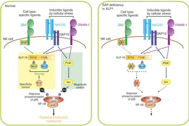
in XLP1 NK cell NK cell DAP10 DAP10 SAP SAP SLP-76 SLP-76 Specificity control Stepwise phosphorylation of p65 NF-κB Magnitude control Synergy Y113 Y113
Vav1 Vav1 Vav1 Vav1
Erk Erk S276 S536 p50 p65 PI3K PI3K Akt Akt 2nd 1st P P P P Y128 Y128 2B4 NKG2D 2B4 NKG2D Cytokine production cytotoxicity Stepwise phosphorylation of p65 NF-κB S276 S536 p50 p65 P DNAM-1 DNAM-1 Cell
type-specific ligands specific ligandsCell
type-Inducible ligands by cellular stress
Inducible ligands by cellular stress
Figure 8 | Proposed mechanism of NF-jB activation via coactivation receptors on NK cells. NF-kB activation in NK cells required the coordinated engagement of coactivation receptors, such as 2B4 and NKG2D or 2B4 and DNAM-1. This combination was required to provide complementary and independent signals leading to Vav1-dependent synergistic signalling involving PLC-g2 and Erk. Further, signals from synergizing receptors converged on NF-kB p65 subunit through selective phosphorylation of p65 serine residues, particularly at serine 276 via Vav1-Erk and at serine 536 via PI3K-Akt pathway, which was crucial to optimal activation of NF-kB. The requisite PI3K-Akt signal was primarily mediated by the engagement of NKG2D or DNAM-1, which recognizes ligands induced by cellular stress. Vav1 controlled downstream p65 phosphorylation and NF-kB activation, suggesting that distinct signalling checkpoints at the level of Vav1 and p65 regulate NF-kB activation. In support, Vav1-dependent synergistic signalling was required for the phosphorylation of p65 at serine 536 by Akt pathway, which was evident in SAP-deficient XLP1 NK cells following coactivation, which exhibited impaired p65